| 657 |
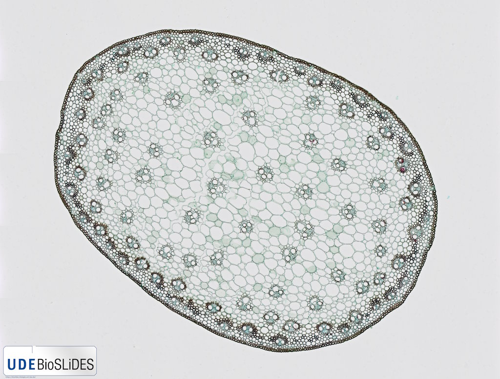
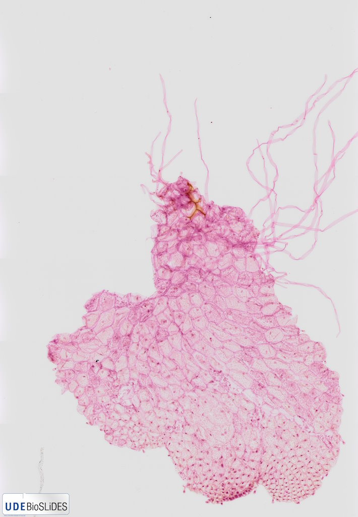
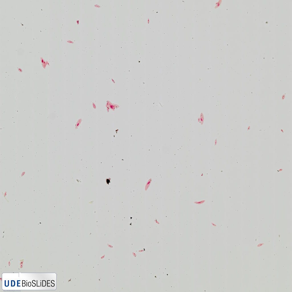
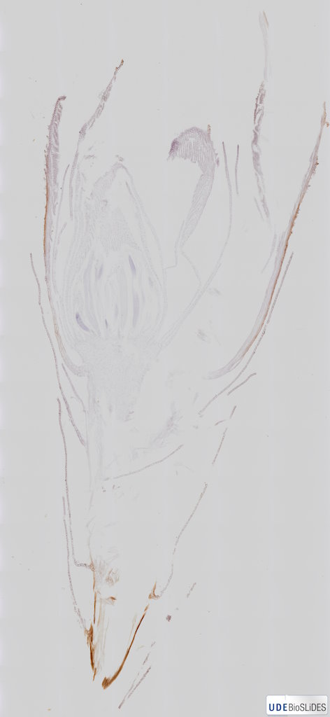
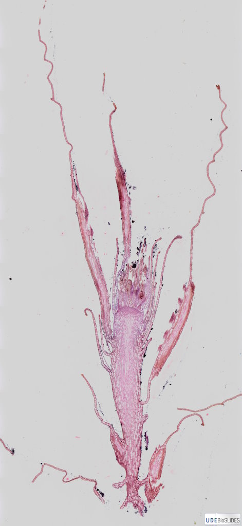
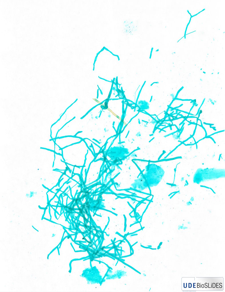
Abies alba - Needle leaves cross section 20x |
 |
Plant |
Abies alba |
Silver fir |
Needle leaf |
Cross section |
|
|
unknown |
unknown |
unknown |
Jungner/microthek |
2025-07-17 19:27:31 |
Olympus VS200 |
UPLXAPO 20x / 0.80 |
20x / 0.80 |
75 % |
|
Throughlight widefield |
iDS VS-264C |
Slide scanning microscope |
19.3 |
12,710 x
15,051 x
101 px³ |
3,471.2 x
4,110.4 x
66.0 µm³ |
✅
|
✅
|
✅
|
❌ |
| 658 |
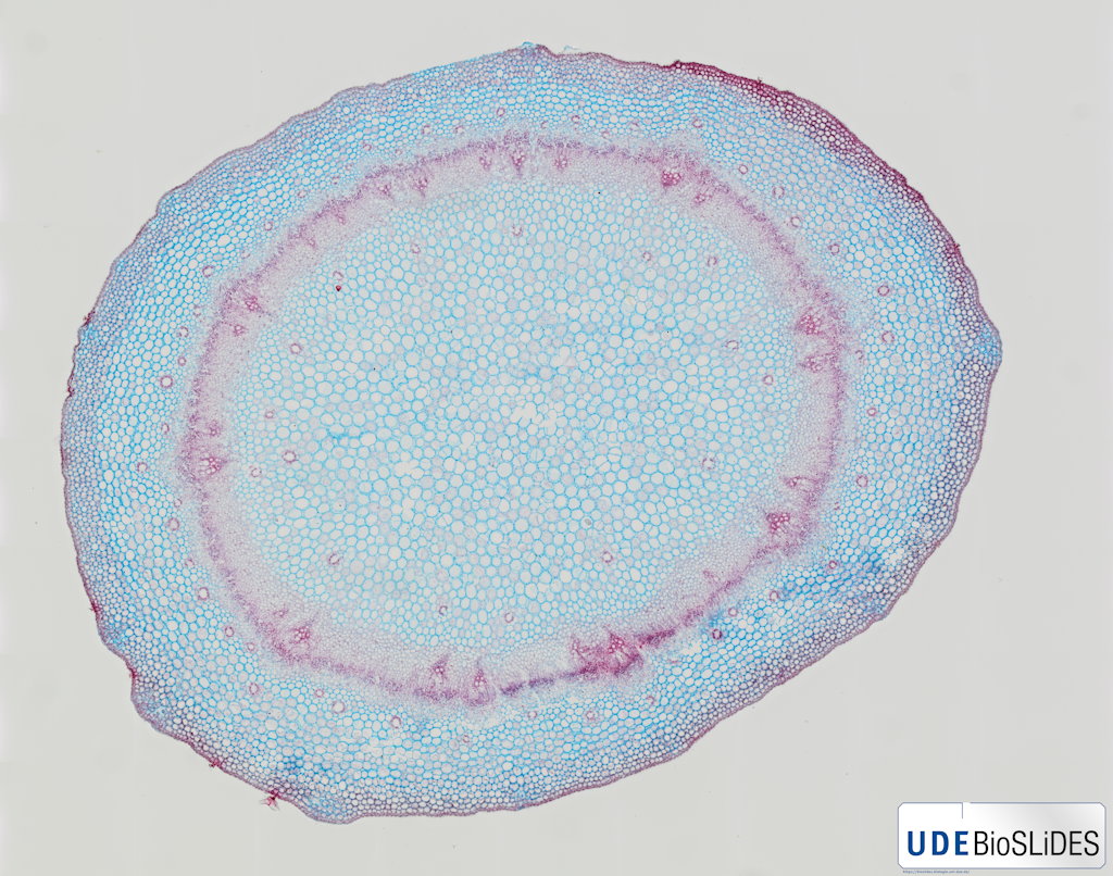
Abies alba - one-year-old branch cross section 20x |
 |
Plant |
Abies alba |
Silver fir |
Branch |
Cross section |
|
|
unknown |
unknown |
unknown |
Jungner/microthek |
2025-07-17 20:42:38 |
Olympus VS200 |
UPLXAPO 20x / 0.80 |
20x / 0.80 |
75 % |
|
Throughlight widefield |
iDS VS-264C |
Slide scanning microscope |
37.3 |
17,878 x
20,638 x
101 px³ |
4,882.5 x
5,636.4 x
100.0 µm³ |
✅
|
✅
|
✅
|
❌ |
| 659 |
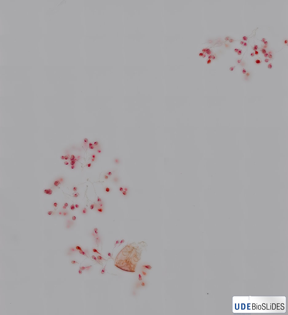
Abies alba - Ovule longitudinal section 20x |
 |
Plant |
Abies alba |
Silver fir |
Ovule |
Longitudinal section |
Wikipedia on Abies alba.
This sample has some minor damage caused by shrinking of the mounting medium due to ageing.
Jungner/microthek micro preparation no. 1208 |
|
unknown |
unknown |
unknown |
Jungner/microthek |
2025-07-17 21:35:40 |
Olympus VS200 |
UPLXAPO 20x / 0.80 |
20x / 0.80 |
75 % |
|
Throughlight widefield |
iDS VS-264C |
Slide scanning microscope |
2.6 |
8,476 x
7,222 x
43 px³ |
2,314.8 x
1,972.2 x
24.8 µm³ |
✅
|
✅
|
✅
|
❌ |
| 630 |
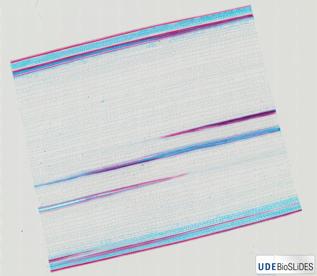
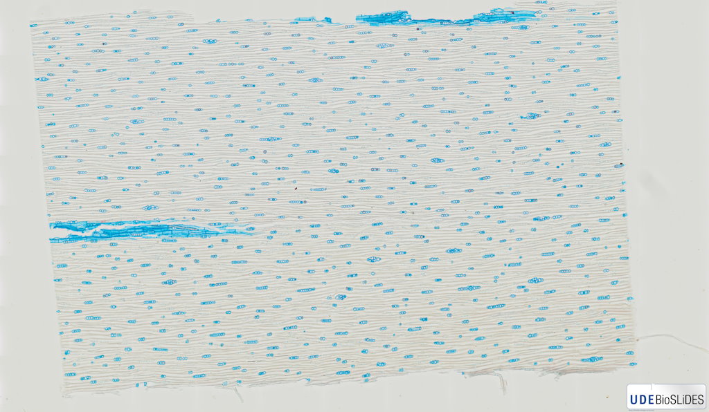
Abies alba - Wood cross section 20x |
 |
Plant |
Abies alba |
Silver fir |
Wood |
Cross section |
Wikipedia on Abies alba.
This sample has some damages (cracks and air bubbles in the mounting medium) due to aging.
Jungner/microthek micro preparation no. 766 |
Annual ring boundary, Bordered pit, Early wood, Fenestriform pit (upper and underside), Late wood, Wood ray parenchyma |
unknown |
unstained |
unknown |
Jungner/microthek |
2025-07-15 16:22:57 |
Olympus VS200 |
UPLXAPO 20x / 0.80 |
20x / 0.80 |
75 % |
|
Throughlight widefield |
iDS VS-264C |
Slide scanning microscope |
42.0 |
19,940 x
20,868 x
101 px³ |
5,445.7 x
5,699.0 x
59.0 µm³ |
✅
|
✅
|
✅
|
✅
|
| 308 |
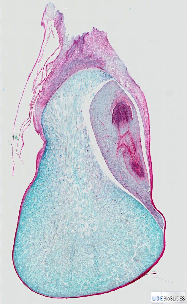
Abies sp. - Shoot apex longitudinal section 20x |
 |
Plant |
Abies sp. |
Fir |
Shoot apex |
Longitudinal section |
|
|
unknown |
|
unknown |
Karl Brügmann |
2023-09-28 13:29:42 |
Olympus VS200 |
UPLXAPO 20x / 0.80 |
20x / 0.80 |
75 % |
|
Throughlight widefield |
iDS VS-264C |
Slide scanning microscope |
5.7 |
7,868 x
19,520 x
37 px³ |
2,148.8 x
5,331.3 x
21.2 µm³ |
✅
|
✅
|
✅
|
❌ |
| 2 |
Acer platanoides - Branch cross section 20x |
 |
Plant |
Acer platanoides |
Norway maple |
Branch |
Cross section |
|
bark parenchyma, cambium, collenchyma, cork, epidermis & cuticle, lenticel, medullary parenchyma, primary xylem, sclerenchyma, sec. phloem/ bast, sec. xylem / wood, xylem vessel or tracheae |
Formol, acetic alcohol |
|
Euparal |
Rainer Teubner |
2021-07-01 14:51:37 |
Olympus VS200 |
UPLXAPO 20x / 0.80 |
20x / 0.80 |
40 % |
|
Throughlight widefield |
iDS VS-264C |
Slide scanning microscope |
23.2 |
19,267 x
19,720 x
61 px³ |
5,259.4 x
5,383.2 x
70.8 µm³ |
✅
|
✅
|
✅
|
✅
|
| 684 |
Acer platanoides - Leaf stalk cross section 20x |
 |
Plant |
Acer platanoides |
Norway maple |
Leaf stalk |
Cross section |
|
|
unknown |
unknown |
unknown |
Jungner/microthek |
2025-08-11 19:06:43 |
Olympus VS200 |
UPLXAPO 20x / 0.80 |
20x / 0.80 |
75 % |
|
Throughlight widefield |
iDS VS-264C |
Slide scanning microscope |
10.5 |
10,338 x
10,080 x
101 px³ |
2,823.4 x
2,752.9 x
59.0 µm³ |
✅
|
✅
|
✅
|
❌ |
| 1 |
Acer platanoides - Leaf stalk cross section 40x |
 |
Plant |
Acer platanoides |
Norway maple |
Leaf stalk |
Cross section |
|
bark parenchyma, cambium, collenchyma, epidermis & cuticle, medullary parenchyma, open collateral vascular bundle, phloem, sclerenchyma, secretory duct, xylem, xylem parenchyma, xylem vessel |
Formol, acetic alcohol |
W3Asim I: Acridine red, acriflavine, astra blue |
Euparal |
Jörg Weiß
www.mikroskopie-bonn.de |
2021-07-27 13:22:49 |
Olympus VS200 |
UPLXAPO O 40x / 1.40 |
40x / 1.40 |
50 % |
Oil |
Throughlight widefield |
iDS VS-264C |
Slide scanning microscope |
30.3 |
22,653 x
21,913 x
61 px³ |
3,116.6 x
3,014.8 x
35.4 µm³ |
✅
|
✅
|
✅
|
✅
|
| 609 |
Acer platanoides - Wood cross section 20x |
 |
Plant |
Acer platanoides |
Norway maple |
Wood |
Cross section |
Wikipedia on Acer platanoides.
This sample was repaired after shrinking of the mounting medium due to aging, and thus has some minor damage.
Jungner/microthek micro preparation no. 746 |
|
unknown |
unstained |
unknown |
Jungner/microthek |
2025-07-03 21:23:51 |
Olympus VS200 |
UPLXAPO 20x / 0.80 |
20x / 0.80 |
75 % |
|
Throughlight widefield |
iDS VS-264C |
Slide scanning microscope |
45.5 |
17,141 x
26,306 x
101 px³ |
4,681.4 x
7,184.3 x
59.0 µm³ |
✅
|
✅
|
✅
|
❌ |
| 672 |
Acer pseudoplatanus - Fruit cross section 20x |
 |
Plant |
Acer pseudoplatanus |
Sycamore |
Fruit |
Cross section |
Wikipedia on Acer pseudoplatanus.
This sample has some damage (partly large bubbles) caused by shrinking of the mounting medium due to ageing.
Jungner/microthek micro preparation no. 1245 |
|
unknown |
unknown |
unknown |
Jungner/microthek |
2025-08-13 16:31:15 |
Olympus VS200 |
UPLXAPO 20x / 0.80 |
20x / 0.80 |
75 % |
|
Throughlight widefield |
iDS VS-264C |
Slide scanning microscope |
7.4 |
10,089 x
16,196 x
45 px³ |
2,755.4 x
4,423.2 x
26.0 µm³ |
✅
|
✅
|
✅
|
❌ |
| 4 |
Acer pseudoplatanus - Leaf cross section 20x |
 |
Plant |
Acer pseudoplatanus |
Sycamore |
Leaf |
Cross section |
|
bark parenchyma, collenchyma, epidermis , guard cells, lower epidermis, medullary parenchyma, palisade parenchyma, phloem, sclerenchyma, secretory duct, sponge parenchyma, upper epidermis, xylem, xylem vessel (tracheae) |
Formol, acetic alcohol |
|
Euparal |
Jörg Weiß
www.mikroskopie-bonn.de |
2021-08-17 11:39:37 |
Olympus VS200 |
UPLXAPO 20x / 0.80 |
20x / 0.80 |
50 % |
|
Throughlight widefield |
iDS VS-264C |
Slide scanning microscope |
13.9 |
34,281 x
6,661 x
61 px³ |
9,358.0 x
1,818.2 x
70.8 µm³ |
✅
|
✅
|
✅
|
✅
|
| 3 |
Acer pseudoplatanus - Samara cross section 20x |
 |
Plant |
Acer pseudoplatanus |
Sycamore |
Fruit |
Cross section |
|
collenchyma, epidermis & cuticle, parenchyma cells, phloem, sclerenchmatic tape, sclerenchyma, sclerenchymatous medullary parenchyma, sponge parenchyma, vascular bundle, xylem |
Formol, acetic alcohol |
|
Euparal |
Jörg Weiß
www.mikroskopie-bonn.de |
2021-07-27 14:29:53 |
Olympus VS200 |
UPLXAPO 20x / 0.80 |
20x / 0.80 |
50 % |
|
Throughlight widefield |
iDS VS-264C |
Slide scanning microscope |
12.9 |
37,949 x
5,563 x
61 px³ |
10,359.3 x
1,518.4 x
70.8 µm³ |
✅
|
✅
|
✅
|
✅
|
| 349 |
Actinia equina - Pharynx cross section 40x |
 |
Animal |
Actinia equina |
Beadlet anemone |
Pharynx |
Cross section |
|
|
unknown |
unknown |
unknown |
Karl Brügmann |
2023-12-01 08:56:43 |
Olympus VS200 |
UPLXAPO O 40x / 1.40 |
40x / 1.40 |
75 % |
Oil |
Throughlight widefield |
iDS VS-264C |
Slide scanning microscope |
54.9 |
32,652 x
27,553 x
61 px³ |
4,474.0 x
3,775.4 x
17.4 µm³ |
✅
|
✅
|
✅
|
❌ |
| 601 |
Adiantum sp. - Rhizome cross section 20x |
 |
Plant |
Adiantum sp. |
Maidenhair fern |
Rhizome |
Cross section |
|
|
unknown |
unknown |
unknown |
Jungner/microthek |
2025-07-02 09:28:01 |
Olympus VS200 |
UPLXAPO 20x / 0.80 |
20x / 0.80 |
75 % |
|
Throughlight widefield |
iDS VS-264C |
Slide scanning microscope |
33.8 |
18,296 x
18,308 x
101 px³ |
4,996.7 x
4,999.9 x
100.0 µm³ |
✅
|
✅
|
✅
|
❌ |
| 688 |
Aesculus hippocastanum - Bud cross section 20x |
 |
Plant |
Aesculus hippocastanum |
Horse chestnut |
Bud |
Cross section |
|
|
unknown |
unknown |
unknown |
Jungner/microthek |
2025-08-13 09:47:21 |
Olympus VS200 |
UPLXAPO 20x / 0.80 |
20x / 0.80 |
75 % |
|
Throughlight widefield |
iDS VS-264C |
Slide scanning microscope |
67.0 |
29,581 x
38,408 x
59 px³ |
8,078.8 x
10,489.5 x
34.2 µm³ |
✅
|
✅
|
✅
|
❌ |
| 627 |
Aesculus hippocastanum - Wood cross section 20x |
 |
Plant |
Aesculus hippocastanum |
Horse chestnut |
Wood |
Cross section |
Wikipedia on Aesculus hippocastanum.
This sample was repaired after shrinking of the mounting medium due to aging, and thus has some minor damage.
Jungner/microthek micro preparation no. 763 |
|
unknown |
unstained |
unknown |
Jungner/microthek |
2025-07-14 20:28:50 |
Olympus VS200 |
UPLXAPO 20x / 0.80 |
20x / 0.80 |
75 % |
|
Throughlight widefield |
iDS VS-264C |
Slide scanning microscope |
56.5 |
24,155 x
23,168 x
101 px³ |
6,596.9 x
6,327.4 x
59.0 µm³ |
✅
|
✅
|
✅
|
❌ |
| 683 |
Aesculus sp. - Leaf stalk cross section 20x |
 |
Plant |
Aesculus sp. |
Chestnut |
Leaf stalk |
Cross section |
Wikipedia on Aesculus sp.
This sample has some damage (partly large bubbles) caused by shrinking of the mounting medium due to ageing.
Jungner/microthek micro preparation no. 1233 |
|
unknown |
unknown |
unknown |
Jungner/microthek |
2025-08-11 16:41:37 |
Olympus VS200 |
UPLXAPO 20x / 0.80 |
20x / 0.80 |
75 % |
|
Throughlight widefield |
iDS VS-264C |
Slide scanning microscope |
16.1 |
18,884 x
17,421 x
49 px³ |
5,157.2 x
4,757.8 x
28.3 µm³ |
✅
|
✅
|
✅
|
❌ |
| 575 |
Agaricus campestris - Fruiting body cross section 20x |
 |
Fungus |
Agaricus campestris |
Meadow mushroom |
Fruiting body |
Cross section |
|
|
unknown |
unknown |
unknown |
Jungner/microthek |
2025-06-24 09:19:48 |
Olympus VS200 |
UPLXAPO 20x / 0.80 |
20x / 0.80 |
75 % |
|
Throughlight widefield |
iDS VS-264C |
Slide scanning microscope |
99.5 |
45,485 x
26,366 x
83 px³ |
12,422.5 x
7,200.6 x
48.4 µm³ |
✅
|
✅
|
✅
|
❌ |
| 5 |
Akebia quinata - Young sprout cross section 20x |
 |
Plant |
Akebia quinata |
Chocolate vine |
Sprout |
Cross section |
|
amyloplasts, bark parenchyma, cambium, companion cells, epidermis & cuticle, medullary parenchyma, open collateral vascular bundle, phloem, primary xylem, sclerenchyma, sive tubes, tracheae, xylem |
Formol, acetic alcohol |
|
Euparal |
Jörg Weiß
www.mikroskopie-bonn.de |
2021-07-27 15:15:46 |
Olympus VS200 |
UPLXAPO 20x / 0.80 |
20x / 0.80 |
50 % |
|
Throughlight widefield |
iDS VS-264C |
Slide scanning microscope |
10.7 |
13,614 x
12,878 x
61 px³ |
3,716.3 x
3,515.4 x
70.8 µm³ |
✅
|
✅
|
✅
|
✅
|
| 569 |
Albugo candida - Conidia cross section 20x |
 |
Oomycete |
Albugo candida |
White rust |
Conidia |
Cross section |
|
|
unknown |
unknown |
unknown |
Jungner/microthek |
2025-06-23 16:42:54 |
Olympus VS200 |
UPLXAPO 20x / 0.80 |
20x / 0.80 |
75 % |
|
Throughlight widefield |
iDS VS-264C |
Slide scanning microscope |
8.3 |
25,659 x
6,335 x
51 px³ |
7,007.6 x
1,730.1 x
29.5 µm³ |
✅
|
✅
|
✅
|
❌ |
| 208 |
Allium cepa - Epidermis 20x |
 |
Plant |
Allium cepa |
Common onion |
Skin of the bulb |
Peel |
|
|
not fixated |
unstained |
Water |
Christiane Wittmann |
2020-10-27 14:32:00 |
Olympus VS200 |
UPLXAPO 20x / 0.80 |
20x / 0.80 |
75 % |
|
Throughlight widefield |
iDS VS-264C |
Slide scanning microscope |
33.3 |
22,169 x
24,596 x
61 px³ |
6,051.6 x
6,714.2 x
70.8 µm³ |
✅
|
✅
|
✅
|
❌ |
| 206 |
Allium cepa - Epidermis red 20x 1 |
 |
Plant |
Allium cepa |
Common onion |
Skin of the bulb |
Peel |
|
|
not fixated |
unstained |
Water |
Christiane Wittmann |
2020-10-27 14:25:38 |
Olympus VS200 |
UPLXAPO 20x / 0.80 |
20x / 0.80 |
75 % |
|
Throughlight widefield |
iDS VS-264C |
Slide scanning microscope |
22.9 |
18,491 x
20,344 x
61 px³ |
5,047.6 x
5,553.5 x
70.8 µm³ |
✅
|
✅
|
✅
|
❌ |
| 207 |
Allium cepa - Epidermis red 20x 2 |
 |
Plant |
Allium cepa |
Common onion |
Skin of the bulb |
Peel |
|
|
not fixated |
unstained |
Water |
Christiane Wittmann |
2020-10-05 13:42:11 |
Olympus VS200 |
UPLXAPO 20x / 0.80 |
20x / 0.80 |
75 % |
|
Throughlight widefield |
iDS VS-264C |
Slide scanning microscope |
36.8 |
21,726 x
27,730 x
61 px³ |
5,930.6 x
7,569.7 x
70.8 µm³ |
✅
|
✅
|
✅
|
❌ |
| 430 |
Allium cepa - Root tip mitosis 40x |
 |
Plant |
Allium cepa |
Common onion |
Root |
Longitudinal section |
|
Anaphase, Anaphase/telophase, Metaphase, Prometaphase, Prophase, Telophase |
unknown |
unknown |
unknown |
Bresser |
2025-04-09 13:33:12 |
Olympus VS200 |
UPLXAPO O 40x / 1.40 |
40x / 1.40 |
75 % |
Oil |
Throughlight widefield |
iDS VS-264C |
Slide scanning microscope |
39.5 |
26,461 x
18,916 x
79 px³ |
3,625.7 x
2,591.9 x
22.6 µm³ |
✅
|
✅
|
✅
|
✅
|
| 233 |
Allium cepa - Root tip mitosis 60x |
 |
Plant |
Allium cepa |
Common onion |
Root |
Squash technique |
|
|
unknown |
unknown |
unknown |
Christian Johannes |
2023-06-16 15:55:17 |
Olympus VS200 |
UPLXAPO OPH 60x / 1.42 |
60x / 1.42 |
75 % |
Oil |
Throughlight widefield |
iDS VS-264C |
Slide scanning microscope |
35.9 |
32,990 x
32,993 x
33 px³ |
2,999.9 x
3,000.2 x
9.0 µm³ |
✅
|
✅
|
✅
|
❌ |
| 478 |
Allium schoenoprasum - Round leaf cross section 20x |
 |
Plant |
Allium schoenoprasum |
Chives |
Leaf |
Cross section |
|
|
unknown |
unknown |
unknown |
Jungner/microthek |
2025-05-06 10:03:38 |
Olympus VS200 |
UPLXAPO 20x / 0.80 |
20x / 0.80 |
75 % |
|
Throughlight widefield |
iDS VS-264C |
Slide scanning microscope |
9.0 |
16,591 x
11,105 x
49 px³ |
4,531.2 x
3,032.8 x
28.3 µm³ |
✅
|
✅
|
✅
|
❌ |
| 680 |
Alnus sp. - Root tubercles cross section 20x |
 |
Plant |
Alnus sp. |
Alder |
Root tubercles |
Cross section |
Wikipedia on Alnus sp.
This sample has some damage (partly large bubbles) caused by shrinking of the mounting medium due to ageing.
Jungner/microthek micro preparation no. 1228 |
Cells with rhizobia, Central cylinder, Endodermis, Pericycle, Rhizodermis, Root parenchyma, Vascular tissue |
unknown |
unknown |
unknown |
Jungner/microthek |
2025-08-11 14:23:11 |
Olympus VS200 |
UPLXAPO 20x / 0.80 |
20x / 0.80 |
75 % |
|
Throughlight widefield |
iDS VS-264C |
Slide scanning microscope |
22.6 |
22,701 x
23,119 x
43 px³ |
6,199.7 x
6,313.8 x
24.8 µm³ |
✅
|
✅
|
✅
|
✅
|
| 410 |
Anabaena sp. - 40x |
 |
Cyanobacterium |
Anabaena sp. |
Anabaena |
Whole organism |
Whole mount |
Anabaena sp. is a cyanobacterium that builds un-branched filaments of concatenated cells and indicates eutrophic waters. Wikipedia on Anabaena sp.
Jungner/microthek micro preparation no. 1446 |
|
unknown |
unknown |
unknown |
Jungner/microthek |
2025-04-01 14:51:28 |
Olympus VS200 |
UPLFLN 40x / 0.75 |
40x / 0.75 |
75 % |
|
Throughlight widefield |
iDS VS-264C |
Slide scanning microscope |
17.4 |
14,908 x
11,550 x
101 px³ |
2,039.8 x
1,580.3 x
67.0 µm³ |
✅
|
✅
|
✅
|
❌ |
| 247 |
Ananas sativa - Pulp cross section 20x |
 |
Plant |
Ananas sativa |
Pineapple |
Fruit |
Cross section |
|
|
unknown |
unknown |
Eukitt |
Jungner/microthek |
2023-09-14 15:52:44 |
Olympus VS200 |
UPLXAPO 20x / 0.80 |
20x / 0.80 |
75 % |
|
Throughlight widefield |
iDS VS-264C |
Slide scanning microscope |
22.9 |
43,516 x
14,240 x
37 px³ |
11,885.3 x
3,889.3 x
21.2 µm³ |
✅
|
✅
|
✅
|
❌ |
| 364 |
Ankylostoma sp. - Body cross section 40x |
 |
Animal |
Ankylostoma sp |
Hookworm |
Whole animal |
Whole mount |
Male and female, Wikipedia on Ankylostoma sp.
The mounting medium on the right hand side has shrunken and become brittle due to ageing.
Jungner/microthek micro preparation no. 464 |
Parasite |
unknown |
unknown |
unknown |
Jungner/microthek |
2023-12-08 13:52:53 |
Olympus VS200 |
UPLXAPO O 40x / 1.40 |
40x / 1.40 |
75 % |
Oil |
Throughlight widefield |
iDS VS-264C |
Slide scanning microscope |
17.3 |
25,963 x
6,611 x
101 px³ |
3,557.5 x
905.8 x
29.0 µm³ |
✅
|
✅
|
✅
|
❌ |
| 516 |
Anthurium sp. - Inflorescence cross section 20x |
 |
Plant |
Anthurium sp. |
Anthurium |
Inflorescence |
Cross section |
|
|
unknown |
unknown |
unknown |
Jungner/microthek |
2025-05-14 20:49:14 |
Olympus VS200 |
UPLXAPO 20x / 0.80 |
20x / 0.80 |
75 % |
|
Throughlight widefield |
iDS VS-264C |
Slide scanning microscope |
133.3 |
41,685 x
31,660 x
101 px³ |
11,385.2 x
8,647.0 x
100.0 µm³ |
✅
|
✅
|
✅
|
❌ |
| 556 |
Aphanizomenon sp. - Whole 20x |
 |
Cyanobacterium |
Aphanizomenon sp. |
Aphanizomenon |
Whole organism |
Whole mount |
|
|
unknown |
unknown |
unknown |
Jungner/microthek |
2025-05-30 11:01:54 |
Olympus VS200 |
UPLXAPO 20x / 0.80 |
20x / 0.80 |
75 % |
|
Throughlight widefield |
iDS VS-264C |
Slide scanning microscope |
20.4 |
18,304 x
18,295 x
61 px³ |
4,999.0 x
4,996.7 x
35.4 µm³ |
✅
|
✅
|
✅
|
❌ |
| 650 |
Aralia sp. - Leaf cross section 20x |
 |
Plant |
Aralia sp. |
Spikenard |
Leaf |
Cross section |
Wikipedia on Aralia sp.
This sample has some damage (partly large bubbles) caused by shrinking of the mounting medium due to ageing.
Jungner/microthek micro preparation no. 1290 |
|
unknown |
unknown |
unknown |
Jungner/microthek |
2025-07-17 09:18:37 |
Olympus VS200 |
UPLXAPO 20x / 0.80 |
20x / 0.80 |
75 % |
|
Throughlight widefield |
iDS VS-264C |
Slide scanning microscope |
10.1 |
11,017 x
20,290 x
45 px³ |
3,008.7 x
5,541.2 x
26.0 µm³ |
✅
|
✅
|
✅
|
❌ |
| 101 |
Araucaria heterophylla - Sprout cross section 20x 1 |
 |
Plant |
Araucaria heterophylla |
Norfolk Island pine |
Sprout |
Cross section |
|
assimilation parenchyma, bark parenchyma, bast, cambium, Diatom (contaminant), Diatoms (contaminant), epidermis & cuticle, guard cells, medullary parenchyma, medullary ray, primary xylem, resin channel, wood |
Formol, acetic alcohol |
|
Euparal |
Rainer Teubner |
2021-10-28 16:22:12 |
Olympus VS200 |
UPLXAPO 20x / 0.80 |
20x / 0.80 |
50 % |
|
Throughlight widefield |
iDS VS-264C |
Slide scanning microscope |
9.5 |
9,916 x
11,276 x
85 px³ |
2,706.7 x
3,078.1 x
99.1 µm³ |
✅
|
✅
|
✅
|
✅
|
| 102 |
Araucaria heterophylla - Sprout cross section 20x 2 |
 |
Plant |
Araucaria heterophylla |
Norfolk Island pine |
Sprout |
Cross section |
|
assimilation parenchyma, bark parenchyma, bast, cambium, epidermis & cuticle, medullary parenchyma, medullary ray, resin channel, wood |
Formol, acetic alcohol |
|
Euparal |
Rainer Teubner |
2021-10-28 16:36:32 |
Olympus VS200 |
UPLXAPO 20x / 0.80 |
20x / 0.80 |
50 % |
|
Throughlight widefield |
iDS VS-264C |
Slide scanning microscope |
10.5 |
10,480 x
11,820 x
85 px³ |
2,860.8 x
3,226.6 x
99.1 µm³ |
✅
|
✅
|
✅
|
✅
|
| 103 |
Araucaria heterophylla - Sprout cross section 20x 3 |
 |
Plant |
Araucaria heterophylla |
Norfolk Island pine |
Sprout |
Cross section |
|
assimilation parenchyma, bark parenchyma, bast, cambium, epidermis & cuticle, hypodermis, medullary parenchyma, primary xylem, resin channel, wood |
Formol, acetic alcohol |
|
Euparal |
Rainer Teubner |
2021-10-28 16:49:42 |
Olympus VS200 |
UPLXAPO 20x / 0.80 |
20x / 0.80 |
50 % |
|
Throughlight widefield |
iDS VS-264C |
Slide scanning microscope |
13.9 |
13,191 x
12,415 x
85 px³ |
3,600.7 x
3,388.9 x
99.1 µm³ |
✅
|
✅
|
✅
|
✅
|
| 104 |
Araucaria heterophylla - Sprout cross section 20x 4 |
 |
Plant |
Araucaria heterophylla |
Norfolk Island pine |
Sprout |
Cross section |
|
assimilation parenchyma, bark parenchyma, bast, cambium, cork, epidermis & cuticle, hypodermis, medullary parenchyma, medullary ray, primary xylem, resin channel, wood |
Formol, acetic alcohol |
|
Euparal |
Rainer Teubner |
2021-10-28 17:04:01 |
Olympus VS200 |
UPLXAPO 20x / 0.80 |
20x / 0.80 |
50 % |
|
Throughlight widefield |
iDS VS-264C |
Slide scanning microscope |
15.3 |
13,332 x
13,483 x
85 px³ |
3,639.2 x
3,680.5 x
99.1 µm³ |
✅
|
✅
|
✅
|
✅
|
| 507 |
Arctium lappa - Stalk cross section 20x |
 |
Plant |
Arctium lappa |
Greater burdock |
Stalk |
Cross section |
|
|
unknown |
unknown |
unknown |
Jungner/microthek |
2025-05-14 09:43:10 |
Olympus VS200 |
UPLXAPO 20x / 0.80 |
20x / 0.80 |
75 % |
|
Throughlight widefield |
iDS VS-264C |
Slide scanning microscope |
134.6 |
39,268 x
33,930 x
101 px³ |
10,724.9 x
9,267.1 x
59.0 µm³ |
✅
|
✅
|
✅
|
❌ |
| 19 |
Aristolochia macrophylla - Sprout cross section 20x |
 |
Plant |
Aristolochia macrophylla |
Dutchman's pipe |
Sprout |
Cross section |
|
bark parenchyma, bast, cambium, cork, epidermis & cuticle, medullary parenchyma, medullary ray, sclerenchyma, stone cell, wood, xylem vessel or tracheae |
Formol, acetic alcohol |
|
Euparal |
Rainer Teubner |
2021-08-17 09:40:15 |
Olympus VS200 |
UPLXAPO 20x / 0.80 |
20x / 0.80 |
50 % |
|
Throughlight widefield |
iDS VS-264C |
Slide scanning microscope |
75.2 |
34,473 x
35,752 x
61 px³ |
9,410.3 x
9,759.6 x
70.8 µm³ |
✅
|
✅
|
✅
|
✅
|
| 17 |
Aristolochia macrophylla - Sprout cross section 20x 1 |
 |
Plant |
Aristolochia macrophylla |
Dutchman's pipe |
Sprout |
Cross section |
|
bark parenchyma, bast, cambium, cork, epidermis & cuticle, medullary parenchyma, medullary ray, sclerenchyma, stone cell, wood, xylem vessel or tracheae |
Formol, acetic alcohol |
unstained |
Euparal |
Rainer Teubner |
2021-09-23 09:00:54 |
Olympus VS200 |
UPLXAPO 20x / 0.80 |
20x / 0.80 |
50 % |
|
Throughlight widefield |
iDS VS-264C |
Slide scanning microscope |
65.6 |
30,110 x
35,722 x
61 px³ |
8,219.2 x
9,751.3 x
99.6 µm³ |
✅
|
✅
|
✅
|
✅
|
| 18 |
Aristolochia macrophylla - Sprout cross section 20x 2 |
 |
Plant |
Aristolochia macrophylla |
Dutchman's pipe |
Sprout |
Cross section |
|
bark parenchyma, bast, cambium, cork, epidermis & cuticle, medullary parenchyma, medullary ray, ring of sclerenchyma, sclerenchyma, wood, xylem vessel or tracheae |
Formol, acetic alcohol |
|
Euparal |
Rainer Teubner |
2021-09-22 15:56:14 |
Olympus VS200 |
UPLXAPO 20x / 0.80 |
20x / 0.80 |
50 % |
|
Throughlight widefield |
iDS VS-264C |
Slide scanning microscope |
66.9 |
32,044 x
34,251 x
61 px³ |
8,747.4 x
9,349.7 x
99.6 µm³ |
✅
|
✅
|
✅
|
✅
|
| 20 |
Aristolochia macrophylla - Sprout cross section 20x 3 |
 |
Plant |
Aristolochia macrophylla |
Dutchman's pipe |
Sprout |
Cross section |
|
bark parenchyma, bast, cambium, collenchyma, epidermis & cuticle, medullary parenchyma, sclerenchyma, xylem, xylem vessel or tracheae |
Formol, acetic alcohol |
|
Euparal |
Rainer Teubner |
2021-09-24 10:07:28 |
Olympus VS200 |
UPLXAPO 20x / 0.80 |
20x / 0.80 |
50 % |
|
Throughlight widefield |
iDS VS-264C |
Slide scanning microscope |
5.9 |
9,775 x
9,946 x
61 px³ |
2,668.2 x
2,715.0 x
78.0 µm³ |
✅
|
✅
|
✅
|
✅
|
| 21 |
Aristolochia macrophylla - Sprout cross section 20x 4 |
 |
Plant |
Aristolochia macrophylla |
Dutchman's pipe |
Sprout |
Cross section |
|
bark parenchyma, bast, cambium, collenchyma, crystal druse, epidermis & cuticle, medullary parenchyma, sclerenchyma, xylem, xylem vessel or tracheae |
Formol, acetic alcohol, sodium hypochlorite |
|
Euparal |
Rainer Teubner |
2021-09-24 10:53:14 |
Olympus VS200 |
UPLXAPO 20x / 0.80 |
20x / 0.80 |
50 % |
|
Throughlight widefield |
iDS VS-264C |
Slide scanning microscope |
6.3 |
10,138 x
10,138 x
61 px³ |
2,767.2 x
2,767.2 x
96.0 µm³ |
✅
|
✅
|
✅
|
✅
|
| 22 |
Aristolochia macrophylla - Sprout cross section 20x 5 |
 |
Plant |
Aristolochia macrophylla |
Dutchman's pipe |
Sprout |
Cross section |
|
bark parenchyma, crystal druse, epidermis & cuticle, guard cells, kollenchyma, sclerenchyma, xylem, xylem vessel or tracheae |
Formol, acetic alcohol |
unstained |
Euparal |
Rainer Teubner |
2021-09-24 11:03:51 |
Olympus VS200 |
UPLXAPO 20x / 0.80 |
20x / 0.80 |
50 % |
|
Throughlight widefield |
iDS VS-264C |
Slide scanning microscope |
6.1 |
9,845 x
10,127 x
61 px³ |
2,687.5 x
2,764.5 x
70.8 µm³ |
✅
|
✅
|
✅
|
✅
|
| 24 |
Aristolochia macrophylla - Stalk cross section 20x |
 |
Plant |
Aristolochia macrophylla |
Dutchman's pipe |
Stalk |
Cross section |
|
bark parenchyma, bast, cambium, collenchyma, epidermis & cuticle, medullary parenchyma, sclerenchyma, xylem, xylem vessel or tracheae |
Formol, acetic alcohol, sodium hypochlorite |
|
Euparal |
Rainer Teubner |
2021-09-24 09:45:19 |
Olympus VS200 |
UPLXAPO 20x / 0.80 |
20x / 0.80 |
50 % |
|
Throughlight widefield |
iDS VS-264C |
Slide scanning microscope |
6.4 |
10,319 x
10,138 x
61 px³ |
2,816.8 x
2,767.2 x
84.0 µm³ |
✅
|
✅
|
✅
|
✅
|
| 23 |
Aristolochia macrophylla - Stalk perennial cross section 20x |
 |
Plant |
Aristolochia macrophylla |
Dutchman's pipe |
Stalk |
Cross section |
|
bark parenchyma, bast, cambium, collenchyma, epidermis & cuticle, medullary parenchyma, medullary ray, sclerenchyma, wood, xylem vessel or tracheae |
Formol, acetic alcohol, sodium hypochlorite |
|
Euparal |
Rainer Teubner |
2021-09-23 11:50:38 |
Olympus VS200 |
UPLXAPO 20x / 0.80 |
20x / 0.80 |
50 % |
|
Throughlight widefield |
iDS VS-264C |
Slide scanning microscope |
21.6 |
18,189 x
19,458 x
61 px³ |
4,965.1 x
5,311.7 x
99.6 µm³ |
✅
|
✅
|
✅
|
✅
|
| 303 |
Aristolochia sp. - Stem one-year-old cross section 20x |
 |
Plant |
Aristolochia sp. |
Dutchman's pipe |
Stem |
Cross section |
|
|
unknown |
unknown |
unknown |
Jungner/microthek |
2023-10-19 09:39:23 |
Olympus VS200 |
UPLXAPO 20x / 0.80 |
20x / 0.80 |
75 % |
|
Throughlight widefield |
iDS VS-264C |
Slide scanning microscope |
24.2 |
15,616 x
15,367 x
101 px³ |
4,264.9 x
4,197.0 x
100.0 µm³ |
✅
|
✅
|
✅
|
❌ |
| 310 |
Aristolochia sp. - Stem perennial cross section 20x |
 |
Plant |
Aristolochia sp. |
Dutchman's pipe |
Stem |
Cross section |
|
|
unknown |
unknown |
unknown |
Jungner/microthek |
2023-10-20 14:00:19 |
Olympus VS200 |
UPLXAPO 20x / 0.80 |
20x / 0.80 |
40 % |
|
Throughlight widefield |
iDS VS-264C |
Slide scanning microscope |
71.9 |
29,267 x
24,320 x
101 px³ |
7,993.5 x
6,642.3 x
100.0 µm³ |
✅
|
✅
|
✅
|
❌ |
| 365 |
Ascaris lumbricoides - Body longitudinal section 20x 1 |
 |
Animal |
Ascaris lumbricoides |
Roundworm |
Body |
Longitudinal section |
Wikipedia on Ascaris lumbricoides.
The mounting medium on the bottom of the sample has shrunken due to ageing. |
Parasite |
unknown |
Cason |
unknown |
Karl Brügmann |
2023-12-11 09:35:22 |
Olympus VS200 |
UPLXAPO 20x / 0.80 |
20x / 0.80 |
75 % |
|
Throughlight widefield |
iDS VS-264C |
Slide scanning microscope |
49.0 |
25,425 x
52,076 x
37 px³ |
6,944.2 x
14,223.0 x
21.2 µm³ |
✅
|
✅
|
✅
|
❌ |
| 366 |
Ascaris lumbricoides - Body longitudinal section 20x 2 |
 |
Animal |
Ascaris lumbricoides |
Roundworm |
Body |
Longitudinal section |
Wikipedia on Ascaris lumbricoides.
The mounting medium on the bottom half of the sample has shrunken due to ageing. |
Parasite |
unknown |
Cason |
unknown |
Karl Brügmann |
2023-12-11 10:20:30 |
Olympus VS200 |
UPLXAPO 20x / 0.80 |
20x / 0.80 |
75 % |
|
Throughlight widefield |
iDS VS-264C |
Slide scanning microscope |
69.4 |
27,238 x
56,627 x
45 px³ |
7,439.2 x
15,466.1 x
26.0 µm³ |
✅
|
✅
|
✅
|
❌ |
| 367 |
Ascaris lumbricoides - Body longitudinal section 20x 3 |
 |
Animal |
Ascaris lumbricoides |
Roundworm |
Body |
Longitudinal section |
Wikipedia on Ascaris lumbricoides.
The mounting medium on the bottom half of the sample has shrunken due to ageing. |
Parasite |
unknown |
Cason |
unknown |
Karl Brügmann |
2023-12-11 13:21:59 |
Olympus VS200 |
UPLXAPO 20x / 0.80 |
20x / 0.80 |
75 % |
|
Throughlight widefield |
iDS VS-264C |
Slide scanning microscope |
21.3 |
19,579 x
21,362 x
51 px³ |
5,347.5 x
5,834.5 x
29.5 µm³ |
✅
|
✅
|
✅
|
❌ |
| 368 |
Ascaris lumbricoides - Body longitudinal section 20x 4 |
 |
Animal |
Ascaris lumbricoides |
Roundworm |
Body |
Longitudinal section |
|
Parasite |
unknown |
Cason |
unknown |
Karl Brügmann |
2023-12-11 13:58:22 |
Olympus VS200 |
UPLXAPO 20x / 0.80 |
20x / 0.80 |
75 % |
|
Throughlight widefield |
iDS VS-264C |
Slide scanning microscope |
67.9 |
18,922 x
51,976 x
69 px³ |
5,168.0 x
14,195.8 x
40.1 µm³ |
✅
|
✅
|
✅
|
❌ |
| 375 |
Ascaris lumbricoides - Body longitudinal section 40x 1 |
 |
Animal |
Ascaris lumbricoides |
Roundworm |
Body |
Longitudinal section |
|
Parasite |
unknown |
unknown |
unknown |
Karl Brügmann |
2023-12-13 10:51:39 |
Olympus VS200 |
UPLXAPO O 40x / 1.40 |
40x / 1.40 |
75 % |
Oil |
Throughlight widefield |
iDS VS-264C |
Slide scanning microscope |
381.1 |
53,974 x
96,733 x
73 px³ |
7,395.7 x
13,254.7 x
20.9 µm³ |
✅
|
✅
|
✅
|
❌ |
| 376 |
Ascaris lumbricoides - Body longitudinal section 40x 2 |
 |
Animal |
Ascaris lumbricoides |
Roundworm |
Body |
Longitudinal section |
Wikipedia on Ascaris lumbricoides.
Some dirt is visible in the upper area of the scan, probably some dust particle got into the immersion oil. |
Parasite |
unknown |
unknown |
unknown |
Karl Brügmann |
2023-12-13 16:55:28 |
Olympus VS200 |
UPLXAPO O 40x / 1.40 |
40x / 1.40 |
75 % |
Oil |
Throughlight widefield |
iDS VS-264C |
Slide scanning microscope |
352.6 |
37,717 x
92,565 x
101 px³ |
5,168.0 x
12,683.5 x
29.0 µm³ |
✅
|
✅
|
✅
|
❌ |
| 359 |
Ascaris sp. - Body cross section 40x |
 |
Animal |
Ascaris sp. |
Roundworm |
Body |
Cross section |
Wikipedia on Ascaris sp.
Jungner/microthek micro preparation no. 473 |
Parasite |
unknown |
unknown |
unknown |
Jungner/microthek |
2023-12-07 10:17:55 |
Olympus VS200 |
UPLXAPO O 40x / 1.40 |
40x / 1.40 |
75 % |
Oil |
Throughlight widefield |
iDS VS-264C |
Slide scanning microscope |
136.2 |
44,899 x
30,035 x
101 px³ |
6,152.2 x
4,115.4 x
50.0 µm³ |
✅
|
✅
|
✅
|
❌ |
| 515 |
Asparagus sp. - Sprout tip longitudinal section 20x |
 |
Plant |
Asparagus sp. |
Asparagus |
Sprout tip |
Longitudinal section |
|
|
unknown |
unknown |
unknown |
Jungner/microthek |
2025-05-16 09:34:13 |
Olympus VS200 |
UPLXAPO 20x / 0.80 |
20x / 0.80 |
75 % |
|
Throughlight widefield |
iDS VS-264C |
Slide scanning microscope |
50.2 |
37,373 x
22,020 x
61 px³ |
10,207.4 x
6,014.0 x
35.4 µm³ |
✅
|
✅
|
✅
|
❌ |
| 600 |
Asplenium sp. - Leaf cross section 20x |
 |
Plant |
Asplenium sp. |
Spleenwort |
Leaf |
Cross section |
|
Assimilation tissue of the frond, Indusium, Lower epidermis, Placenta, Sorus, Sporangia, Spores, Stoma, Upper epidermis, Vascular tissue |
unknown |
unknown |
unknown |
Jungner/microthek |
2025-07-01 15:56:55 |
Olympus VS200 |
UPLXAPO 20x / 0.80 |
20x / 0.80 |
75 % |
|
Throughlight widefield |
iDS VS-264C |
Slide scanning microscope |
11.5 |
31,556 x
9,811 x
37 px³ |
8,618.1 x
2,679.5 x
21.2 µm³ |
✅
|
✅
|
✅
|
✅
|
| 599 |
Asplenium sp. - Rhizome cross section 20x |
 |
Plant |
Asplenium sp. |
Spleenwort |
Rhizome |
Cross section |
|
|
unknown |
unknown |
unknown |
Jungner/microthek |
2025-07-01 12:41:57 |
Olympus VS200 |
UPLXAPO 20x / 0.80 |
20x / 0.80 |
75 % |
|
Throughlight widefield |
iDS VS-264C |
Slide scanning microscope |
7.0 |
10,468 x
9,423 x
71 px³ |
2,858.8 x
2,573.4 x
41.3 µm³ |
✅
|
✅
|
✅
|
❌ |
| 622 |
Aucoumea klaineana - Wood cross section 20x |
 |
Plant |
Aucoumea klaineana |
Angouma |
Wood |
Cross section |
|
|
unknown |
unstained |
unknown |
Jungner/microthek |
2025-07-14 15:38:37 |
Olympus VS200 |
UPLXAPO 20x / 0.80 |
20x / 0.80 |
75 % |
|
Throughlight widefield |
iDS VS-264C |
Slide scanning microscope |
53.6 |
25,717 x
20,638 x
101 px³ |
7,023.4 x
5,636.4 x
63.0 µm³ |
✅
|
✅
|
✅
|
❌ |
| 361 |
Bdellonyssus sp. - total 40x |
 |
Animal |
Bdellonyssus sp. |
Mite |
Whole animal |
Whole mount |
An ectoparasitic blood-sucking arachnid.
Jungner/microthek micro preparation no. 460 |
Parasite |
unknown |
unstained |
unknown |
Jungner/microthek |
2023-12-08 14:44:03 |
Olympus VS200 |
UPLXAPO O 40x / 1.40 |
40x / 1.40 |
50 % |
Oil |
Throughlight widefield |
iDS VS-264C |
Slide scanning microscope |
1.4 |
3,950 x
3,613 x
101 px³ |
541.2 x
495.0 x
100.0 µm³ |
✅
|
✅
|
✅
|
❌ |
| 209 |
Begonia rex - Leaf stalk 20x 2 |
 |
Plant |
Begonia rex |
Begonia |
Leaf stalk |
Cross section |
|
Collenchyma |
Alcohol |
Astra blue, safranin |
Water |
Christiane Wittmann |
2020-10-05 14:14:28 |
Olympus VS200 |
UPLXAPO 20x / 0.80 |
20x / 0.80 |
75 % |
|
Throughlight widefield |
iDS VS-264C |
Slide scanning microscope |
28.5 |
23,731 x
19,689 x
61 px³ |
6,478.0 x
5,374.7 x
99.0 µm³ |
✅
|
✅
|
✅
|
❌ |
| 210 |
Begonia rex - Leaf stalk 20x 3 |
 |
Plant |
Begonia rex |
Begonia |
Leaf stalk |
Cross section |
|
Collenchyma |
Alcohol |
Astra blue, safranin |
Water |
Christiane Wittmann |
2020-10-05 14:42:20 |
Olympus VS200 |
UPLXAPO 20x / 0.80 |
20x / 0.80 |
75 % |
|
Throughlight widefield |
iDS VS-264C |
Slide scanning microscope |
30.5 |
22,270 x
22,480 x
61 px³ |
6,079.2 x
6,136.6 x
70.8 µm³ |
✅
|
✅
|
✅
|
❌ |
| 105 |
Bellis perennis - Peduncle cross section 20x |
 |
Plant |
Bellis perennis |
Daisy |
Peduncle |
Cross section |
|
bark parenchyma, collenchyma, epidermis & cuticle, medullary parenchyma, phloem, sclerenchyma, triomes, vascular bundle, xylem |
Formol, acetic alcohol, Chlorix |
|
Euparal |
Rainer Teubner |
2021-09-22 09:27:38 |
Olympus VS200 |
UPLXAPO 20x / 0.80 |
20x / 0.80 |
50 % |
|
Throughlight widefield |
iDS VS-264C |
Slide scanning microscope |
6.1 |
10,077 x
9,946 x
61 px³ |
2,750.7 x
2,715.0 x
99.6 µm³ |
✅
|
✅
|
✅
|
✅
|
| 695 |
Berberis vulgaris - Leaf cross section 20x |
 |
Plant |
Berberis vulgaris |
Common barberry |
Leaf |
Cross section |
Wikipedia on Berberis vulgaris.
This sample has some damage caused by shrinking of the mounting medium due to ageing.
Jungner/microthek micro preparation no. 1256 |
|
unknown |
unknown |
unknown |
Jungner/microthek |
2025-08-15 10:51:15 |
Olympus VS200 |
UPLXAPO 20x / 0.80 |
20x / 0.80 |
75 % |
|
Throughlight widefield |
iDS VS-264C |
Slide scanning microscope |
24.8 |
53,463 x
11,873 x
39 px³ |
14,601.2 x
3,242.6 x
22.4 µm³ |
✅
|
✅
|
✅
|
❌ |
| 455 |
Beta vulgaris - Storage root cross section 20x |
 |
Plant |
Beta vulgaris |
Beet |
Root |
Cross section |
|
|
unknown |
unknown |
unknown |
Jungner/microthek |
2025-05-02 15:22:54 |
Olympus VS200 |
UPLXAPO 20x / 0.80 |
20x / 0.80 |
75 % |
|
Throughlight widefield |
iDS VS-264C |
Slide scanning microscope |
147.1 |
43,856 x
33,203 x
101 px³ |
11,978.0 x
9,068.6 x
59.0 µm³ |
✅
|
✅
|
✅
|
❌ |
| 611 |
Betula pendula - Wood cross section 20x |
 |
Plant |
Betula pendula |
Silver birch |
Wood |
Cross section |
|
|
unknown |
unstained |
unknown |
Jungner/microthek |
2025-07-04 08:37:01 |
Olympus VS200 |
UPLXAPO 20x / 0.80 |
20x / 0.80 |
75 % |
|
Throughlight widefield |
iDS VS-264C |
Slide scanning microscope |
70.6 |
33,090 x
21,126 x
101 px³ |
9,037.0 x
5,769.7 x
59.0 µm³ |
✅
|
✅
|
✅
|
❌ |
| 574 |
Boletus edulis - Fruiting body cross section 20x |
 |
Fungus |
Boletus edulis |
Porcino |
Fruiting body |
Cross section |
|
|
unknown |
unknown |
unknown |
Jungner/microthek |
2025-06-23 21:43:17 |
Olympus VS200 |
UPLXAPO 20x / 0.80 |
20x / 0.80 |
75 % |
|
Throughlight widefield |
iDS VS-264C |
Slide scanning microscope |
37.7 |
23,826 x
25,957 x
61 px³ |
6,506.9 x
7,089.1 x
35.4 µm³ |
✅
|
✅
|
✅
|
❌ |
| 256 |
Bos taurus - Cerebellum 40x |
 |
Animal |
Bos taurus |
Cow |
Cerebellum |
Sagittal thin section |
|
|
unknown |
Nuclear ruby combination stain |
unknown |
Karl Brügmann |
2023-09-15 09:56:12 |
Olympus VS200 |
UPLXAPO O 40x / 1.40 |
40x / 1.40 |
75 % |
Oil |
Throughlight widefield |
iDS VS-264C |
Slide scanning microscope |
220.2 |
57,943 x
50,680 x
75 px³ |
7,939.5 x
6,944.2 x
21.5 µm³ |
✅
|
✅
|
✅
|
❌ |
| 257 |
Bos taurus - Ganglion cross section 60x |
 |
Animal |
Bos taurus |
Cow |
Ganglion |
Cross section |
|
|
unknown |
unknown |
Eukitt |
Jungner/microthek |
2023-09-18 14:03:30 |
Olympus VS200 |
UPLXAPO OPH 60x / 1.42 |
60x / 1.42 |
75 % |
Oil |
Throughlight widefield |
iDS VS-264C |
Slide scanning microscope |
112.2 |
37,147 x
39,214 x
77 px³ |
3,378.0 x
3,566.0 x
21.3 µm³ |
✅
|
✅
|
✅
|
❌ |
| 8 |
Brassica oleracea var. gongylodes L. - Leaf stalk cross section 20x |
 |
Plant |
Brassica oleracea |
German turnip |
Leaf stalk |
Cross section |
|
bark parenchyma, cambium, collenchyma, epidermis & cuticle, medullary parenchyma, phloem, sclerenchyma, vascular bundle, xylem |
Formol, acetic, alcohol |
|
Euparal |
Rainer Teubner |
2021-06-30 16:00:14 |
Olympus VS200 |
UPLXAPO 20x / 0.80 |
20x / 0.80 |
50 % |
|
Throughlight widefield |
iDS VS-264C |
Slide scanning microscope |
71.1 |
36,478 x
31,953 x
61 px³ |
9,957.7 x
8,722.6 x
70.8 µm³ |
✅
|
✅
|
✅
|
✅
|
| 12 |
Buxus sempervirens - Leaf cross section 20x |
 |
Plant |
Buxus sempervirens |
Boxwood |
Leaf |
Cross section |
|
guard cells, lower epidermis & cuticle, palisade parenchyma, phloem, sclerenchyma, sponge parenchyma, upper epidermis & cuticle, xylem |
Formol, acetic alcohol |
|
Euparal |
Rainer Teubner |
2021-08-27 14:30:25 |
Olympus VS200 |
UPLXAPO 20x / 0.80 |
20x / 0.80 |
50 % |
|
Throughlight widefield |
iDS VS-264C |
Slide scanning microscope |
7.9 |
31,732 x
4,092 x
61 px³ |
8,662.1 x
1,116.8 x
70.8 µm³ |
✅
|
✅
|
✅
|
✅
|
| 9 |
Buxus sempervirens - Leaf cross section 20x 2 |
 |
Plant |
Buxus sempervirens |
Boxwood |
Leaf |
Cross section |
|
cuticle, lower epidermis & cuticle, palisade parenchyma, phloem, sclerenchyma, sponge parenchyma, upper epidermis & cuticle, xylem |
Formol, acetic alcohol |
|
Euparal |
Rainer Teubner |
2021-08-27 14:57:34 |
Olympus VS200 |
UPLXAPO 20x / 0.80 |
20x / 0.80 |
50 % |
|
Throughlight widefield |
iDS VS-264C |
Slide scanning microscope |
8.1 |
32,629 x
4,071 x
61 px³ |
8,906.9 x
1,111.3 x
70.8 µm³ |
✅
|
✅
|
✅
|
✅
|
| 10 |
Buxus sempervirens - Leaf cross section 20x 3 |
 |
Plant |
Buxus sempervirens |
Boxwood |
Leaf |
Cross section |
|
lower epidermis & cuticle, palisade parenchyma, phloem, sclerenchyma, sponge parenchyma, upper epidermis & cuticle, xylem |
Formol, acetic alcohol |
W3A: Acridine red, acriflavine, astra blue |
Euparal |
Rainer Teubner |
2021-08-27 15:07:00 |
Olympus VS200 |
UPLXAPO 20x / 0.80 |
20x / 0.80 |
50 % |
|
Throughlight widefield |
iDS VS-264C |
Slide scanning microscope |
10.2 |
35,198 x
4,747 x
61 px³ |
9,608.4 x
1,295.6 x
70.8 µm³ |
✅
|
✅
|
✅
|
✅
|
| 11 |
Buxus sempervirens - Leaf cross section 20x 4 |
 |
Plant |
Buxus sempervirens |
Boxwood |
Leaf |
Cross section |
|
epidermis & cuticle, lower epidermis & cuticle, palisade parenchyma, phloem, sclerenchyma, sponge parenchyma, xylem |
Formol, acetic alcohol |
unstained |
Euparal |
Rainer Teubner |
2021-08-27 15:18:18 |
Olympus VS200 |
UPLXAPO 20x / 0.80 |
20x / 0.80 |
50 % |
|
Throughlight widefield |
iDS VS-264C |
Slide scanning microscope |
8.6 |
32,850 x
4,303 x
61 px³ |
8,967.4 x
1,174.6 x
70.8 µm³ |
✅
|
✅
|
✅
|
✅
|
| 648 |
Calathea sp. - Leaf cross section 20x |
 |
Plant |
Calathea sp. |
Calathea |
Leaf |
Cross section |
Wikipedia on Calathea sp.
This sample has some damage caused by shrinking of the mounting medium due to ageing.
Jungner/microthek micro preparation no. 1287 |
|
unknown |
unknown |
unknown |
Jungner/microthek |
2025-07-16 21:37:04 |
Olympus VS200 |
UPLXAPO 20x / 0.80 |
20x / 0.80 |
75 % |
|
Throughlight widefield |
iDS VS-264C |
Slide scanning microscope |
45.3 |
41,382 x
12,292 x
89 px³ |
11,301.8 x
3,356.8 x
51.9 µm³ |
✅
|
✅
|
✅
|
❌ |
| 448 |
Caltha palustris - tetrarch root cross section 20x |
 |
Plant |
Caltha palustris |
Marsh-marigold |
Root |
Cross section |
|
|
unknown |
unknown |
unknown |
Jungner/microthek |
2025-04-30 17:11:47 |
Olympus VS200 |
UPLXAPO 20x / 0.80 |
20x / 0.80 |
75 % |
|
Throughlight widefield |
iDS VS-264C |
Slide scanning microscope |
21.5 |
15,635 x
13,624 x
101 px³ |
4,270.1 x
3,721.0 x
83.0 µm³ |
✅
|
✅
|
✅
|
❌ |
| 577 |
Calvatia gigantea - Fruiting body cross section 20x |
 |
Fungus |
Calvatia gigantea |
Giant puffball |
Fruiting body |
Cross section |
|
|
unknown |
unknown |
unknown |
Jungner/microthek |
2025-06-24 14:51:31 |
Olympus VS200 |
UPLXAPO 20x / 0.80 |
20x / 0.80 |
75 % |
|
Throughlight widefield |
iDS VS-264C |
Slide scanning microscope |
104.3 |
34,673 x
29,792 x
101 px³ |
9,469.5 x
8,136.4 x
74.0 µm³ |
✅
|
✅
|
✅
|
❌ |
| 652 |
Canna indica - Flower stalk cross section 20x |
 |
Plant |
Canna indica |
Purple arrowroot |
Flower stalk |
Cross section |
|
|
unknown |
unknown |
unknown |
Jungner/microthek |
2025-07-17 11:22:13 |
Olympus VS200 |
UPLXAPO 20x / 0.80 |
20x / 0.80 |
75 % |
|
Throughlight widefield |
iDS VS-264C |
Slide scanning microscope |
12.9 |
13,137 x
11,853 x
83 px³ |
3,587.7 x
3,237.1 x
48.4 µm³ |
✅
|
✅
|
✅
|
❌ |
| 14 |
Cannabis sativa - sprout cross section 20x |
 |
Plant |
Cannabis sativa |
Hemp |
Sprout |
Cross section |
|
bast? phloem?, cambium, collenchyma, epidermis & cuticle, medullary parenchyma, medullary ray, primary xylem, secretory duct, wood, xylem vessel or tracheae |
Formol, acetic alcohol |
W3Asim I: Acridine red, acriflavine, astra blue |
Euparal |
Jörg Weiß
www.mikroskopie-bonn.de |
2021-07-27 12:57:21 |
Olympus VS200 |
UPLXAPO 20x / 0.80 |
20x / 0.80 |
50 % |
|
Throughlight widefield |
iDS VS-264C |
Slide scanning microscope |
17.6 |
17,453 x
16,536 x
61 px³ |
4,764.3 x
4,513.9 x
70.8 µm³ |
✅
|
✅
|
✅
|
✅
|
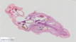
| 541 |
Capsella sp. - Seed pod longitudinal section 20x |
 |
Plant |
Capsella sp. |
Shepherd's purse |
Seed pod |
Longitudinal section |
|
|
unknown |
unknown |
unknown |
Jungner/mikrothek |
2025-05-22 09:56:25 |
Olympus VS200 |
UPLXAPO 20x / 0.80 |
20x / 0.80 |
75 % |
|
Throughlight widefield |
iDS VS-264C |
Slide scanning microscope |
6.9 |
14,889 x
8,366 x
55 px³ |
4,066.4 x
2,284.8 x
31.9 µm³ |
✅
|
✅
|
✅
|
❌ |
| 15 |
Carpinus betulus - Branch cross section 20x |
 |
Plant |
Carpinus betulus |
Common hornbeam |
Branch |
Cross section |
|
bark parenchyma, bast, cambium, cork, medullary parenchyma, medullary ray, primary xylem, stone cell, wood, xylem vessel or tracheae |
Formol, acetic alcohol, Chlorix |
|
Euparal |
Rainer Teubner |
2021-07-01 19:33:07 |
Olympus VS200 |
UPLXAPO 20x / 0.80 |
20x / 0.80 |
40 % |
|
Throughlight widefield |
iDS VS-264C |
Slide scanning microscope |
25.2 |
21,282 x
19,388 x
61 px³ |
5,809.6 x
5,292.4 x
70.8 µm³ |
✅
|
✅
|
✅
|
✅
|
| 108 |
Castanea sativa - Stalk cross section 20x |
 |
Plant |
Castanea sativa |
Sweet chestnut |
Stalk |
Cross section |
|
bark parenchyma, cambium, collenchyma, cork? hypodermis?, epidermis & cuticle, medullary parenchyma, phloem, sclerenchyma, triomes, xylem |
Formol, acetic alcohol |
|
Euparal |
Rainer Teubner |
2021-11-03 14:02:41 |
Olympus VS200 |
UPLXAPO 20x / 0.80 |
20x / 0.80 |
50 % |
|
Throughlight widefield |
iDS VS-264C |
Slide scanning microscope |
33.6 |
20,335 x
19,428 x
85 px³ |
5,551.0 x
5,303.4 x
99.1 µm³ |
✅
|
✅
|
✅
|
✅
|
| 332 |
Ceratophyllum sp. - Stem cross section 20x |
 |
Plant |
Ceratophyllum sp. |
unknown |
Stem |
Cross section |
|
Hydrophyte |
unknown |
unknown |
unknown |
Jungner/microthek |
2023-10-26 09:25:46 |
Olympus VS200 |
UPLXAPO 20x / 0.80 |
20x / 0.80 |
75 % |
|
Throughlight widefield |
iDS VS-264C |
Slide scanning microscope |
2.1 |
14,949 x
3,506 x
41 px³ |
4,082.7 x
957.5 x
23.6 µm³ |
✅
|
✅
|
✅
|
❌ |
| 707 |
Ceratophyllum sp. - Stem cross section 20x |
 |
Plant |
Ceratophyllum sp. |
Hornwort |
Stem |
Cross section |
|
Cell with crystals, Epidermis, Large intercellular space |
unknown |
unknown |
unknown |
Jungner/microthek |
2025-08-21 08:56:56 |
Olympus VS200 |
UPLXAPO 20x / 0.80 |
20x / 0.80 |
75 % |
|
Throughlight widefield |
iDS VS-264C |
Slide scanning microscope |
1.6 |
9,263 x
3,995 x
43 px³ |
2,529.6 x
1,090.8 x
24.8 µm³ |
✅
|
✅
|
✅
|
✅
|
| 565 |
Chaetophora sp. - Whole 20x |
 |
Algae |
Chaetophora sp. |
Chaetophora |
Whole organism |
Whole mount |
|
|
unknown |
unknown |
unknown |
Jungner/microthek |
2025-06-23 12:06:22 |
Olympus VS200 |
UPLXAPO 20x / 0.80 |
20x / 0.80 |
75 % |
|
Throughlight widefield |
iDS VS-264C |
Slide scanning microscope |
71.5 |
23,077 x
30,659 x
101 px³ |
6,302.4 x
8,373.1 x
100.0 µm³ |
✅
|
✅
|
✅
|
❌ |
| 559 |
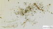
Chroococcus sp. - Whole 20x |
 |
Cyanobacterium |
Chroococcus sp. |
Chroococcus |
Whole Organism |
Whole mount |
|
|
unknown |
unknown |
unknown |
Jungner/microthek |
2025-05-30 12:05:12 |
Olympus VS200 |
UPLXAPO 20x / 0.80 |
20x / 0.80 |
75 % |
|
Throughlight widefield |
iDS VS-264C |
Slide scanning microscope |
74.4 |
36,487 x
20,187 x
101 px³ |
9,965.3 x
5,513.5 x
100.0 µm³ |
✅
|
✅
|
✅
|
❌ |
| 550 |
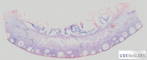
Citrus x limon - Fruit peel cross section 20x |
 |
Plant |
Citrus x limon |
Lemon |
Fruit peel |
Cross section |
|
Exocarp, Glandular cavity, Mesocarp, Vesicle of the endocarp |
unknown |
unknown |
unknown |
Jungner/microthek |
2025-05-23 10:43:30 |
Olympus VS200 |
UPLXAPO 20x / 0.80 |
20x / 0.80 |
75 % |
|
Throughlight widefield |
iDS VS-264C |
Slide scanning microscope |
72.0 |
55,531 x
22,747 x
57 px³ |
15,166.9 x
6,212.5 x
33.0 µm³ |
✅
|
✅
|
✅
|
✅
|
| 578 |
Cladonia sp. - Thallus cross section 20x |
 |
Lichen |
Cladonia sp. |
Cladonia |
Thallus |
Cross section |
|
|
unknown |
unknown |
unknown |
Jungner/microthek |
2025-06-24 16:17:19 |
Olympus VS200 |
UPLXAPO 20x / 0.80 |
20x / 0.80 |
75 % |
|
Throughlight widefield |
iDS VS-264C |
Slide scanning microscope |
24.4 |
17,191 x
16,664 x
85 px³ |
4,695.0 x
4,551.1 x
49.6 µm³ |
✅
|
✅
|
✅
|
❌ |
| 272 |
Cladophora sp. - 20x |
 |
Algae |
Cladophora sp. |
Cladophora |
Whole organism |
Whole mount |
Cladophora is a branched filamentous green alge. Wikipedia on Cladophora.
Jungner/microthek micro preparation no. 952 |
|
unknown |
unknown |
unknown |
Jungner/microthek |
2023-09-19 13:21:12 |
Olympus VS200 |
UPLXAPO 20x / 0.80 |
20x / 0.80 |
75 % |
|
Throughlight widefield |
iDS VS-264C |
Slide scanning microscope |
30.7 |
19,180 x
18,813 x
85 px³ |
5,238.3 x
5,138.1 x
100.0 µm³ |
✅
|
✅
|
✅
|
❌ |
| 404 |
Cladophora sp. 2 - 40x |
 |
Algae |
Cladophora sp. |
Cladophora |
Whole organism |
Whole mount |
|
|
unknown |
unknown |
unknown |
Jungner/microthek |
2025-03-31 14:21:24 |
Olympus VS200 |
UPLFLN 40x / 0.75 |
40x / 0.75 |
75 % |
|
Throughlight widefield |
iDS VS-264C |
Slide scanning microscope |
65.9 |
31,009 x
21,032 x
101 px³ |
4,242.9 x
2,877.8 x
70.0 µm³ |
✅
|
✅
|
✅
|
❌ |
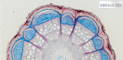
| 509 |
Clematis sp. - older stalk cross section 20x |
 |
Plant |
Clematis sp. |
Clematis |
Stalk |
Cross section |
|
|
unknown |
unknown |
unknown |
Jungner/microthek |
2025-05-14 12:40:56 |
Olympus VS200 |
UPLXAPO 20x / 0.80 |
20x / 0.80 |
75 % |
|
Throughlight widefield |
iDS VS-264C |
Slide scanning microscope |
25.6 |
22,814 x
11,105 x
101 px³ |
6,231.0 x
3,032.8 x
100.0 µm³ |
✅
|
✅
|
✅
|
❌ |
| 508 |
Clematis sp. - young stalk cross section 20x |
 |
Plant |
Clematis sp. |
Clematis |
Stalk |
Cross section |
|
|
unknown |
unknown |
unknown |
Jungner/microthek |
2025-05-14 11:43:19 |
Olympus VS200 |
UPLXAPO 20x / 0.80 |
20x / 0.80 |
75 % |
|
Throughlight widefield |
iDS VS-264C |
Slide scanning microscope |
40.2 |
20,733 x
19,221 x
101 px³ |
5,662.6 x
5,249.7 x
100.0 µm³ |
✅
|
✅
|
✅
|
❌ |
| 109 |
Clematis vitalba - Stalk cross section 20x |
 |
Plant |
Clematis vitalba |
Old man's beard |
Stalk |
Cross section |
|
bark parenchyma, cambium, collenchyma, epidermis & cuticle, medullary parenchyma, phloem/bast, primary xylem, triomes, vascular bundle, xylem, xylem vessel or tracheae |
Not fixated |
|
Euparal |
Rainer Teubner |
2021-10-12 15:18:25 |
Olympus VS200 |
UPLXAPO 20x / 0.80 |
20x / 0.80 |
50 % |
|
Throughlight widefield |
iDS VS-264C |
Slide scanning microscope |
10.4 |
13,030 x
13,130 x
61 px³ |
3,556.7 x
3,584.2 x
70.8 µm³ |
✅
|
✅
|
✅
|
✅
|
| 212 |
Clivia miniata - Root Wurzel 20x |
 |
Plant |
Clivia miniata |
Clivia |
Root |
Cross section |
|
|
Alcohol |
Astra blue, safranin |
Water |
Christiane Wittmann |
2020-10-05 13:16:04 |
Olympus VS200 |
UPLXAPO 20x / 0.80 |
20x / 0.80 |
75 % |
|
Throughlight widefield |
iDS VS-264C |
Slide scanning microscope |
45.1 |
28,780 x
25,715 x
61 px³ |
7,856.1 x
7,019.6 x
70.8 µm³ |
✅
|
✅
|
✅
|
❌ |
| 304 |
Clivia sp. - Root cross section 20x |
 |
Plant |
Clivia sp. |
Clivia |
Root |
Cross section |
Wikipedia on Clivia sp.
Due to shrinking of the mounting media caused by ageing, air got into the specimen.
Jungner/microthek micro preparation no. 1085 |
Air bubbles |
unknown |
unknown |
unknown |
Jungner/microthek |
2023-10-19 10:32:28 |
Olympus VS200 |
UPLXAPO 20x / 0.80 |
20x / 0.80 |
75 % |
|
Throughlight widefield |
iDS VS-264C |
Slide scanning microscope |
63.8 |
26,051 x
24,240 x
101 px³ |
7,115.0 x
6,620.6 x
74.0 µm³ |
✅
|
✅
|
✅
|
✅
|
| 202 |
Closterium lunula - Living sample 60x 1 |
 |
Protist |
Closterium lunula |
Closterium lunula |
Whole cell |
Whole organism |
|
|
not fixated |
not stained |
Agarose 2% |
Nadine Graupner |
2022-02-23 13:23:57 |
Olympus VS200 |
UPLXAPO OPH 60x / 1.42 |
60x / 1.42 |
40 % |
Oil |
Throughlight widefield |
iDS VS-264C |
Slide scanning microscope |
5.0 |
10,181 x
8,121 x
61 px³ |
924.2 x
737.2 x
51.0 µm³ |
✅
|
✅
|
✅
|
❌ |
| 201 |
Closterium lunula - Living sample 60x 2 |
 |
Protist |
Closterium lunula |
Closterium lunula |
Whole cell |
Whole organism |
|
|
not fixated |
not stained |
Agarose 2% |
Nadine Graupner |
2022-02-23 13:28:11 |
Olympus VS200 |
UPLXAPO OPH 60x / 1.42 |
60x / 1.42 |
40 % |
Oil |
Throughlight widefield |
iDS VS-264C |
Slide scanning microscope |
3.1 |
5,333 x
9,575 x
61 px³ |
484.1 x
869.2 x
51.0 µm³ |
✅
|
✅
|
✅
|
❌ |
| 200 |
Closterium lunula - Living sample 60x 3 |
 |
Protist |
Closterium lunula |
Closterium lunula |
Whole cell |
Whole organism |
|
|
not fixated |
not stained |
Agarose 2% |
Nadine Graupner |
2022-02-23 13:41:18 |
Olympus VS200 |
UPLXAPO OPH 60x / 1.42 |
60x / 1.42 |
40 % |
Oil |
Throughlight widefield |
iDS VS-264C |
Slide scanning microscope |
4.9 |
7,151 x
11,272 x
61 px³ |
649.2 x
1,023.2 x
51.0 µm³ |
✅
|
✅
|
✅
|
❌ |
| 199 |
Closterium lunula - Living sample 60x 4 |
 |
Protist |
Closterium lunula |
Closterium lunula |
Whole cell |
Whole organism |
|
|
not fixated |
not stained |
Agarose 2% |
Nadine Graupner |
2022-02-23 13:46:48 |
Olympus VS200 |
UPLXAPO OPH 60x / 1.42 |
60x / 1.42 |
40 % |
Oil |
Throughlight widefield |
iDS VS-264C |
Slide scanning microscope |
4.2 |
12,120 x
5,697 x
61 px³ |
1,100.3 x
517.1 x
51.0 µm³ |
✅
|
✅
|
✅
|
❌ |
| 198 |
Closterium lunula - Living sample 60x 5 |
 |
Protist |
Closterium lunula |
Closterium lunula |
Whole cell |
Whole organism |
|
|
not fixated |
not stained |
Agarose 2% |
Nadine Graupner |
2022-02-23 13:52:53 |
Olympus VS200 |
UPLXAPO OPH 60x / 1.42 |
60x / 1.42 |
40 % |
Oil |
Throughlight widefield |
iDS VS-264C |
Slide scanning microscope |
5.5 |
8,242 x
11,029 x
61 px³ |
748.2 x
1,001.2 x
51.0 µm³ |
✅
|
✅
|
✅
|
❌ |
| 203 |
Closterium lunula - Living sample 60x 6 |
 |
Protist |
Closterium lunula |
Closterium lunula |
Whole cell |
Whole organism |
|
|
not fixated |
not stained |
Agarose 2% |
Nadine Graupner |
2022-02-23 12:14:37 |
Olympus VS200 |
UPLXAPO OPH 60x / 1.42 |
60x / 1.42 |
40 % |
Oil |
Throughlight widefield |
iDS VS-264C |
Slide scanning microscope |
4.0 |
6,181 x
10,544 x
61 px³ |
561.1 x
957.2 x
51.0 µm³ |
✅
|
✅
|
✅
|
❌ |
| 653 |
Cobaea scandens - Stalk cross section 20x |
 |
Plant |
Cobaea scandens |
Cup-and-saucer vine |
Stalk |
Cross section |
Wikipedia on Cobaea scandens.
This sample has some damage (partly large bubbles) caused by shrinking of the mounting medium due to ageing.
Jungner/microthek micro preparation no. 1296 |
|
unknown |
unknown |
unknown |
Jungner/microthek |
2025-07-17 12:09:33 |
Olympus VS200 |
UPLXAPO 20x / 0.80 |
20x / 0.80 |
75 % |
|
Throughlight widefield |
iDS VS-264C |
Slide scanning microscope |
11.7 |
19,900 x
14,373 x
41 px³ |
5,434.7 x
3,925.4 x
23.6 µm³ |
✅
|
✅
|
✅
|
❌ |
| 536 |
Cocos nucifera - Fruit pulp cross section 20x |
 |
Plant |
Cocos nucifera |
Coconut |
Fruit |
Cross section |
|
|
unknown |
unknown |
unknown |
Jungner/microthek |
2025-05-21 19:13:29 |
Olympus VS200 |
UPLXAPO 20x / 0.80 |
20x / 0.80 |
75 % |
|
Throughlight widefield |
iDS VS-264C |
Slide scanning microscope |
64.1 |
27,216 x
23,324 x
101 px³ |
7,433.2 x
6,370.3 x
59.0 µm³ |
✅
|
✅
|
✅
|
❌ |
| 633 |
Cocos nucifera - Leaf stalk cross section 20x |
 |
Plant |
Cocos nucifera |
Coconut |
Leaf stalk |
Cross section |
|
|
unknown |
unknown |
unknown |
Jungner/microthek |
2025-07-15 20:27:58 |
Olympus VS200 |
UPLXAPO 20x / 0.80 |
20x / 0.80 |
75 % |
|
Throughlight widefield |
iDS VS-264C |
Slide scanning microscope |
126.6 |
38,127 x
32,870 x
101 px³ |
10,412.7 x
8,977.0 x
69.0 µm³ |
✅
|
✅
|
✅
|
❌ |
| 638 |
Codiaeum variegatum - Leaf cross section 20x |
 |
Plant |
Codiaeum variegatum |
Fire croton |
Leaf |
Cross section |
|
|
unknown |
unknown |
unknown |
Jungner/microthek |
2025-07-16 13:52:19 |
Olympus VS200 |
UPLXAPO 20x / 0.80 |
20x / 0.80 |
75 % |
|
Throughlight widefield |
iDS VS-264C |
Slide scanning microscope |
19.7 |
20,886 x
20,947 x
45 px³ |
5,703.9 x
5,720.8 x
26.0 µm³ |
✅
|
✅
|
✅
|
❌ |
| 634 |
Coffea sp. - one-year-old branch cross section 20x |
 |
Plant |
Coffea sp. |
Coffee plant |
Branch |
Cross section |
Wikipedia on Coffea sp.
This sample has some minor damage (cracks and bubbles) due aging of the mounting medium.
Jungner/microthek micro preparation no. 1275 |
|
unknown |
unknown |
unknown |
Jungner/microthek |
2025-07-16 09:36:06 |
Olympus VS200 |
UPLXAPO 20x / 0.80 |
20x / 0.80 |
75 % |
|
Throughlight widefield |
iDS VS-264C |
Slide scanning microscope |
8.5 |
9,004 x
9,323 x
101 px³ |
2,458.9 x
2,546.2 x
59.0 µm³ |
✅
|
✅
|
✅
|
❌ |
| 649 |
Coleus blumei - Stalk cross section 20x |
 |
Plant |
Coleus blumei |
Painted nettle |
Stalk |
Cross section |
|
|
unknown |
unknown |
unknown |
Jungner/microthek |
2025-07-17 08:48:33 |
Olympus VS200 |
UPLXAPO 20x / 0.80 |
20x / 0.80 |
75 % |
|
Throughlight widefield |
iDS VS-264C |
Slide scanning microscope |
26.7 |
15,806 x
16,734 x
101 px³ |
4,316.7 x
4,570.1 x
73.0 µm³ |
✅
|
✅
|
✅
|
❌ |
| 333 |
Collybia maculata - Cap cross section 20x |
 |
Fungus |
Collybia maculata |
Spotted toughshank |
Cap |
Cross section |
|
|
unknown |
Astra blue, fuchsine |
unknown |
Karl Brügmann |
2023-09-28 14:30:49 |
Olympus VS200 |
UPLXAPO 20x / 0.80 |
20x / 0.80 |
75 % |
|
Throughlight widefield |
iDS VS-264C |
Slide scanning microscope |
73.2 |
53,598 x
20,396 x
67 px³ |
14,638.8 x
5,570.6 x
38.9 µm³ |
✅
|
✅
|
✅
|
❌ |
| 446 |
Convallaria majalis - Root tip cross section 20x |
 |
Plant |
Convallaria majalis |
Lily of the valley |
Root |
Cross section |
|
|
unknown |
unknown |
unknown |
Jungner/microthek |
2025-04-30 16:40:54 |
Olympus VS200 |
UPLXAPO 20x / 0.80 |
20x / 0.80 |
75 % |
|
Throughlight widefield |
iDS VS-264C |
Slide scanning microscope |
4.8 |
13,803 x
6,085 x
57 px³ |
3,769.9 x
1,661.9 x
33.0 µm³ |
✅
|
✅
|
✅
|
❌ |
| 497 |
Convallaria sp. - Root cross section 20x |
 |
Plant |
Convallaria sp. |
Lily-of-the-valley |
Root |
Cross section |
|
|
unknown |
unknown |
unknown |
Jungner/microthek |
2025-05-13 09:12:20 |
Olympus VS200 |
UPLXAPO 20x / 0.80 |
20x / 0.80 |
75 % |
|
Throughlight widefield |
iDS VS-264C |
Slide scanning microscope |
15.1 |
13,165 x
11,393 x
101 px³ |
3,595.6 x
3,111.7 x
59.0 µm³ |
✅
|
✅
|
✅
|
❌ |
| 576 |
Coprinus comatus - Fruiting body cross section 20x |
 |
Fungus |
Coprinus comatus |
Shaggy ink cap |
Fruiting body |
Cross section |
|
|
unknown |
unknown |
unknown |
Jungner/microthek |
2025-06-24 13:45:34 |
Olympus VS200 |
UPLXAPO 20x / 0.80 |
20x / 0.80 |
75 % |
|
Throughlight widefield |
iDS VS-264C |
Slide scanning microscope |
50.4 |
31,383 x
28,168 x
57 px³ |
8,570.9 x
7,693.0 x
33.0 µm³ |
✅
|
✅
|
✅
|
❌ |
| 699 |
Cornus mas - Leaf cross section 20x |
 |
Plant |
Cornus mas |
Cornel |
Leaf |
Cross section |
Wikipedia on Cornus mas.
This sample has some damage (partly large bubbles) caused by shrinking of the mounting medium due to ageing.
Jungner/microthek micro preparation no. 1260 |
|
unknown |
unknown |
unknown |
Jungner/microthek |
2025-08-19 18:24:01 |
Olympus VS200 |
UPLXAPO 20x / 0.80 |
20x / 0.80 |
75 % |
|
Throughlight widefield |
iDS VS-264C |
Slide scanning microscope |
56.1 |
57,427 x
13,019 x
75 px³ |
15,683.8 x
3,555.4 x
43.7 µm³ |
✅
|
✅
|
✅
|
❌ |
| 701 |
Cornus sanguinea - Branch cross section 20x |
 |
Plant |
Cornus sanguinea |
Bloody dogwood |
Branch |
Cross section |
|
|
unknown |
unknown |
unknown |
Jungner/microthek |
2025-08-19 21:30:23 |
Olympus VS200 |
UPLXAPO 20x / 0.80 |
20x / 0.80 |
75 % |
|
Throughlight widefield |
iDS VS-264C |
Slide scanning microscope |
39.6 |
20,786 x
18,856 x
101 px³ |
5,676.7 x
5,149.5 x
100.0 µm³ |
✅
|
✅
|
✅
|
❌ |
| 698 |
Corylus avellana - male flower longitudinal section |
 |
Plant |
Corylus avellana |
Common hazel |
Flower |
Longitudinal section |
Wikipedia on Corylus avellana.
This sample has some damage (small bubbles) caused by shrinking of the mounting medium due to ageing.
Jungner/microthek micro preparation no. 1259 |
|
unknown |
unknown |
unknown |
Jungner/microthek |
2025-08-19 15:46:25 |
Olympus VS200 |
UPLXAPO 20x / 0.80 |
20x / 0.80 |
75 % |
|
Throughlight widefield |
iDS VS-264C |
Slide scanning microscope |
75.1 |
41,990 x
32,531 x
55 px³ |
11,467.7 x
8,884.5 x
31.9 µm³ |
✅
|
✅
|
✅
|
❌ |
| 554 |
Corylus avellana - Seed 20x |
 |
Plant |
Corylus avellana |
Common hazel |
Seed |
unknown |
|
|
unknown |
unknown |
unknown |
Jungner/microthek |
2025-05-16 12:15:36 |
Olympus VS200 |
UPLXAPO 20x / 0.80 |
20x / 0.80 |
75 % |
|
Throughlight widefield |
iDS VS-264C |
Slide scanning microscope |
36.3 |
27,206 x
18,295 x
73 px³ |
7,430.4 x
4,996.7 x
42.5 µm³ |
✅
|
✅
|
✅
|
❌ |
| 538 |
Corylus sp. - young fruit cross section 20x |
 |
Plant |
Corylus sp. |
Hazelnut |
Fruit |
Cross section |
|
|
unknown |
unknown |
unknown |
Jungner/mikrothek |
2025-05-22 06:21:31 |
Olympus VS200 |
UPLXAPO 20x / 0.80 |
20x / 0.80 |
75 % |
|
Throughlight widefield |
iDS VS-264C |
Slide scanning microscope |
35.8 |
24,778 x
19,271 x
75 px³ |
6,767.3 x
5,263.3 x
43.7 µm³ |
✅
|
✅
|
✅
|
❌ |
| 639 |
Cosmarium sp. 20x |
 |
Protist |
Cosmarium sp. |
Cosmarium |
Whole cell |
Whole organism |
|
|
not fixated |
not stained |
Water |
Michael Kloster / Eva Papendick |
2025-07-18 13:19:11 |
Olympus VS200 |
UPLXAPO 20x / 0.80 |
20x / 0.80 |
75 % |
|
Throughlight widefield |
iDS VS-264C |
Slide scanning microscope |
20.0 |
12,958 x
17,332 x
89 px³ |
3,538.8 x
4,733.3 x
51.9 µm³ |
✅
|
✅
|
✅
|
❌ |
| 694 |
Cotoneaster sp. - Leaf cross section 20x |
 |
Plant |
Cotoneaster sp. |
Cotoneaster |
Leaf |
Cross section |
Wikipedia on Cotoneaster sp.
This sample has some damage (partly large bubbles) caused by shrinking of the mounting medium due to ageing.
Jungner/microthek micro preparation no. 1255 |
|
unknown |
unknown |
unknown |
Jungner/microthek |
2025-08-15 10:02:39 |
Olympus VS200 |
UPLXAPO 20x / 0.80 |
20x / 0.80 |
75 % |
|
Throughlight widefield |
iDS VS-264C |
Slide scanning microscope |
15.3 |
34,729 x
11,325 x
39 px³ |
9,484.8 x
3,093.0 x
22.4 µm³ |
✅
|
✅
|
✅
|
❌ |
| 607 |
Crataegus sp. - Leaf cross section 20x |
 |
Plant |
Crataegus sp. |
Hawthorn |
Leaf |
Cross section |
|
|
unknown |
unknown |
unknown |
Jungner/microthek |
2025-07-03 17:56:18 |
Olympus VS200 |
UPLXAPO 20x / 0.80 |
20x / 0.80 |
75 % |
|
Throughlight widefield |
iDS VS-264C |
Slide scanning microscope |
27.7 |
41,790 x
15,399 x
43 px³ |
11,413.3 x
4,205.6 x
24.8 µm³ |
✅
|
✅
|
✅
|
❌ |
| 676 |
Crataegus sp. - Root cross section 20x |
 |
Plant |
Crataegus sp. |
Hawthorn |
Root |
Cross section |
Wikipedia on Crataegus sp.
This sample has some damage (partly large bubbles) caused by shrinking of the mounting medium due to ageing.
Jungner/microthek micro preparation no. 1249 |
|
unknown |
unknown |
unknown |
Jungner/microthek |
2025-08-13 19:56:12 |
Olympus VS200 |
UPLXAPO 20x / 0.80 |
20x / 0.80 |
75 % |
|
Throughlight widefield |
iDS VS-264C |
Slide scanning microscope |
26.4 |
16,623 x
15,708 x
101 px³ |
4,539.8 x
4,289.9 x
59.0 µm³ |
✅
|
✅
|
✅
|
❌ |
| 16 |
Cricetulus griseus - sagittal 40x |
 |
Animal |
Cricetulus griseus |
Chinese hamster |
Body |
Sagittal thin section |
|
|
unknown |
|
unknown |
|
2021-07-23 10:37:39 |
Olympus VS200 |
UPLXAPO O 40x / 1.40 |
40x / 1.40 |
75 % |
Oil |
Throughlight widefield |
iDS VS-264C |
Slide scanning microscope |
223.9 |
66,258 x
116,540 x
29 px³ |
9,116.0 x
16,034.0 x
16.4 µm³ |
✅
|
✅
|
✅
|
❌ |
| 496 |
Cucurbita pepo - Stalk cross section 20x |
 |
Plant |
Cucurbita pepo |
Squash |
Stalk |
Cross section |
|
Bicollateral vascular bundle, Collenchyma, Epidermis, Phloem, Pith cavity, Sclerenchyma, Trichome, Xylem |
unknown |
unknown |
unknown |
Jungner/microthek |
2025-05-13 16:35:33 |
Olympus VS200 |
UPLXAPO 20x / 0.80 |
20x / 0.80 |
75 % |
|
Throughlight widefield |
iDS VS-264C |
Slide scanning microscope |
34.9 |
18,861 x
18,295 x
101 px³ |
5,151.3 x
4,996.7 x
100.0 µm³ |
✅
|
✅
|
✅
|
✅
|
| 34 |
Cydonia oblonga - Leaf cross section 20x |
 |
Plant |
Cydonia oblonga |
Quince |
Leaf |
Cross section |
|
bark parenchyma, collenchyma, epidermis & cuticle, guard cells, lower epidermis, palisade parenchyma, phloem, secretory duct, sponge parenchyma, upper epidermis & cuticle, vascular bundle, xylem |
Formol, acetic alcohol |
|
Euparal |
Jörg Weiß
www.mikroskopie-bonn.de |
2021-08-17 11:52:46 |
Olympus VS200 |
UPLXAPO 20x / 0.80 |
20x / 0.80 |
50 % |
|
Throughlight widefield |
iDS VS-264C |
Slide scanning microscope |
8.4 |
32,447 x
4,223 x
61 px³ |
8,857.4 x
1,152.6 x
70.8 µm³ |
✅
|
✅
|
✅
|
✅
|
| 35 |
Cydonia oblonga - Leaf stalk cross section 20x |
 |
Plant |
Cydonia oblonga |
Quince |
Leaf stalk |
Cross section |
|
bark parenchyma, collenchyma, epidermis & cuticle, intercellular spaces, phloem, sclerenchyma, secretory duct, trichome, xylem |
Formol, acetic alcohol |
|
Euparal |
Jörg Weiß
www.mikroskopie-bonn.de |
2021-08-17 13:00:12 |
Olympus VS200 |
UPLXAPO 20x / 0.80 |
20x / 0.80 |
50 % |
|
Throughlight widefield |
iDS VS-264C |
Slide scanning microscope |
5.4 |
9,775 x
9,039 x
61 px³ |
2,668.2 x
2,467.4 x
70.8 µm³ |
✅
|
✅
|
✅
|
✅
|
| 36 |
Cydonia oblonga - Leaf stalk cross section AP40 20x |
 |
Plant |
Cydonia oblonga |
Quince |
Leaf stalk |
Cross section |
|
bark parenchyma, collenchyma, cyrstal druse, epidermis & cuticle, intercellular spaces, phloem, sclerenchyma, triome, xylem |
Formol, acetic alcohol |
|
Euparal |
Rainer Teubner |
2021-06-30 16:53:55 |
Olympus VS200 |
UPLXAPO 20x / 0.80 |
20x / 0.80 |
40 % |
|
Throughlight widefield |
iDS VS-264C |
Slide scanning microscope |
11.2 |
13,090 x
13,977 x
61 px³ |
3,573.2 x
3,815.3 x
70.8 µm³ |
✅
|
✅
|
✅
|
✅
|
| 196 |
Cymbella sp. - Living sample 60x 1 |
 |
Protist |
Cymbella sp. |
Cymbella |
Whole cell |
Whole organism |
|
|
not fixated |
not stained |
Agarose 2% |
Nadine Graupner |
2022-02-22 19:10:34 |
Olympus VS200 |
UPLXAPO OPH 60x / 1.42 |
60x / 1.42 |
40 % |
Oil |
Throughlight widefield |
iDS VS-264C |
Slide scanning microscope |
48.2 |
22,179 x
35,632 x
61 px³ |
2,013.5 x
3,234.8 x
68.4 µm³ |
✅
|
✅
|
✅
|
❌ |
| 194 |
Cymbella sp. - Living sample 60x 2 |
 |
Protist |
Cymbella sp. |
Cymbella |
Whole cell |
Whole organism |
|
|
not fixated |
not stained |
Agarose 2% |
Nadine Graupner |
2022-02-22 14:54:13 |
Olympus VS200 |
UPLXAPO OPH 60x / 1.42 |
60x / 1.42 |
40 % |
Oil |
Throughlight widefield |
iDS VS-264C |
Slide scanning microscope |
1.8 |
4,788 x
6,030 x
61 px³ |
434.6 x
547.4 x
68.4 µm³ |
✅
|
✅
|
✅
|
❌ |
| 424 |
Cymbidium sp. - Aerial root cross section 20x |
 |
Plant |
Cymbidium sp. |
Boat orchid |
Root |
Cross section |
|
|
unknown |
unknown |
unknown |
Bresser |
2025-04-08 16:52:36 |
Olympus VS200 |
UPLXAPO 20x / 0.80 |
20x / 0.80 |
75 % |
|
Throughlight widefield |
iDS VS-264C |
Slide scanning microscope |
2.1 |
7,977 x
7,280 x
37 px³ |
2,178.7 x
1,988.1 x
21.2 µm³ |
✅
|
✅
|
✅
|
❌ |
| 111 |
Cyperus alternifolius - Stalk cross section 20x I |
 |
Plant |
Cyperus alternifolius |
Umbrella papyrus |
Stalk |
Cross section |
|
assimilation parenchyma, closed collateral vascular bundle, epidermis & cuticle, guard cells, intercellular spaces, parenchyma cells, phloem, sclerenchyma, sive tubes, triomes, vascular sheath, xylem, xylem vessel or tracheae |
Formol, acetic alcohol |
unstained |
Euparal |
Rainer Teubner |
2021-09-08 14:21:51 |
Olympus VS200 |
UPLXAPO 20x / 0.80 |
20x / 0.80 |
40 % |
|
Throughlight widefield |
iDS VS-264C |
Slide scanning microscope |
22.7 |
19,529 x
19,015 x
61 px³ |
5,330.9 x
5,190.6 x
70.8 µm³ |
✅
|
✅
|
✅
|
✅
|
| 110 |
Cyperus alternifolius - Stalk cross section 20x II |
 |
Plant |
Cyperus alternifolius |
Umbrella papyrus |
Stalk |
Cross section |
|
assimilation parenchyma, closed collateral vascular bundle, companion cells, epidermis & cuticle, intercellular spaces, parenchyma cells, phloem, sclerenchyma, sive tubes, triomes, vascular sheath, xylem, xylem vessel or tracheae |
Formol, acetic alcohol |
|
Euparal |
Rainer Teubner |
2021-09-08 14:48:01 |
Olympus VS200 |
UPLXAPO 20x / 0.80 |
20x / 0.80 |
50 % |
|
Throughlight widefield |
iDS VS-264C |
Slide scanning microscope |
21.7 |
19,650 x
18,138 x
61 px³ |
5,364.0 x
4,951.3 x
99.6 µm³ |
✅
|
✅
|
✅
|
✅
|
| 112 |
Cyperus longus - Leaf cross section 20x 1 |
 |
Plant |
Cyperus longus |
Galingale |
Leaf |
Cross section |
|
assimilation parenchyma, closed collateral vascular bundle, epidermis & cuticle, guard cells, intercellular spaces, parenchyma cells, phloem, sclerenchyma, xylem, xylem vessel or tracheae |
Formol, acetic alcohol |
|
Euparal |
Rainer Teubner |
2021-10-21 13:24:23 |
Olympus VS200 |
UPLXAPO 20x / 0.80 |
20x / 0.80 |
50 % |
|
Throughlight widefield |
iDS VS-264C |
Slide scanning microscope |
12.5 |
15,982 x
11,367 x
69 px³ |
4,362.7 x
3,102.8 x
80.2 µm³ |
✅
|
✅
|
✅
|
✅
|
| 113 |
Cyperus longus - Leaf cross section 20x 2 |
 |
Plant |
Cyperus longus |
Galingale |
Leaf |
Cross section |
|
assimilation parenchyma, closed collateral vascular bundle, epidermis & cuticle, guard cells, intercellular spaces, parenchyma cells, phloem, sclerenchyma, xylem, xylem vessel or tracheae |
Formol, acetic alcohol |
unstained |
Euparal |
Rainer Teubner |
2021-10-21 13:42:11 |
Olympus VS200 |
UPLXAPO 20x / 0.80 |
20x / 0.80 |
50 % |
|
Throughlight widefield |
iDS VS-264C |
Slide scanning microscope |
17.6 |
19,368 x
13,191 x
69 px³ |
5,286.9 x
3,600.7 x
80.2 µm³ |
✅
|
✅
|
✅
|
✅
|
| 114 |
Cyperus longus - Stalk cross section 20x 1 |
 |
Plant |
Cyperus longus |
Galingale |
Stalk |
Cross section |
|
assimilation parenchyma, closed collateral vascular bundle, companion cells, epidermis & cuticle, guard cells, intercellular spaces, parenchyma cells, phloem, sclerenchyma, sive tubes, vascular sheath, xylem, xylem vessel or tracheae |
Formol, acetic alcohol |
|
Euparal |
Rainer Teubner |
2021-10-21 11:08:27 |
Olympus VS200 |
UPLXAPO 20x / 0.80 |
20x / 0.80 |
50 % |
|
Throughlight widefield |
iDS VS-264C |
Slide scanning microscope |
15.5 |
15,428 x
16,476 x
61 px³ |
4,211.4 x
4,497.4 x
70.8 µm³ |
✅
|
✅
|
✅
|
✅
|
| 115 |
Cyperus longus - Stalk cross section 20x 1 |
 |
Plant |
Cyperus longus |
Galingale |
Stalk |
Cross section |
|
assimilation parenchyma, closed collateral vascular bundle, epidermis & cuticle, guard cells, intercellular spaces, parenchyma cells, phloem, sclerenchyma, sive tubes, vascular sheath, xylem, xylem vessel or tracheae |
Formol, acetic alcohol |
unstained |
Euparal |
Rainer Teubner |
2021-10-21 11:24:02 |
Olympus VS200 |
UPLXAPO 20x / 0.80 |
20x / 0.80 |
50 % |
|
Throughlight widefield |
iDS VS-264C |
Slide scanning microscope |
14.4 |
15,770 x
15,015 x
61 px³ |
4,304.9 x
4,098.6 x
70.8 µm³ |
✅
|
✅
|
✅
|
✅
|
| 220 |
Daphnia pulex 20x - whole mount 1/3 (300-200µm) |
 |
Animal |
Daphnia pulex |
Water flea |
Whole animal |
Whole mount |
|
|
unknown |
unknown |
unknown |
|
2023-03-15 13:15:01 |
Olympus VS200 |
UPLXAPO 20x / 0.80 |
20x / 0.80 |
75 % |
|
Throughlight widefield |
iDS VS-264C |
Slide scanning microscope |
8.9 |
10,616 x
9,909 x
85 px³ |
2,899.3 x
2,706.4 x
99.1 µm³ |
✅
|
✅
|
✅
|
❌ |
| 221 |
Daphnia pulex - whole mount 20x 2/3 (210-110µm) |
 |
Animal |
Daphnia pulex |
Water flea |
Whole animal |
Whole mount |
|
|
unknown |
unknown |
unknown |
|
2023-03-15 13:19:34 |
Olympus VS200 |
UPLXAPO 20x / 0.80 |
20x / 0.80 |
75 % |
|
Throughlight widefield |
iDS VS-264C |
Slide scanning microscope |
8.9 |
10,616 x
9,910 x
85 px³ |
2,899.3 x
2,706.4 x
99.1 µm³ |
✅
|
✅
|
✅
|
❌ |
| 222 |
Daphnia pulex - whole mount 20x 3/3 (120-70µm) |
 |
Animal |
Daphnia pulex |
Water flea |
Whole animal |
Whole mount |
|
|
unknown |
unknown |
unknown |
|
2023-03-15 13:24:46 |
Olympus VS200 |
UPLXAPO 20x / 0.80 |
20x / 0.80 |
75 % |
|
Throughlight widefield |
iDS VS-264C |
Slide scanning microscope |
4.3 |
10,616 x
9,910 x
41 px³ |
2,899.3 x
2,706.4 x
47.2 µm³ |
✅
|
✅
|
✅
|
❌ |
| 458 |
Daucus carota - Storage root cross section 20x |
 |
Plant |
Daucus carota |
Wild carrot |
Root |
Cross section |
|
|
unknown |
unknown |
unknown |
Jungner/microthek |
2025-05-02 18:53:29 |
Olympus VS200 |
UPLXAPO 20x / 0.80 |
20x / 0.80 |
75 % |
|
Throughlight widefield |
iDS VS-264C |
Slide scanning microscope |
118.6 |
48,526 x
44,447 x
55 px³ |
13,253.5 x
12,139.5 x
31.9 µm³ |
✅
|
✅
|
✅
|
❌ |
| 40 |
Davidia involucrata 20x |
 |
Plant |
Davidia involucrata |
Dove-tree |
? |
Cross section |
|
bark parenchyma, bast, collenchyma, cork, medullary parenchyma, stone cell, wood |
Formol, acetic alcohol |
|
Euparal |
Rainer Teubner |
2021-07-01 15:27:36 |
Olympus VS200 |
UPLXAPO 20x / 0.80 |
20x / 0.80 |
40 % |
|
Throughlight widefield |
iDS VS-264C |
Slide scanning microscope |
20.6 |
17,463 x
19,317 x
61 px³ |
4,767.0 x
5,273.1 x
70.8 µm³ |
✅
|
✅
|
✅
|
✅
|
| 462 |
Dendrobium sp. - Aerial root cross section 20x |
 |
Plant |
Dendrobium sp. |
Dendrobium |
Root |
Cross section |
|
|
unknown |
unknown |
unknown |
Jungner/microthek |
2025-05-02 10:46:53 |
Olympus VS200 |
UPLXAPO 20x / 0.80 |
20x / 0.80 |
75 % |
|
Throughlight widefield |
iDS VS-264C |
Slide scanning microscope |
17.3 |
13,872 x
12,330 x
101 px³ |
3,788.7 x
3,367.4 x
59.0 µm³ |
✅
|
✅
|
✅
|
❌ |
| 645 |
Dendrobium sp. - Aerial root cross section 20x 2 |
 |
Plant |
Dendrobium sp. |
Dendrobium |
Aerial root |
Cross section |
|
|
unknown |
unknown |
unknown |
Jungner/microthek |
2025-07-16 19:24:53 |
Olympus VS200 |
UPLXAPO 20x / 0.80 |
20x / 0.80 |
75 % |
|
Throughlight widefield |
iDS VS-264C |
Slide scanning microscope |
23.8 |
16,286 x
15,409 x
95 px³ |
4,447.8 x
4,208.3 x
55.5 µm³ |
✅
|
✅
|
✅
|
❌ |
| 602 |
Dennstaedtia sp. - Rhizome cross section 20x |
 |
Plant |
Dennstaedtia sp. |
Hayscented fern |
Rhizome |
Cross section |
|
|
unknown |
unknown |
unknown |
Jungner/microthek |
2025-07-02 10:06:41 |
Olympus VS200 |
UPLXAPO 20x / 0.80 |
20x / 0.80 |
75 % |
|
Throughlight widefield |
iDS VS-264C |
Slide scanning microscope |
10.8 |
16,762 x
12,112 x
53 px³ |
4,577.8 x
3,307.9 x
30.7 µm³ |
✅
|
✅
|
✅
|
❌ |
| 77 |
Dichanthelium clandestinum - Sprout cross section quer 20x |
 |
Plant |
Dichanthelium clandestinum |
Deer-tongue witchgrass |
Sprout |
Cross section |
|
assimilation parenchyma, closed collateral vascular bundle, companion cells, epidermis & cuticle, intercellular corridor, lacune, medullary cavity, medullary parenchyma, phloem, sclerenchyma, sieve tubes, tracheae, xylem parenchyma, xylem vessel or tracheae |
Formol, acetic alcohol |
W3Asim II: Acridine red, acriflavine, alcian blue |
Euparal |
Jörg Weiß
www.mikroskopie-bonn.de |
2021-08-17 13:07:52 |
Olympus VS200 |
UPLXAPO 20x / 0.80 |
20x / 0.80 |
50 % |
|
Throughlight widefield |
iDS VS-264C |
Slide scanning microscope |
5.4 |
9,221 x
9,593 x
61 px³ |
2,516.9 x
2,618.7 x
70.8 µm³ |
✅
|
✅
|
✅
|
✅
|
| 646 |
Dieffenbachia sp. - Leaf stalk cross section 20x |
 |
Plant |
Dieffenbachia sp. |
Dumb cane |
Leaf stalk |
Cross section |
|
|
unknown |
unknown |
unknown |
Jungner/microthek |
2025-07-16 16:23:26 |
Olympus VS200 |
UPLXAPO 20x / 0.80 |
20x / 0.80 |
75 % |
|
Throughlight widefield |
iDS VS-264C |
Slide scanning microscope |
75.6 |
41,551 x
30,838 x
59 px³ |
11,348.0 x
8,422.0 x
34.2 µm³ |
✅
|
✅
|
✅
|
❌ |
| 406 |
Dinobryon sp. - 40x |
 |
Algae |
Dinobryon sp. |
Dinobryon |
Whole organism |
Whole mount |
Dinobryon sp. is a colony-forming golden-brown algae (Chrysophyceae). Wikipedia über Dinobryon sp.
Jungner/microthek micro preparation no. 954 |
|
unknown |
unknown |
unknown |
Jungner/microthek |
2025-03-31 17:05:50 |
Olympus VS200 |
UPLXAPO O 40x / 1.40 |
40x / 1.40 |
75 % |
Oil |
Throughlight widefield |
iDS VS-264C |
Slide scanning microscope |
50.0 |
17,269 x
28,665 x
101 px³ |
2,366.3 x
3,927.7 x
29.0 µm³ |
✅
|
✅
|
✅
|
❌ |
| 44 |
Dracaena draco - Leaf cross section 20x 1 |
 |
Plant |
Dracaena draco |
Canary Islands dragon tree |
Leaf |
Cross section |
|
assimilation parenchyma, closed collateral vascular bundle, epidermis & cuticle, guard cells, intercellular spaces, phloem, sclerenchyma, vascular sheath, xylem |
Formol, acetic alcohol |
|
Euparal |
Rainer Teubner |
2021-08-26 13:36:13 |
Olympus VS200 |
UPLXAPO 20x / 0.80 |
20x / 0.80 |
50 % |
|
Throughlight widefield |
iDS VS-264C |
Slide scanning microscope |
25.3 |
35,087 x
11,820 x
61 px³ |
9,578.1 x
3,226.6 x
70.8 µm³ |
✅
|
✅
|
✅
|
✅
|
| 41 |
Dracaena draco - Leaf cross section 20x 2 |
 |
Plant |
Dracaena draco |
Canary Islands dragon tree |
Leaf |
Cross section |
|
assimilation parenchyma, closed collateral vascular bundle, epidermis & cuticle, phloem, sclerenchyma, stomata with guard cells, vascular sheath, xylem |
Formol, acetic alcohol |
|
Euparal |
Rainer Teubner |
2021-08-25 13:35:57 |
Olympus VS200 |
UPLXAPO 20x / 0.80 |
20x / 0.80 |
50 % |
|
Throughlight widefield |
iDS VS-264C |
Slide scanning microscope |
30.2 |
32,820 x
15,065 x
61 px³ |
8,959.2 x
4,112.3 x
70.8 µm³ |
✅
|
✅
|
✅
|
✅
|
| 42 |
Dracaena draco - Leaf cross section 20x 3 |
 |
Plant |
Dracaena draco |
Canary Islands dragon tree |
Leaf |
Cross section |
|
assimilation parenchyma, chloroplasts, closed collateral vascular bundle, epidermis & cuticle, guard cells |
Formol, acetic alcohol |
unstained |
Euparal |
Rainer Teubner |
2021-08-25 14:04:57 |
Olympus VS200 |
UPLXAPO 20x / 0.80 |
20x / 0.80 |
40 % |
|
Throughlight widefield |
iDS VS-264C |
Slide scanning microscope |
19.9 |
34,533 x
9,432 x
61 px³ |
9,426.8 x
2,574.7 x
70.8 µm³ |
✅
|
✅
|
✅
|
✅
|
| 45 |
Dracaena draco - Leaf cross section 20x 4 |
 |
Plant |
Dracaena draco |
Canary Islands dragon tree |
Leaf |
Cross section |
|
assimilation parenchyma, closed collateral vascular bundle, epidermis & cuticle, phloem, sclerenchyma, stomata with guard cells, vascular sheath, xylem |
Formol, acetic alcohol |
|
Euparal |
Rainer Teubner |
2021-08-26 15:04:13 |
Olympus VS200 |
UPLXAPO 20x / 0.80 |
20x / 0.80 |
50 % |
|
Throughlight widefield |
iDS VS-264C |
Slide scanning microscope |
27.7 |
34,825 x
13,030 x
61 px³ |
9,506.6 x
3,556.7 x
70.8 µm³ |
✅
|
✅
|
✅
|
✅
|
| 46 |
Dracaena draco - Leaf cross section 20x 5 |
 |
Plant |
Dracaena draco |
Canary Islands dragon tree |
Leaf |
Cross section |
|
chloroplasts, epidermis & cuticle, stomata with guard cells, vascular bundles |
Formol, acetic alcohol |
unstained |
Euparal |
Rainer Teubner |
2021-08-26 15:29:18 |
Olympus VS200 |
UPLXAPO 20x / 0.80 |
20x / 0.80 |
40 % |
|
Throughlight widefield |
iDS VS-264C |
Slide scanning microscope |
32.8 |
35,813 x
15,025 x
61 px³ |
9,776.2 x
4,101.3 x
70.8 µm³ |
✅
|
✅
|
✅
|
✅
|
| 43 |
Dracaena draco - Leaf cross section 20x 6 |
 |
Plant |
Dracaena draco |
Canary Islands dragon tree |
Leaf |
Cross section |
|
assimilation parenchyma, closed collateral vascular bundle, epidermis & cuticle, phloem, sclerenchyma, stomata with guard cells, vascular sheath, xylem |
Formol, acetic alcohol |
|
Euparal |
Rainer Teubner |
2021-08-25 13:13:23 |
Olympus VS200 |
UPLXAPO 20x / 0.80 |
20x / 0.80 |
50 % |
|
Throughlight widefield |
iDS VS-264C |
Slide scanning microscope |
30.1 |
28,739 x
17,191 x
61 px³ |
7,845.1 x
4,692.7 x
70.8 µm³ |
✅
|
✅
|
✅
|
✅
|
| 595 |
Dryopteris sp. - Leaf cross section 20x |
 |
Plant |
Dryopteris sp. |
Wood fern |
Leaf |
Cross section |
|
Anulus, Indusium, Lower epidermis, Mesophyll, Peristomium cells, Placenta, Sorus, Sporangium, Spores, Stoma, Stomium, Upper epidermis, Vascular tissue |
unknown |
unknown |
unknown |
Jungner/microthek |
2025-07-01 09:18:34 |
Olympus VS200 |
UPLXAPO 20x / 0.80 |
20x / 0.80 |
75 % |
|
Throughlight widefield |
iDS VS-264C |
Slide scanning microscope |
38.2 |
40,032 x
13,069 x
73 px³ |
10,933.1 x
3,569.0 x
42.5 µm³ |
✅
|
✅
|
✅
|
✅
|
| 596 |
Dryopteris sp. - Sporangia 20x 1 |
 |
Plant |
Dryopteris sp. |
Wood fern |
Sporangium |
Whole mount |
|
|
unknown |
unknown |
unknown |
Jungner/microthek |
2025-07-01 16:12:22 |
Olympus VS200 |
UPLXAPO 20x / 0.80 |
20x / 0.80 |
75 % |
|
Throughlight widefield |
iDS VS-264C |
Slide scanning microscope |
7.1 |
7,919 x
9,224 x
97 px³ |
2,162.5 x
2,519.0 x
56.6 µm³ |
✅
|
✅
|
✅
|
❌ |
| 597 |
Dryopteris sp. - Sporangia 20x 2 |
 |
Plant |
Dryopteris sp. |
Wood fern |
Sporangium |
Whole mount |
|
|
unknown |
unknown |
unknown |
Jungner/microthek |
2025-07-01 16:21:54 |
Olympus VS200 |
UPLXAPO 20x / 0.80 |
20x / 0.80 |
75 % |
|
Throughlight widefield |
iDS VS-264C |
Slide scanning microscope |
4.9 |
8,197 x
5,877 x
101 px³ |
2,238.7 x
1,605.0 x
59.0 µm³ |
✅
|
✅
|
✅
|
❌ |
| 598 |
Dryopteris sp. - Sporangia 20x 3 |
 |
Plant |
Dryopteris sp. |
Wood fern |
Sporangium |
Whole mount |
|
Anulus |
unknown |
unknown |
unknown |
Jungner/microthek |
2025-07-01 16:28:56 |
Olympus VS200 |
UPLXAPO 20x / 0.80 |
20x / 0.80 |
75 % |
|
Throughlight widefield |
iDS VS-264C |
Slide scanning microscope |
2.4 |
7,251 x
3,297 x
101 px³ |
1,980.3 x
900.4 x
59.0 µm³ |
✅
|
✅
|
✅
|
✅
|
| 594 |
Dryopteris sp. - Stem cross section 20x |
 |
Plant |
Dryopteris sp. |
Wood fern |
Stem |
Cross section |
|
Bundle sheath, Concentric vascular bundle, Phloem, Xylem |
unknown |
unknown |
unknown |
Jungner/microthek |
2025-06-25 17:03:12 |
Olympus VS200 |
UPLXAPO 20x / 0.80 |
20x / 0.80 |
75 % |
|
Throughlight widefield |
iDS VS-264C |
Slide scanning microscope |
34.6 |
23,378 x
14,652 x
101 px³ |
6,384.6 x
4,001.5 x
100.0 µm³ |
✅
|
✅
|
✅
|
✅
|
| 370 |
Echinococcus sp. - Cyst 40x |
 |
Animal |
Echinococcus sp. |
Echinococcus |
Body |
Cross section |
|
Parasite |
unknown |
unknown |
unknown |
Jungner/microthek |
2023-12-08 10:41:09 |
Olympus VS200 |
UPLXAPO O 40x / 1.40 |
40x / 1.40 |
75 % |
Oil |
Throughlight widefield |
iDS VS-264C |
Slide scanning microscope |
83.2 |
35,592 x
23,147 x
101 px³ |
4,876.9 x
3,171.6 x
45.0 µm³ |
✅
|
✅
|
✅
|
❌ |
| 480 |
Elaeagnus sp. - Leaf trichome 20x 1 |
 |
Plant |
Elaeagnus sp. |
Oleaster |
Leaf trichome |
Whole mount |
|
|
unknown |
unknown |
unknown |
Jungner/microthek |
2025-05-06 10:56:36 |
Olympus VS200 |
UPLXAPO 20x / 0.80 |
20x / 0.80 |
75 % |
|
Throughlight widefield |
iDS VS-264C |
Slide scanning microscope |
13.9 |
12,329 x
11,164 x
101 px³ |
3,367.1 x
3,049.2 x
59.0 µm³ |
✅
|
✅
|
✅
|
❌ |
| 481 |
Elaeagnus sp. - Leaf trichome 20x 2 |
 |
Plant |
Elaeagnus sp. |
Oleaster |
Leaf trichome |
Whole mount |
|
|
unknown |
unknown |
unknown |
Jungner/microthek |
2025-05-06 11:13:25 |
Olympus VS200 |
UPLXAPO 20x / 0.80 |
20x / 0.80 |
75 % |
|
Throughlight widefield |
iDS VS-264C |
Slide scanning microscope |
1.0 |
3,376 x
2,879 x
101 px³ |
922.0 x
786.1 x
59.0 µm³ |
✅
|
✅
|
✅
|
❌ |
| 482 |
Elaeagnus sp. - Leaf trichome 20x 3 |
 |
Plant |
Elaeagnus sp. |
Oleaster |
Leaf trichome |
Whole mount |
|
|
unknown |
unknown |
unknown |
Jungner/microthek |
2025-05-06 11:23:24 |
Olympus VS200 |
UPLXAPO 20x / 0.80 |
20x / 0.80 |
75 % |
|
Throughlight widefield |
iDS VS-264C |
Slide scanning microscope |
4.6 |
6,165 x
8,814 x
85 px³ |
1,683.6 x
2,407.2 x
49.6 µm³ |
✅
|
✅
|
✅
|
❌ |
| 483 |
Elaeagnus sp. - Leaf trichome 20x 4 |
 |
Plant |
Elaeagnus sp. |
Oleaster |
Leaf trichome |
Whole mount |
|
|
unknown |
unknown |
unknown |
Jungner/microthek |
2025-05-06 11:35:38 |
Olympus VS200 |
UPLXAPO 20x / 0.80 |
20x / 0.80 |
75 % |
|
Throughlight widefield |
iDS VS-264C |
Slide scanning microscope |
20.5 |
13,703 x
14,829 x
101 px³ |
3,742.4 x
4,050.1 x
59.0 µm³ |
✅
|
✅
|
✅
|
❌ |
| 475 |
Elodea canadensis - Leaf bud longitudinal section |
 |
Plant |
Elodea canadensis |
Canadian waterweed |
Leaf bud |
Cross section |
|
|
unknown |
unknown |
unknown |
Jungner/microthek |
2025-05-05 14:44:08 |
Olympus VS200 |
UPLXAPO 20x / 0.80 |
20x / 0.80 |
75 % |
|
Throughlight widefield |
iDS VS-264C |
Slide scanning microscope |
20.6 |
21,651 x
12,041 x
79 px³ |
5,913.3 x
3,288.5 x
46.0 µm³ |
✅
|
✅
|
✅
|
❌ |
| 249 |
Elodea canadensis - Leaf cross section 40x |
 |
Plant |
Elodea canadensis |
Canadian waterweed |
Leaf |
Cross section |
|
|
unknown |
unknown |
Eukitt |
Jungner/microthek |
2023-09-14 10:42:54 |
Olympus VS200 |
UPLFLN 40x / 0.75 |
40x / 0.75 |
75 % |
|
Throughlight widefield |
iDS VS-264C |
Slide scanning microscope |
19.3 |
20,434 x
16,538 x
57 px³ |
2,795.9 x
2,262.8 x
37.5 µm³ |
✅
|
✅
|
✅
|
❌ |
| 464 |
Elodea canadensis - Root cross section 20x |
 |
Plant |
Elodea canadensis |
Canadian waterweed |
Root |
Cross section |
|
|
unknown |
unknown |
unknown |
Jungner/microthek |
2025-05-02 11:07:24 |
Olympus VS200 |
UPLXAPO 20x / 0.80 |
20x / 0.80 |
75 % |
|
Throughlight widefield |
iDS VS-264C |
Slide scanning microscope |
7.1 |
20,225 x
6,414 x
55 px³ |
5,523.9 x
1,751.7 x
31.9 µm³ |
✅
|
✅
|
✅
|
❌ |
| 495 |
Elodea canadensis - Stalk cross section 20x |
 |
Plant |
Elodea canadensis |
Canadian waterweed |
Stalk |
Cross section |
|
|
unknown |
unknown |
unknown |
Jungner/microthek |
2025-05-13 08:09:12 |
Olympus VS200 |
UPLXAPO 20x / 0.80 |
20x / 0.80 |
75 % |
|
Throughlight widefield |
iDS VS-264C |
Slide scanning microscope |
3.0 |
18,562 x
4,950 x
33 px³ |
5,069.7 x
1,351.9 x
18.9 µm³ |
✅
|
✅
|
✅
|
❌ |
| 710 |
Elodea sp. - Leaf cross section 20x |
 |
Plant |
Elodea sp. |
Waterweed |
Leaf |
Cross section |
|
|
unknown |
unknown |
unknown |
Jungner/microthek |
2025-08-21 10:24:04 |
Olympus VS200 |
UPLXAPO 20x / 0.80 |
20x / 0.80 |
75 % |
|
Throughlight widefield |
iDS VS-264C |
Slide scanning microscope |
0.7 |
5,010 x
2,989 x
45 px³ |
1,368.2 x
816.1 x
26.0 µm³ |
✅
|
✅
|
✅
|
❌ |
| 711 |
Elodea sp. - Root cross section 20x |
 |
Plant |
Elodea sp. |
Waterweed |
Root |
Cross section |
|
|
unknown |
unknown |
unknown |
Jungner/microthek |
2025-08-21 11:41:28 |
Olympus VS200 |
UPLXAPO 20x / 0.80 |
20x / 0.80 |
75 % |
|
Throughlight widefield |
iDS VS-264C |
Slide scanning microscope |
5.1 |
19,800 x
5,100 x
51 px³ |
5,407.5 x
1,392.8 x
29.5 µm³ |
✅
|
✅
|
✅
|
❌ |
| 248 |
Elodea sp.- Leaf 40x |
 |
Plant |
Elodea sp. |
Waterweed |
Leaf |
Whole mount |
|
|
unknown |
unknown |
Eukitt |
Jungner/microthek |
2023-09-14 11:11:33 |
Olympus VS200 |
UPLFLN 40x / 0.75 |
40x / 0.75 |
75 % |
|
Throughlight widefield |
iDS VS-264C |
Slide scanning microscope |
52.7 |
45,837 x
13,517 x
85 px³ |
6,271.7 x
1,849.4 x
98.3 µm³ |
✅
|
✅
|
✅
|
❌ |
| 358 |
Entamoeba histolytica - within tissue 40x |
 |
Protist |
Entamoeba histolytica |
Cause of amoebiasis |
Whole cell |
Thin section |
|
Parasite |
unknown |
unknown |
unknown |
Jungner/microthek |
2023-12-06 15:32:38 |
Olympus VS200 |
UPLXAPO O 40x / 1.40 |
40x / 1.40 |
75 % |
Oil |
Throughlight widefield |
iDS VS-264C |
Slide scanning microscope |
37.1 |
22,767 x
16,139 x
101 px³ |
3,119.6 x
2,211.4 x
29.0 µm³ |
✅
|
✅
|
✅
|
❌ |
| 517 |
Epipactis sp. - Ovary cross section 20x |
 |
Plant |
Epipactis sp. |
Helleborine |
Ovary |
Cross section |
|
|
unknown |
unknown |
unknown |
Jungner/microthek |
2025-05-15 16:37:35 |
Olympus VS200 |
UPLXAPO 20x / 0.80 |
20x / 0.80 |
75 % |
|
Throughlight widefield |
iDS VS-264C |
Slide scanning microscope |
7.9 |
12,429 x
10,447 x
61 px³ |
3,394.5 x
2,853.3 x
35.4 µm³ |
✅
|
✅
|
✅
|
❌ |
| 593 |
Equisetum sp. - Sporophyll cross section 20x |
 |
Plant |
Equisetum sp. |
Horsetail |
Sporophyll |
Cross section |
|
Peltate disc, Sporangiophore, Sporangium, Spore, Stalk, Strobilus axis |
unknown |
unknown |
unknown |
Jungner/microthek |
2025-06-25 16:15:31 |
Olympus VS200 |
UPLXAPO 20x / 0.80 |
20x / 0.80 |
75 % |
|
Throughlight widefield |
iDS VS-264C |
Slide scanning microscope |
16.6 |
15,390 x
15,210 x
71 px³ |
4,202.9 x
4,153.9 x
41.3 µm³ |
✅
|
✅
|
✅
|
✅
|
| 116 |
Equisetum sp. - Sprout cross section 20x |
 |
Plant |
Equisetum sp. |
Horsetail |
Sprout |
Cross section |
|
bark parenchyma?, bast?, epidermis & cuticle, palisade parenchyma ?, xylem? |
Formol, acetic alcohol |
|
Euparal |
Rainer Teubner |
2021-09-08 11:44:54 |
Olympus VS200 |
UPLXAPO 20x / 0.80 |
20x / 0.80 |
50 % |
|
Throughlight widefield |
iDS VS-264C |
Slide scanning microscope |
45.7 |
27,863 x
26,875 x
61 px³ |
7,605.8 x
7,336.2 x
70.8 µm³ |
✅
|
✅
|
✅
|
✅
|
| 488 |
Erica sp. - Leaf cross section 20x |
 |
Plant |
Erica sp. |
Heath |
Leaf |
Cross section |
|
|
unknown |
unknown |
unknown |
Jungner/microthek |
2025-05-08 18:43:26 |
Olympus VS200 |
UPLXAPO 20x / 0.80 |
20x / 0.80 |
75 % |
|
Throughlight widefield |
iDS VS-264C |
Slide scanning microscope |
1.7 |
9,591 x
4,243 x
43 px³ |
2,619.4 x
1,158.7 x
24.8 µm³ |
✅
|
✅
|
✅
|
❌ |
| 640 |
Euastrum sp. 20x |
 |
Protist |
Euastrum sp. |
Euastrum |
Whole cell |
Whole organism |
|
|
not fixated |
not stained |
Water |
Michael Kloster / Eva Papendick |
2025-07-18 13:53:46 |
Olympus VS200 |
UPLXAPO 20x / 0.80 |
20x / 0.80 |
75 % |
|
Throughlight widefield |
iDS VS-264C |
Slide scanning microscope |
5.8 |
13,766 x
7,600 x
55 px³ |
3,759.5 x
2,075.6 x
31.9 µm³ |
✅
|
✅
|
✅
|
❌ |
| 49 |
Eucalyptus globulus - Sprout cross section 20x 1 |
 |
Plant |
Eucalyptus globulus |
Blue gum |
Sprout |
Cross section |
|
bark parenchyma, cambium, epidermis, medullary parenchyma, sec. phloem / bast, wood, xylem vessel or tracheae |
Formol, acetic alcohol |
W3A: Acridine red, acriflavine, astra blue |
Euparal |
Rainer Teubner |
2021-07-01 14:13:39 |
Olympus VS200 |
UPLXAPO 20x / 0.80 |
20x / 0.80 |
40 % |
|
Throughlight widefield |
iDS VS-264C |
Slide scanning microscope |
10.2 |
11,881 x
14,037 x
61 px³ |
3,243.1 x
3,831.8 x
70.8 µm³ |
✅
|
✅
|
✅
|
✅
|
| 50 |
Eucalyptus globulus - Sprout cross section 20x 2 |
 |
Plant |
Eucalyptus globulus |
Blue gum |
Sprout |
Cross section |
|
bark parenchyma, cambium, collenchyma, crystal druse, epidermis & cuticle, medullary parenchyma, pholem/bast, Tracheae, wood |
Formol, acetic alcohol |
|
Euparal |
Rainer Teubner |
2021-07-01 13:15:53 |
Olympus VS200 |
UPLXAPO 20x / 0.80 |
20x / 0.80 |
40 % |
|
Throughlight widefield |
iDS VS-264C |
Slide scanning microscope |
10.7 |
11,941 x
14,642 x
61 px³ |
3,259.6 x
3,996.8 x
70.8 µm³ |
✅
|
✅
|
✅
|
✅
|
| 51 |
Eucalyptus globulus - Sprout cross section 20x 3 |
 |
Plant |
Eucalyptus globulus |
Blue gum |
Sprout |
Cross section |
|
bark parenchyma, cambium, crystal druse, epidermis & cuticle, medullary parenchyma, phloem/ bast, tracheae, wood |
Formol, acetic alcohol |
|
Euparal |
Rainer Teubner |
2021-07-01 13:38:34 |
Olympus VS200 |
UPLXAPO 20x / 0.80 |
20x / 0.80 |
40 % |
|
Throughlight widefield |
iDS VS-264C |
Slide scanning microscope |
9.9 |
11,589 x
13,977 x
61 px³ |
3,163.4 x
3,815.3 x
70.8 µm³ |
✅
|
✅
|
✅
|
✅
|
| 52 |
Eucalyptus globulus - Sprout cross section 20x 4 |
 |
Plant |
Eucalyptus globulus |
Blue gum |
Sprout |
Cross section |
|
cambium, crystal druse, epidermis & cuticle, phloem/bast, tracheae, wood |
Formol, acetic alcohol |
unstained |
Euparal |
Rainer Teubner |
2021-07-01 14:04:08 |
Olympus VS200 |
UPLXAPO 20x / 0.80 |
20x / 0.80 |
40 % |
|
Throughlight widefield |
iDS VS-264C |
Slide scanning microscope |
10.5 |
11,528 x
14,894 x
61 px³ |
3,146.9 x
4,065.6 x
70.8 µm³ |
✅
|
✅
|
✅
|
✅
|
| 48 |
Eucalyptus globulus - Stalk longitudinal section längs 20x |
 |
Plant |
Eucalyptus globulus |
Blue gum |
Stalk |
Longitudinal section |
|
bark parenchyma, bast, epidermis & cuticle, medullary parenchyma, resin channel, wood |
Formol, acetic alcohol |
|
Euparal |
Rainer Teubner |
2021-08-27 11:52:48 |
Olympus VS200 |
UPLXAPO 20x / 0.80 |
20x / 0.80 |
50 % |
|
Throughlight widefield |
iDS VS-264C |
Slide scanning microscope |
112.3 |
38,342 x
48,026 x
61 px³ |
10,466.6 x
13,109.9 x
70.8 µm³ |
✅
|
✅
|
✅
|
✅
|
| 613 |
Euglena gracilis - Living sample 40x |
 |
Protist |
Euglena gracilis |
Euglena gracilis |
Whole cell |
Whole organism |
|
Chloroplasts and paramylon granules, Elongated Form, Spherical form, Stigma |
Formol 1% |
not stained |
Water |
UDE BioSLiDES |
2025-07-04 14:18:39 |
Olympus VS200 |
UPLFLN 40x / 0.75 |
40x / 0.75 |
75 % |
|
Throughlight widefield |
iDS VS-264C |
Slide scanning microscope |
7.8 |
16,856 x
18,408 x
25 px³ |
2,306.6 x
2,519.0 x
16.1 µm³ |
✅
|
✅
|
✅
|
✅
|
| 195 |
Euglena gracilis - Living sample 60x |
 |
Protist |
Euglena gracilis |
Euglena gracilis |
Whole cell |
Whole organism |
|
|
not fixated |
not stained |
Agarose 2% |
Nadine Graupner |
2022-02-22 18:54:47 |
Olympus VS200 |
UPLXAPO OPH 60x / 1.42 |
60x / 1.42 |
40 % |
Oil |
Throughlight widefield |
iDS VS-264C |
Slide scanning microscope |
22.3 |
26,058 x
21,361 x
40 px³ |
2,365.6 x
1,939.2 x
22.2 µm³ |
✅
|
✅
|
✅
|
❌ |
| 71 |
Euonymus eruopaeus - Stalk cross section 20x |
 |
Plant |
Euonymus eruopaeus |
Common spindle |
Stalk |
Cross section |
|
bark parenchyma, bast, cambium, collenchyma, epidermis, medullary parenchyma, primary xylem, wood, xylem vessel or tracheae |
not fixated |
|
Euparal |
Rainer Teubner |
2021-09-02 16:10:40 |
Olympus VS200 |
UPLXAPO 20x / 0.80 |
20x / 0.80 |
50 % |
|
Throughlight widefield |
iDS VS-264C |
Slide scanning microscope |
6.7 |
10,400 x
11,246 x
57 px³ |
2,838.8 x
3,069.8 x
99.1 µm³ |
✅
|
✅
|
✅
|
✅
|
| 250 |
Euphorbia sp. - Latex tubes 20x |
 |
Plant |
Euphorbia sp. |
Euphorbia |
unknown |
Thin section |
|
Laticiferous tubes |
unknown |
unknown |
Eukitt |
Jungner/microthek |
2023-09-14 15:18:16 |
Olympus VS200 |
UPLXAPO 20x / 0.80 |
20x / 0.80 |
75 % |
|
Throughlight widefield |
iDS VS-264C |
Slide scanning microscope |
34.7 |
19,239 x
21,221 x
85 px³ |
5,254.6 x
5,795.8 x
99.1 µm³ |
✅
|
✅
|
✅
|
✅
|
| 251 |
Euphorbia sp. - Stem cross section 20x |
 |
Plant |
Euphorbia sp. |
Euphorbia |
Stem |
Thin section |
|
Xerophyte,CAM plant |
unknown |
unknown |
Eukitt |
Jungner/microthek |
2023-09-14 16:19:40 |
Olympus VS200 |
UPLXAPO 20x / 0.80 |
20x / 0.80 |
75 % |
|
Throughlight widefield |
iDS VS-264C |
Slide scanning microscope |
26.4 |
32,134 x
13,035 x
63 px³ |
8,776.6 x
3,560.2 x
36.6 µm³ |
✅
|
✅
|
✅
|
❌ |
| 476 |
Fagus silvatica - Leaf bud cross section 20x |
 |
Plant |
Fagus sylvatica |
European beech |
Leaf |
Cross section |
|
|
unknown |
unknown |
unknown |
Jungner/microthek |
2025-05-05 15:30:14 |
Olympus VS200 |
UPLXAPO 20x / 0.80 |
20x / 0.80 |
75 % |
|
Throughlight widefield |
iDS VS-264C |
Slide scanning microscope |
13.0 |
10,836 x
11,862 x
101 px³ |
2,959.3 x
3,239.6 x
59.0 µm³ |
✅
|
✅
|
✅
|
❌ |
| 686 |
Fagus sylvatica - Leaf bud cross section 20x |
 |
Plant |
Fagus sylvatica |
European beech |
Leaf bud |
Cross section |
Wikipedia on Fagus sylvatica.
This sample has some damage (partly large bubbles) caused by shrinking of the mounting medium due to ageing.
Jungner/microthek micro preparation no. 1236 |
|
unknown |
unknown |
unknown |
Jungner/microthek |
2025-08-11 21:41:16 |
Olympus VS200 |
UPLXAPO 20x / 0.80 |
20x / 0.80 |
75 % |
|
Throughlight widefield |
iDS VS-264C |
Slide scanning microscope |
29.1 |
20,497 x
21,864 x
65 px³ |
5,597.9 x
5,971.1 x
37.8 µm³ |
✅
|
✅
|
✅
|
❌ |
| 314 |
Fagus sylvatica - Sun and shade leaf cross section 20x (AP 50%) |
 |
Plant |
Fagus sylvatica |
European beech |
Leaf |
Cross section |
Wikipedia on Fagus sylvatica.
Images with the aperture diaphragm opened to 50%, causing an excessive contrast.
This sample has some minor damage caused by shrinking of the mounting medium due to ageing, mostly outside of the specimen.
Jungner/microthek micro preparation no. 530 |
Shade leaf, Sun leaf |
unknown |
unknown |
unknown |
Jungner/microthek |
2023-10-24 09:23:22 |
Olympus VS200 |
UPLXAPO 20x / 0.80 |
20x / 0.80 |
50 % |
|
Throughlight widefield |
iDS VS-264C |
Slide scanning microscope |
26.2 |
41,100 x
13,545 x
47 px³ |
11,225.4 x
3,699.2 x
27.1 µm³ |
✅
|
✅
|
✅
|
✅
|
| 313 |
Fagus sylvatica - Sun and shade leaf cross section 20x (AP 75%) |
 |
Plant |
Fagus sylvatica |
European beech |
Leaf |
Cross section |
Wikipedia on Fagus sylvatica.
Images with the aperture diaphragm opened to 75%.
This sample has some minor damage caused by shrinking of the mounting medium due to ageing, mostly outside of the specimen.
Jungner/microthek micro preparation no. 530 |
Shade leaf, Sun leaf |
unknown |
unknown |
unknown |
Jungner/microthek |
2023-10-24 10:20:52 |
Olympus VS200 |
UPLXAPO 20x / 0.80 |
20x / 0.80 |
75 % |
|
Throughlight widefield |
iDS VS-264C |
Slide scanning microscope |
26.1 |
41,160 x
13,505 x
47 px³ |
11,241.7 x
3,688.4 x
27.1 µm³ |
✅
|
✅
|
✅
|
✅
|
| 628 |
Fagus sylvatica - Wood cross section 20x |
 |
Plant |
Fagus sylvatica |
European beech |
Wood |
Cross section |
|
|
unknown |
unstained |
unknown |
Jungner/microthek |
2025-07-14 21:33:59 |
Olympus VS200 |
UPLXAPO 20x / 0.80 |
20x / 0.80 |
75 % |
|
Throughlight widefield |
iDS VS-264C |
Slide scanning microscope |
53.7 |
23,824 x
22,322 x
101 px³ |
6,506.6 x
6,096.2 x
68.0 µm³ |
✅
|
✅
|
✅
|
❌ |
| 357 |
Fasciola hepatica - Body cross section 40x |
 |
Animal |
Fasciola hepatica |
Common liver fluke |
Body |
Cross section |
|
Parasite |
unknown |
unknown |
unknown |
Jungner/microthek |
2023-12-06 11:49:07 |
Olympus VS200 |
UPLXAPO O 40x / 1.40 |
40x / 1.40 |
75 % |
Oil |
Throughlight widefield |
iDS VS-264C |
Slide scanning microscope |
49.7 |
33,109 x
14,849 x
101 px³ |
4,536.6 x
2,034.6 x
35.0 µm³ |
✅
|
✅
|
✅
|
❌ |
| 253 |
Felis catus - Cornea eye cross section |
 |
Animal |
Felis catus |
Domestic cat |
Eye |
Cross section |
|
|
unknown |
unknown |
Eukitt |
Jungner/microthek |
2023-09-15 15:12:06 |
Olympus VS200 |
UPLXAPO O 40x / 1.40 |
40x / 1.40 |
75 % |
Oil |
Throughlight widefield |
iDS VS-264C |
Slide scanning microscope |
71.6 |
44,284 x
21,003 x
77 px³ |
6,067.8 x
2,877.8 x
22.0 µm³ |
✅
|
✅
|
✅
|
❌ |
| 252 |
Felis catus - Retina cross section 60x |
 |
Animal |
Felis catus |
Domestic cat |
Eye |
Cross section |
|
|
unknown |
unknown |
Eukitt |
Jungner/microthek |
2023-09-15 16:09:29 |
Olympus VS200 |
UPLXAPO OPH 60x / 1.42 |
60x / 1.42 |
75 % |
Oil |
Throughlight widefield |
iDS VS-264C |
Slide scanning microscope |
55.0 |
60,416 x
11,516 x
79 px³ |
5,494.0 x
1,047.2 x
21.8 µm³ |
✅
|
✅
|
✅
|
❌ |
| 39 |
Ficus carica - Stalk cross section 20x |
 |
Plant |
Ficus carica |
Fig |
Stalk |
Cross section |
|
bark parenchyma, bast, cambium, collenchyma, epidermis & cuticle, medullary parenchyma, wood, xylem vessel or tracheae |
Formol, acetic alcohol |
|
Euparal |
Rainer Teubner |
2021-07-01 19:02:25 |
Olympus VS200 |
UPLXAPO 20x / 0.80 |
20x / 0.80 |
40 % |
|
Throughlight widefield |
iDS VS-264C |
Slide scanning microscope |
33.6 |
23,137 x
23,832 x
61 px³ |
6,315.7 x
6,505.5 x
70.8 µm³ |
✅
|
✅
|
✅
|
✅
|
| 484 |
Ficus elastica - Leaf cross section 20x 1 |
 |
Plant |
Ficus elastica |
Rubber fig |
Leaf |
Cross section |
|
Cuticula, Hypodermis, Laticifer, Laticifers, Lithocyst wit cystolith, Lower Epidermis, Palisade parenchyma, Spongy parenchyma, Stoma and intercellular space, Stomatal apparatus, Upper Epidermis, Vascular bundle cross, Vascular bundle longitudinal |
unknown |
unknown |
unknown |
Jungner/microthek |
2025-05-06 08:55:35 |
Olympus VS200 |
UPLXAPO 20x / 0.80 |
20x / 0.80 |
75 % |
|
Throughlight widefield |
iDS VS-264C |
Slide scanning microscope |
9.9 |
50,746 x
4,133 x
47 px³ |
13,860.1 x
1,128.8 x
27.1 µm³ |
✅
|
✅
|
✅
|
✅
|
| 485 |
Ficus elastica - Leaf cross section 20x 2 |
 |
Plant |
Ficus elastica |
Rubber fig |
Leaf |
Cross section |
|
|
unknown |
unknown |
unknown |
Jungner/microthek |
2025-05-06 09:08:32 |
Olympus VS200 |
UPLXAPO 20x / 0.80 |
20x / 0.80 |
75 % |
|
Throughlight widefield |
iDS VS-264C |
Slide scanning microscope |
24.3 |
56,990 x
6,982 x
61 px³ |
15,565.3 x
1,906.7 x
35.4 µm³ |
✅
|
✅
|
✅
|
❌ |
| 486 |
Ficus elastica - Leaf cross section 20x 3 |
 |
Plant |
Ficus elastica |
Rubber fig |
Leaf |
Cross section |
|
|
unknown |
unknown |
unknown |
Jungner/microthek |
2025-05-06 09:43:38 |
Olympus VS200 |
UPLXAPO 20x / 0.80 |
20x / 0.80 |
75 % |
|
Throughlight widefield |
iDS VS-264C |
Slide scanning microscope |
14.0 |
50,378 x
4,721 x
59 px³ |
13,759.4 x
1,289.3 x
34.2 µm³ |
✅
|
✅
|
✅
|
❌ |
| 54 |
Foeniculum vulgare var. dulce - Stalk cross section 20x 1 |
 |
Plant |
Foeniculum vulgare var. dulce |
Sweet fennel |
Stalk |
Cross section |
|
cambium, collenchyma, epidermis & cuticle, medullary parenchyma, palisade parenchyma, phloem, secretory duct, sklerenchyma, xylem / Holz |
Formol, acetic alcohol |
|
Euparal |
Rainer Teubner |
2021-06-30 11:03:41 |
Olympus VS200 |
UPLXAPO 20x / 0.80 |
20x / 0.80 |
40 % |
|
Throughlight widefield |
iDS VS-264C |
Slide scanning microscope |
12.9 |
15,035 x
14,037 x
61 px³ |
4,104.1 x
3,831.8 x
70.8 µm³ |
✅
|
✅
|
✅
|
✅
|
| 53 |
Foeniculum vulgare var. dulce - Stalk cross section 20x 2 |
 |
Plant |
Foeniculum vulgare var. dulce |
Sweet fennel |
Stalk |
Cross section |
|
cambium, collenchyma, epidermis & cuticle, medullary parenchyma, palisade parenchyma, phloem, sclerenchyma, secretory duct, xylem |
Formol, acetic alcohol |
|
Euparal |
Rainer Teubner |
2021-06-30 15:04:43 |
Olympus VS200 |
UPLXAPO 20x / 0.80 |
20x / 0.80 |
40 % |
|
Throughlight widefield |
iDS VS-264C |
Slide scanning microscope |
12.5 |
14,531 x
14,148 x
61 px³ |
3,966.6 x
3,862.0 x
70.8 µm³ |
✅
|
✅
|
✅
|
✅
|
| 55 |
Fomes fomentarius - Fruiting body pores cross section 20x |
 |
Fungus |
Fomes fomentarius |
Tinder fungus |
Fruiting body |
Cross section |
|
pore layer |
Formol, acetic alcohol |
unstained |
Euparal |
Jörg Weiß
www.mikroskopie-bonn.de |
2021-07-27 11:39:10 |
Olympus VS200 |
UPLXAPO 20x / 0.80 |
20x / 0.80 |
50 % |
|
Throughlight widefield |
iDS VS-264C |
Slide scanning microscope |
44.0 |
30,261 x
23,862 x
61 px³ |
8,260.5 x
6,513.7 x
70.8 µm³ |
✅
|
✅
|
✅
|
✅
|
| 712 |
Forsythia sp. - Shoot cross section 20x |
 |
Plant |
Forsythia sp. |
Forsythia |
Shoot |
Cross section |
Wikipedia on Forsythia sp.
This sample has some damage (partly large bubbles) caused by shrinking of the mounting medium due to ageing.
Jungner/microthek micro preparation no. 1267 |
|
unknown |
unknown |
unknown |
Jungner/microthek |
2025-08-20 09:05:36 |
Olympus VS200 |
UPLXAPO 20x / 0.80 |
20x / 0.80 |
75 % |
|
Throughlight widefield |
iDS VS-264C |
Slide scanning microscope |
38.2 |
19,143 x
19,782 x
101 px³ |
5,227.9 x
5,402.5 x
100.0 µm³ |
✅
|
✅
|
✅
|
❌ |
| 547 |
Fragaria sp. - Fruit longitudinal section 20x |
 |
Plant |
Fragaria sp. |
Strawberry |
Fruit |
Longitudinal section |
|
Broken trichomes, Hull leaf, Pedicel
, Pericarp, Receptacle, Seed, Vascular tissue |
unknown |
unknown |
unknown |
Jungner/microthek |
2025-05-23 09:04:58 |
Olympus VS200 |
UPLXAPO 20x / 0.80 |
20x / 0.80 |
75 % |
|
Throughlight widefield |
iDS VS-264C |
Slide scanning microscope |
96.7 |
33,898 x
28,234 x
101 px³ |
9,258.3 x
7,711.3 x
59.0 µm³ |
✅
|
✅
|
✅
|
✅
|
| 616 |
Fraxinus excelsior - Wood cross section 20x |
 |
Plant |
Fraxinus excelsior |
European ash |
Wood |
Cross section |
|
|
unknown |
unstained |
unknown |
Jungner/microthek |
2025-07-14 10:17:52 |
Olympus VS200 |
UPLXAPO 20x / 0.80 |
20x / 0.80 |
75 % |
|
Throughlight widefield |
iDS VS-264C |
Slide scanning microscope |
18.1 |
15,756 x
11,345 x
101 px³ |
4,303.1 x
3,098.4 x
59.0 µm³ |
✅
|
✅
|
✅
|
❌ |
| 669 |
Fraxinus sp. - Fruit cross section 20x |
 |
Plant |
Fraxinus sp. |
Ash |
Fruit |
Cross section |
Wikipedia on Fraxinus sp.
This sample has some minor damage caused by shrinking of the mounting medium due to ageing.
Jungner/microthek micro preparation no. 1243 |
|
unknown |
unknown |
unknown |
Jungner/microthek |
2025-08-13 14:59:18 |
Olympus VS200 |
UPLXAPO 20x / 0.80 |
20x / 0.80 |
75 % |
|
Throughlight widefield |
iDS VS-264C |
Slide scanning microscope |
14.6 |
26,871 x
13,278 x
41 px³ |
7,338.7 x
3,626.2 x
23.6 µm³ |
✅
|
✅
|
✅
|
❌ |
| 309 |
Fuchsia sp. - Ovary cross section 20x |
 |
Plant |
Fuchsia sp. |
Fuchsia |
Ovary |
Cross section |
|
|
unknown |
unknown |
unknown |
Karl Brügmann |
2023-09-28 13:40:37 |
Olympus VS200 |
UPLXAPO 20x / 0.80 |
20x / 0.80 |
75 % |
|
Throughlight widefield |
iDS VS-264C |
Slide scanning microscope |
11.8 |
15,207 x
14,690 x
53 px³ |
4,153.4 x
4,012.0 x
30.7 µm³ |
✅
|
✅
|
✅
|
❌ |
| 567 |
Fucus vesiculosus - cross section 20x |
 |
Algae |
Fucus vesiculosus |
Bladderwrack |
Thallus |
Cross section |
|
Conceptacle, Cortex, Medulla, Meristoderm |
unknown |
unknown |
unknown |
Jungner/microthek |
2025-06-23 14:26:01 |
Olympus VS200 |
UPLXAPO 20x / 0.80 |
20x / 0.80 |
75 % |
|
Throughlight widefield |
iDS VS-264C |
Slide scanning microscope |
17.4 |
26,744 x
14,463 x
45 px³ |
7,304.0 x
3,949.9 x
26.0 µm³ |
✅
|
✅
|
✅
|
✅
|
| 568 |
Fuligo septica - cross section 20x |
 |
Slime mold |
Fuligo septica |
Scrambled egg slime |
unknown |
Cross section |
|
|
unknown |
unknown |
unknown |
Jungner/microthek |
2025-06-23 14:46:33 |
Olympus VS200 |
UPLXAPO 20x / 0.80 |
20x / 0.80 |
75 % |
|
Throughlight widefield |
iDS VS-264C |
Slide scanning microscope |
36.2 |
28,019 x
21,903 x
59 px³ |
7,652.2 x
5,981.9 x
34.2 µm³ |
✅
|
✅
|
✅
|
❌ |
| 522 |
Ginkgo biloba - male flower cross section 20x |
 |
Plant |
Ginkgo biloba |
Ginkgo |
Flower |
Cross section |
|
|
unknown |
unknown |
unknown |
Jungner/microthek |
2025-05-15 19:30:16 |
Olympus VS200 |
UPLXAPO 20x / 0.80 |
20x / 0.80 |
75 % |
|
Throughlight widefield |
iDS VS-264C |
Slide scanning microscope |
14.3 |
15,983 x
12,987 x
69 px³ |
4,365.3 x
3,546.9 x
40.1 µm³ |
✅
|
✅
|
✅
|
❌ |
| 493 |
Ginkgo biloba, Leaf cross section 20x |
 |
Plant |
Ginkgo biloba |
Ginkgo |
Leaf |
Cross section |
|
|
unknown |
unknown |
unknown |
Jungner/microthek |
2025-05-09 08:38:57 |
Olympus VS200 |
UPLXAPO 20x / 0.80 |
20x / 0.80 |
75 % |
|
Throughlight widefield |
iDS VS-264C |
Slide scanning microscope |
15.5 |
28,333 x
9,621 x
57 px³ |
7,738.4 x
2,627.6 x
33.0 µm³ |
✅
|
✅
|
✅
|
❌ |
| 444 |
Gladiolus sp. - Root tip longitudinal section 20x |
 |
Plant |
Gladiolus sp. |
Gladiolus |
Root |
Longitudinal section |
|
Apical meristem, Central cylinder, Corpus, Kalyptra, Tunica |
unknown |
unknown |
unknown |
Jungner/microthek |
2025-04-30 15:55:53 |
Olympus VS200 |
UPLXAPO 20x / 0.80 |
20x / 0.80 |
75 % |
|
Throughlight widefield |
iDS VS-264C |
Slide scanning microscope |
18.5 |
25,883 x
11,015 x
65 px³ |
7,069.2 x
3,008.4 x
37.8 µm³ |
✅
|
✅
|
✅
|
✅
|
| 560 |
Gloeotrichia sp. - Whole 20x |
 |
Cyanobacterium |
Gloeotrichia sp. |
Gloeotrichia |
Whole organism |
Whole mount |
|
|
unknown |
unknown |
unknown |
Jungner/microthek |
2025-06-02 09:20:02 |
Olympus VS200 |
UPLXAPO 20x / 0.80 |
20x / 0.80 |
75 % |
|
Throughlight widefield |
iDS VS-264C |
Slide scanning microscope |
4.4 |
7,927 x
5,508 x
101 px³ |
2,165.0 x
1,504.2 x
100.0 µm³ |
✅
|
✅
|
✅
|
❌ |
| 336 |
Glycine max - Leaf cross section 20x |
 |
Plant |
Glycine max |
Soybean |
Leaf |
Cross section |
Wikipedia on Glycine max.
Jungner/microthek micro preparation no. 1405 |
C3 plant,dicotyledonous |
unknown |
unknown |
unknown |
Jungner/microthek |
2023-10-26 13:23:01 |
Olympus VS200 |
UPLXAPO 20x / 0.80 |
20x / 0.80 |
75 % |
|
Throughlight widefield |
iDS VS-264C |
Slide scanning microscope |
11.4 |
41,157 x
4,870 x
57 px³ |
11,240.9 x
1,330.1 x
33.0 µm³ |
✅
|
✅
|
✅
|
❌ |
| 56 |
Glycyrrhiza glabra - Root cross section 20x |
 |
Plant |
Glycyrrhiza glabra |
Liquorice |
Root |
Cross section |
|
cambium, crystallization nuclei, dysfunctional phloem, medullary ray, periderm, phloem, sclerenchyma, xylem parenchyma, xylem vessel or tracheae |
Formol, acetic alcohol |
|
Euparal |
Jörg Weiß
www.mikroskopie-bonn.de |
2021-07-27 09:19:35 |
Olympus VS200 |
UPLXAPO 20x / 0.80 |
20x / 0.80 |
50 % |
|
Throughlight widefield |
iDS VS-264C |
Slide scanning microscope |
76.4 |
39,078 x
32,034 x
61 px³ |
10,667.4 x
8,744.6 x
70.8 µm³ |
✅
|
✅
|
✅
|
✅
|
| 205 |
Gomphonema acuminatum - Living sample dying 60x |
 |
Protist |
Gomphonema acuminatum |
Gomphonema acuminatum |
Whole cell |
Whole organism |
|
|
not fixated |
not stained |
Agarose 2% |
Nadine Graupner |
2022-02-25 11:33:15 |
Olympus VS200 |
UPLXAPO OPH 60x / 1.42 |
60x / 1.42 |
40 % |
Oil |
Throughlight widefield |
iDS VS-264C |
Slide scanning microscope |
0.6 |
3,030 x
3,242 x
61 px³ |
275.1 x
294.3 x
51.0 µm³ |
✅
|
✅
|
✅
|
❌ |
| 204 |
Gomphonema acuminatum - Living sample vital 60x |
 |
Protist |
Gomphonema acuminatum |
Gomphonema acuminatum |
Whole cell |
Whole organism |
|
|
not fixated |
not stained |
Agarose 2% |
Nadine Graupner |
2022-02-25 11:34:28 |
Olympus VS200 |
UPLXAPO OPH 60x / 1.42 |
60x / 1.42 |
40 % |
Oil |
Throughlight widefield |
iDS VS-264C |
Slide scanning microscope |
1.2 |
5,182 x
3,697 x
61 px³ |
470.4 x
335.6 x
51.0 µm³ |
✅
|
✅
|
✅
|
❌ |
| 369 |
Gordius aquaticus - Body cross section 40x |
 |
Animal |
Gordius aquaticus |
Gordius aquaticus |
Body |
Cross section |
|
Parasite |
unknown |
unknown |
unknown |
Jungner/microthek |
2023-12-08 13:47:56 |
Olympus VS200 |
UPLXAPO O 40x / 1.40 |
40x / 1.40 |
75 % |
Oil |
Throughlight widefield |
iDS VS-264C |
Slide scanning microscope |
1.0 |
3,018 x
3,296 x
101 px³ |
413.4 x
451.5 x
29.0 µm³ |
✅
|
✅
|
✅
|
❌ |
| 350 |
Gregarina sp. - within the a meal worm colon |
 |
Protist |
Gregarina sp. |
Gregarine |
Whole cell |
Thin section |
Wikipedia on Gregarina sp.
Jungner/microthek micro preparation no. 129 |
Parasite |
unknown |
unknown |
unknown |
Jungner/microthek |
2023-12-05 14:40:31 |
Olympus VS200 |
UPLXAPO O 40x / 1.40 |
40x / 1.40 |
75 % |
Oil |
Throughlight widefield |
iDS VS-264C |
Slide scanning microscope |
20.3 |
19,909 x
10,105 x
101 px³ |
2,728.0 x
1,384.5 x
29.0 µm³ |
✅
|
✅
|
✅
|
❌ |
| 331 |
Guzmania sp. - Leaf cross section 20x |
 |
Plant |
Guzmania sp. |
Tufted airplant |
Leaf |
Cross section |
Wikipedia on Guzmania sp..
This sample has some minor damage caused by shrinking of the mounting medium due to ageing.
Jungner/microthek micro preparation no. 1280 |
Epiphyte |
unknown |
unknown |
unknown |
Jungner/microthek |
2023-10-26 15:27:19 |
Olympus VS200 |
UPLXAPO 20x / 0.80 |
20x / 0.80 |
75 % |
|
Throughlight widefield |
iDS VS-264C |
Slide scanning microscope |
9.3 |
20,932 x
11,423 x
39 px³ |
5,717.0 x
3,119.9 x
22.4 µm³ |
✅
|
✅
|
✅
|
❌ |
| 330 |
Gymnosporangium sabinae - within leaf cross section 20x |
 |
Fungus |
Gymnosporangium sabinae |
Pear rust |
Leaf |
Cross section |
|
Parasite |
unknown |
Astra blue, fuchsine |
unknown |
Karl Brügmann |
2023-09-28 14:10:13 |
Olympus VS200 |
UPLXAPO 20x / 0.80 |
20x / 0.80 |
75 % |
|
Throughlight widefield |
iDS VS-264C |
Slide scanning microscope |
11.8 |
28,631 x
10,049 x
41 px³ |
7,819.8 x
2,744.5 x
23.6 µm³ |
✅
|
✅
|
✅
|
❌ |
| 461 |
Hedera helix - Anchoring root cross section 20x |
 |
Plant |
Hedera helix |
Common ivy |
Root |
Cross section |
|
|
unknown |
unknown |
unknown |
Jungner/microthek |
2025-05-02 10:25:59 |
Olympus VS200 |
UPLXAPO 20x / 0.80 |
20x / 0.80 |
75 % |
|
Throughlight widefield |
iDS VS-264C |
Slide scanning microscope |
18.0 |
15,575 x
11,463 x
101 px³ |
4,253.8 x
3,130.8 x
100.0 µm³ |
✅
|
✅
|
✅
|
❌ |
| 63 |
Hedera helix - Leaf stalk cross section 20x 1 |
 |
Plant |
Hedera helix |
Common ivy |
Leaf stalk |
Cross section |
|
bark parenchyma, collenchyma, epidermis & cuticle, medullary parenchyma, phloem, sclerenchyma, secretory duct, Starch? Fat? Crystals?, xylem |
Formol, acetic alcohol |
|
Euparal |
Jörg Weiß
www.mikroskopie-bonn.de |
2021-07-27 15:53:30 |
Olympus VS200 |
UPLXAPO 20x / 0.80 |
20x / 0.80 |
50 % |
|
Throughlight widefield |
iDS VS-264C |
Slide scanning microscope |
8.5 |
11,599 x
11,961 x
61 px³ |
3,166.1 x
3,265.1 x
70.8 µm³ |
✅
|
✅
|
✅
|
✅
|
| 64 |
Hedera helix - Leaf stalk cross section 20x 2 |
 |
Plant |
Hedera helix |
Common ivy |
Leaf stalk |
Cross section |
|
bark parenchyma, Cells with large calcium oxalate crystals, collenchyma, crystal druse, epidermis & cuticle, medullary parenchyma, phloem, sclerenchyma, secretory duct, xylem, xylem vessel or tracheae |
Formol, acetic alcohol |
Johannsen 4 |
Euparal |
Jörg Weiß
www.mikroskopie-bonn.de |
2021-07-27 15:31:54 |
Olympus VS200 |
UPLXAPO 20x / 0.80 |
20x / 0.80 |
50 % |
|
Throughlight widefield |
iDS VS-264C |
Slide scanning microscope |
11.1 |
13,544 x
13,382 x
61 px³ |
3,697.0 x
3,653.0 x
70.8 µm³ |
✅
|
✅
|
✅
|
✅
|
| 386 |
Hedera helix - Leaf stalk cross section 20x 3 |
 |
Plant |
Hedera helix |
Common ivy |
Leaf stalk |
Cross section |
|
Cell with Calcium oxalate crystal, Cell with calcium oxalate crystal, Secretory duct |
Formol, acetic alcohol |
Hamburg green: New fuchsine, chrysoidine, alcian blue, titan yellow |
Euparal |
Jörg Weiß
www.mikroskopie-bonn.de |
2023-11-08 16:25:06 |
Olympus VS200 |
UPLXAPO 20x / 0.80 |
20x / 0.80 |
75 % |
|
Throughlight widefield |
iDS VS-264C |
Slide scanning microscope |
15.8 |
13,275 x
11,802 x
101 px³ |
3,625.5 x
3,223.2 x
59.0 µm³ |
✅
|
✅
|
✅
|
✅
|
| 69 |
Hedera Helix - Sprout 20x 1 |
 |
Plant |
Hedera helix |
Common ivy |
Sprout |
Cross section |
|
bark parenchyma, cambium, collenchyma, crystal druse, epidermis & cuticle, medullary parenchyma, medullary ray, primary xylem, sclerenchyma, sclernchyma, sec. phloem/ bast, secretory duct, side vascular bundle, xylem/ wood |
Formol, acetic alcohol, sodium hypochlorite |
|
Euparal |
Rainer Teubner |
2021-06-30 12:56:56 |
Olympus VS200 |
UPLXAPO 20x / 0.80 |
20x / 0.80 |
40 % |
|
Throughlight widefield |
iDS VS-264C |
Slide scanning microscope |
14.3 |
15,216 x
15,387 x
61 px³ |
4,153.6 x
4,200.4 x
70.8 µm³ |
✅
|
✅
|
✅
|
✅
|
| 70 |
Hedera Helix - Sprout 20x 2 |
 |
Plant |
Hedera helix |
Common ivy |
Sprout |
Cross section |
|
bark parenchyma, bast, cambium, collenchyma, epidermis & cuticle, medullary parenchyma, ring of sclerenchyma, sclerenchyma, secretory duct, wood, xylem |
Formol, acetic alcohol, sodium hypochlorite |
|
Euparal |
Rainer Teubner |
2021-06-30 13:30:11 |
Olympus VS200 |
UPLXAPO 20x / 0.80 |
20x / 0.80 |
40 % |
|
Throughlight widefield |
iDS VS-264C |
Slide scanning microscope |
13.5 |
15,025 x
14,722 x
61 px³ |
4,101.4 x
4,018.8 x
70.8 µm³ |
✅
|
✅
|
✅
|
✅
|
| 65 |
Hedera Helix - Stalk of shade-leaf 20x 1 |
 |
Plant |
Hedera helix |
Common ivy |
Leaf stalk |
Cross section |
|
bark parenchyma, collenchyma, crystal druse, epidermis & cuticle, medullary parenchyma, phloem, sclerenchyma, secretory duct with glandular epithelium, xylem, xylem parenchyma, xylem vessel or tracheae |
Formol, acetic alcohol |
|
Euparal |
Rainer Teubner |
2021-07-01 15:51:31 |
Olympus VS200 |
UPLXAPO 20x / 0.80 |
20x / 0.80 |
40 % |
|
Throughlight widefield |
iDS VS-264C |
Slide scanning microscope |
14.5 |
15,478 x
15,347 x
61 px³ |
4,225.1 x
4,189.4 x
70.8 µm³ |
✅
|
✅
|
✅
|
✅
|
| 66 |
Hedera Helix - Stalk of shade-leaf 20x 2 |
 |
Plant |
Hedera helix |
Common ivy |
Leaf stalk |
Cross section |
|
bark parenchyma, collenchyma, cristallization nuclei, epidermis & cuticle, medullary parenchyma, phloem, sclerenchyma, secretory duct with glandular epithelium, xylem |
Formol, acetic alcohol |
unstained |
Euparal |
Rainer Teubner |
2021-07-01 16:11:44 |
Olympus VS200 |
UPLXAPO 20x / 0.80 |
20x / 0.80 |
40 % |
|
Throughlight widefield |
iDS VS-264C |
Slide scanning microscope |
14.1 |
15,478 x
14,964 x
61 px³ |
4,225.1 x
4,084.8 x
70.8 µm³ |
✅
|
✅
|
✅
|
✅
|
| 67 |
Hedera Helix - Stalk of shade-leaf 20x 3 |
 |
Plant |
Hedera helix |
Common ivy |
Leaf stalk |
Cross section |
|
bark parenchyma, collenchyma, epidermis & cuticle, medullary parenchyma, phloem, sclerenchyma, secretory duct with glandular epithelium, xylem, xylem vessel or tracheae |
Formol, acetic alcohol |
|
Euparal |
Rainer Teubner |
2021-07-01 16:42:54 |
Olympus VS200 |
UPLXAPO 20x / 0.80 |
20x / 0.80 |
40 % |
|
Throughlight widefield |
iDS VS-264C |
Slide scanning microscope |
7.3 |
11,236 x
10,621 x
61 px³ |
3,067.1 x
2,899.3 x
70.8 µm³ |
✅
|
✅
|
✅
|
✅
|
| 68 |
Hedera Helix - Stalk of shade-leaf 20x 4 |
 |
Plant |
Hedera helix |
Common ivy |
Leaf stalk |
Cross section |
|
bark parenchyma, collenchyma, epidermis & cuticle, medullary parenchyma, phloem, sclerenchyma, secretory duct with glandular epithelium, xylem, xylem vessel or tracheae |
Formol, acetic alcohol |
unstained |
Euparal |
Rainer Teubner |
2021-07-01 16:53:10 |
Olympus VS200 |
UPLXAPO 20x / 0.80 |
20x / 0.80 |
40 % |
|
Throughlight widefield |
iDS VS-264C |
Slide scanning microscope |
7.6 |
11,095 x
11,256 x
61 px³ |
3,028.6 x
3,072.6 x
70.8 µm³ |
✅
|
✅
|
✅
|
✅
|
| 288 |
Helianthus annuus - Bud longitudinal section 20x |
 |
Plant |
Helianthus annuus |
Common sunflower |
Bud |
Longitudinal section |
|
|
unknown |
|
unknown |
Karl Brügmann |
2023-09-22 12:46:00 |
Olympus VS200 |
UPLXAPO 20x / 0.80 |
20x / 0.80 |
75 % |
|
Throughlight widefield |
iDS VS-264C |
Slide scanning microscope |
252.1 |
74,125 x
61,845 x
55 px³ |
20,245.3 x
16,891.4 x
31.9 µm³ |
✅
|
✅
|
✅
|
❌ |
| 287 |
Helianthus annuus - Flower head cross section 20x |
 |
Plant |
Helianthus annuus |
Common sunflower |
Flower head |
Cross section |
|
|
unknown |
|
unknown |
Karl Brügmann |
2023-09-22 11:39:51 |
Olympus VS200 |
UPLXAPO 20x / 0.80 |
20x / 0.80 |
75 % |
|
Throughlight widefield |
iDS VS-264C |
Slide scanning microscope |
52.2 |
54,255 x
13,953 x
69 px³ |
14,818.3 x
3,810.8 x
40.1 µm³ |
✅
|
✅
|
✅
|
❌ |
| 289 |
Helianthus annuus - Flower head longitudinal section 20x |
 |
Plant |
Helianthus annuus |
Common sunflower |
Flower head |
Longitudinal section |
|
|
unknown |
|
unknown |
Karl Brügmann |
2023-09-27 17:03:40 |
Olympus VS200 |
UPLXAPO 20x / 0.80 |
20x / 0.80 |
75 % |
|
Throughlight widefield |
iDS VS-264C |
Slide scanning microscope |
536.7 |
85,716 x
73,666 x
85 px³ |
23,411.1 x
20,120.1 x
75.6 µm³ |
✅
|
✅
|
✅
|
❌ |
| 450 |
Helianthus annuus - older root cross section 20x |
 |
Plant |
Helianthus annuus |
Common sunflower |
Root |
Cross section |
|
|
unknown |
unknown |
unknown |
Jungner/microthek |
2025-04-30 20:03:26 |
Olympus VS200 |
UPLXAPO 20x / 0.80 |
20x / 0.80 |
75 % |
|
Throughlight widefield |
iDS VS-264C |
Slide scanning microscope |
52.0 |
22,446 x
22,946 x
101 px³ |
6,130.4 x
6,266.9 x
100.0 µm³ |
✅
|
✅
|
✅
|
❌ |
| 539 |
Helianthus annuus - Seed cross section 20x |
 |
Plant |
Helianthus annuus |
Common sunflower |
Seed |
Cross section |
|
|
unknown |
unknown |
unknown |
Jungner/mikrothek |
2025-05-22 08:25:22 |
Olympus VS200 |
UPLXAPO 20x / 0.80 |
20x / 0.80 |
75 % |
|
Throughlight widefield |
iDS VS-264C |
Slide scanning microscope |
33.6 |
26,132 x
14,760 x
87 px³ |
7,137.3 x
4,031.1 x
50.7 µm³ |
✅
|
✅
|
✅
|
❌ |
| 291 |
Helianthus annuus - Stalk cross section 20x |
 |
Plant |
Helianthus annuus |
Common sunflower |
Stalk |
Cross section |
|
C3 plant,dicotyledonous |
unknown |
unknown |
unknown |
Karl Brügmann |
2023-09-28 08:55:54 |
Olympus VS200 |
UPLXAPO 20x / 0.80 |
20x / 0.80 |
75 % |
|
Throughlight widefield |
iDS VS-264C |
Slide scanning microscope |
24.7 |
21,013 x
19,898 x
59 px³ |
5,739.2 x
5,434.6 x
34.2 µm³ |
✅
|
✅
|
✅
|
❌ |
| 290 |
Helianthus annuus - Stalk longitudinal section 20x |
 |
Plant |
Helianthus annuus |
Common sunflower |
Stalk |
Longitudinal section |
|
C3 plant,dicotyledonous |
unknown |
unknown |
unknown |
Karl Brügmann |
2023-09-28 09:22:13 |
Olympus VS200 |
UPLXAPO 20x / 0.80 |
20x / 0.80 |
75 % |
|
Throughlight widefield |
iDS VS-264C |
Slide scanning microscope |
12.9 |
19,241 x
12,618 x
53 px³ |
5,255.0 x
3,446.3 x
30.7 µm³ |
✅
|
✅
|
✅
|
❌ |
| 499 |
Helianthus sp. - Stalk cross section 2 20x |
 |
Plant |
Helianthus sp. |
Sunflower |
Stalk |
Cross section |
|
|
unknown |
unknown |
unknown |
Jungner/microthek |
2025-05-13 14:59:49 |
Olympus VS200 |
UPLXAPO 20x / 0.80 |
20x / 0.80 |
75 % |
|
Throughlight widefield |
iDS VS-264C |
Slide scanning microscope |
102.7 |
35,085 x
28,971 x
101 px³ |
9,582.5 x
7,912.6 x
100.0 µm³ |
✅
|
✅
|
✅
|
❌ |
| 47 |
Helleborus niger - Leaf cross section 20x |
 |
Plant |
Helleborus niger |
Christmas rose |
Leaf |
Cross section |
|
collenchyma, cuticle, epidermis, guard cells, palisade parenchyma, parenchyma cells, phloem, sponge parenchyma, xylem |
Formol, acetic alcohol |
|
Euparal |
Rainer Teubner |
2021-06-30 10:44:26 |
Olympus VS200 |
UPLXAPO 20x / 0.80 |
20x / 0.80 |
40 % |
|
Throughlight widefield |
iDS VS-264C |
Slide scanning microscope |
19.4 |
30,321 x
10,500 x
61 px³ |
8,277.0 x
2,866.3 x
70.8 µm³ |
✅
|
✅
|
✅
|
✅
|
| 216 |
Helleborus niger - Leaf cross section 20x |
 |
Plant |
Helleborus niger |
Christmas rose |
Leaf stalk |
Cross section |
|
Cuticle, Epidermis of the underside, Intercellular space, Palisade parenchyma, Spongy parenchyma, Stabilizing tissue, Stoma, Upper epidermis, Vascular bundle, Vascular tissue of the central leaf vein |
not fixated |
unstained |
Water |
Christiane Wittmann |
2020-10-05 15:17:30 |
Olympus VS200 |
UPLXAPO 20x / 0.80 |
20x / 0.80 |
75 % |
|
Throughlight widefield |
iDS VS-264C |
Slide scanning microscope |
31.1 |
17,796 x
28,607 x
61 px³ |
4,857.8 x
7,809.0 x
70.8 µm³ |
✅
|
✅
|
✅
|
✅
|
| 213 |
Helleborus niger - Leaf top view 20x 1 |
 |
Plant |
Helleborus niger |
Christmas rose |
Leaf |
Surface cut |
|
|
not fixated |
unstained |
Water |
Christiane Wittmann |
2020-10-27 14:49:44 |
Olympus VS200 |
UPLXAPO 20x / 0.80 |
20x / 0.80 |
75 % |
|
Throughlight widefield |
iDS VS-264C |
Slide scanning microscope |
4.8 |
7,820 x
10,036 x
61 px³ |
2,134.6 x
2,739.6 x
70.8 µm³ |
✅
|
✅
|
✅
|
❌ |
| 214 |
Helleborus niger - Leaf top view 20x 2 |
 |
Plant |
Helleborus niger |
Christmas rose |
Leaf |
Surface cut |
|
|
not fixated |
unstained |
Water |
Christiane Wittmann |
2020-10-27 14:57:33 |
Olympus VS200 |
UPLXAPO 20x / 0.80 |
20x / 0.80 |
75 % |
|
Throughlight widefield |
iDS VS-264C |
Slide scanning microscope |
3.2 |
8,344 x
6,288 x
61 px³ |
2,277.6 x
1,716.4 x
70.8 µm³ |
✅
|
✅
|
✅
|
❌ |
| 215 |
Helleborus niger - Leaf top view 20x 3 |
 |
Plant |
Helleborus niger |
Christmas rose |
Leaf |
Surface cut |
|
|
not fixated |
unstained |
Water |
Christiane Wittmann |
2020-10-27 14:51:23 |
Olympus VS200 |
UPLXAPO 20x / 0.80 |
20x / 0.80 |
75 % |
|
Throughlight widefield |
iDS VS-264C |
Slide scanning microscope |
2.4 |
6,127 x
6,288 x
61 px³ |
1,672.5 x
1,716.4 x
70.8 µm³ |
✅
|
✅
|
✅
|
❌ |
| 117 |
Helleborus sp. - Leaf cross section 20x 1 |
 |
Plant |
Helleborus sp. |
Hellebore |
Leaf |
Cross section |
|
epidermis & cuticle, palisade parenchyma, sponge parenchyma, vascular bundle |
Formol, acetic alcohol |
unstained |
Euparal |
Rainer Teubner |
2021-09-22 11:51:53 |
Olympus VS200 |
UPLXAPO 20x / 0.80 |
20x / 0.80 |
50 % |
|
Throughlight widefield |
iDS VS-264C |
Slide scanning microscope |
26.4 |
36,740 x
11,790 x
61 px³ |
10,029.2 x
3,218.4 x
99.6 µm³ |
✅
|
✅
|
✅
|
✅
|
| 118 |
Helleborus sp. - Leaf cross section 20x 2 |
 |
Plant |
Helleborus sp. |
Hellebore |
Leaf |
Cross section |
|
collenchyma, cuticle, epidermis, lower epidermis & cuticle, palisade parenchyma, parenchyma cells, phloem, sclerenchyma, sponge parenchyma, upper epidermis & cuticle, xylem |
Formol, acetic alcohol |
|
Euparal |
Rainer Teubner |
2021-09-22 12:55:39 |
Olympus VS200 |
UPLXAPO 20x / 0.80 |
20x / 0.80 |
50 % |
|
Throughlight widefield |
iDS VS-264C |
Slide scanning microscope |
23.6 |
31,984 x
12,103 x
61 px³ |
8,730.9 x
3,303.6 x
99.6 µm³ |
✅
|
✅
|
✅
|
✅
|
| 119 |
Helleborus sp. - Leaf cross section 20x 3 |
 |
Plant |
Helleborus sp. |
Hellebore |
Leaf |
Cross section |
|
collenchyma, cuticle, guard cells, lower epidermis (leaf underside), palisade parenchyma, parenchyma cells, phloem, sclerenchyma, sclerenchyma cells, sponge parenchyma, upper epidermis (leaf top), vascular bundle, xylem |
Formol, acetic alcohol |
|
Euparal |
Rainer Teubner |
2021-09-22 13:22:05 |
Olympus VS200 |
UPLXAPO 20x / 0.80 |
20x / 0.80 |
50 % |
|
Throughlight widefield |
iDS VS-264C |
Slide scanning microscope |
27.9 |
32,488 x
14,078 x
61 px³ |
8,868.4 x
3,842.8 x
99.6 µm³ |
✅
|
✅
|
✅
|
✅
|
| 311 |
Helleborus viridis - Young leaf cross section 20x 1 |
 |
Plant |
Helleborus viridis |
Green hellebore |
Leaf |
Cross section |
|
|
unknown |
|
unknown |
Karl Brügmann |
2023-10-24 08:32:17 |
Olympus VS200 |
UPLXAPO 20x / 0.80 |
20x / 0.80 |
75 % |
|
Throughlight widefield |
iDS VS-264C |
Slide scanning microscope |
25.6 |
20,943 x
36,998 x
33 px³ |
5,719.9 x
10,104.9 x
18.9 µm³ |
✅
|
✅
|
✅
|
❌ |
| 312 |
Helleborus viridis - Young leaf cross section 20x 2 |
 |
Plant |
Helleborus viridis |
Green hellebore |
Leaf |
Cross section |
|
|
unknown |
|
unknown |
Karl Brügmann |
2023-10-24 09:00:01 |
Olympus VS200 |
UPLXAPO 20x / 0.80 |
20x / 0.80 |
75 % |
|
Throughlight widefield |
iDS VS-264C |
Slide scanning microscope |
23.4 |
18,911 x
37,565 x
33 px³ |
5,165.1 x
10,259.9 x
18.9 µm³ |
✅
|
✅
|
✅
|
❌ |
| 636 |
Hevea brasiliensis - Stem cross section 20x |
 |
Plant |
Hevea brasiliensis |
Rubber tree |
Stem |
Cross section |
Wikipedia on Hevea brasiliensis.
This sample has some damage (partly large bubbles) caused by shrinking of the mounting medium due to ageing.
Jungner/microthek micro preparation no. 1277 |
|
unknown |
unknown |
unknown |
Jungner/microthek |
2025-07-16 10:26:31 |
Olympus VS200 |
UPLXAPO 20x / 0.80 |
20x / 0.80 |
75 % |
|
Throughlight widefield |
iDS VS-264C |
Slide scanning microscope |
59.9 |
23,913 x
24,782 x
101 px³ |
6,530.8 x
6,768.1 x
100.0 µm³ |
✅
|
✅
|
✅
|
❌ |
| 381 |
Hibiscus syriacus - Sprout cross section 20x |
 |
Plant |
Hibiscus syriacus |
Shrub althea |
Sprout |
Cross section |
|
Cells with calcium oxalate crystals, Cell with calcium oxalate crystal, Cortical parenchyma, Medullary parenchyma, Periderm with cork layer, Phloem ray, Sclereids, Trichome, Trichomes, Vascular tissue |
Formol, acetic alcohol |
W3Asim I: Acridine red, acriflavine, astra blue |
Euparal |
Jörg Weiß
www.mikroskopie-bonn.de |
2023-11-08 13:54:06 |
Olympus VS200 |
UPLXAPO 20x / 0.80 |
20x / 0.80 |
75 % |
|
Throughlight widefield |
iDS VS-264C |
Slide scanning microscope |
36.0 |
29,755 x
11,981 x
101 px³ |
8,126.7 x
3,272.2 x
59.0 µm³ |
✅
|
✅
|
✅
|
✅
|
| 121 |
Hieracium sp. - Stalk cross section 20x |
 |
Plant |
Hieracium sp. |
Hawkweed |
Stalk |
Cross section |
|
bast, cambium, collenchyma, cork, epidermis & cuticle, medullary parenchyma, medullary ray, primary xylem, sclerenchyma, wood |
Formol, acetic alcohol |
|
Euparal |
Rainer Teubner |
2021-09-22 14:10:09 |
Olympus VS200 |
UPLXAPO 20x / 0.80 |
20x / 0.80 |
50 % |
|
Throughlight widefield |
iDS VS-264C |
Slide scanning microscope |
38.8 |
24,870 x
25,585 x
61 px³ |
6,788.8 x
6,984.1 x
70.8 µm³ |
✅
|
✅
|
✅
|
✅
|
| 693 |
Hippophae sp. - Trichomes leaf underside 20x |
 |
Plant |
Hippophae sp. |
Sea buckthorn |
Trichomes |
Whole mount |
|
|
unknown |
unknown |
unknown |
Jungner/microthek |
2025-08-15 10:24:10 |
Olympus VS200 |
UPLXAPO 20x / 0.80 |
20x / 0.80 |
75 % |
|
Throughlight widefield |
iDS VS-264C |
Slide scanning microscope |
25.2 |
24,909 x
10,887 x
93 px³ |
6,802.8 x
2,973.3 x
54.3 µm³ |
✅
|
✅
|
✅
|
❌ |
| 351 |
Hirudo medicinalis - Body cross section 40x |
 |
Animal |
Hirudo medicinalis |
European medicinal leech |
Body |
Cross section |
|
Parasite |
unknown |
unknown |
unknown |
Jungner/microthek |
2023-12-06 10:20:42 |
Olympus VS200 |
UPLXAPO O 40x / 1.40 |
40x / 1.40 |
75 % |
Oil |
Throughlight widefield |
iDS VS-264C |
Slide scanning microscope |
84.3 |
45,157 x
24,238 x
77 px³ |
6,187.5 x
3,321.2 x
22.0 µm³ |
✅
|
✅
|
✅
|
❌ |
| 360 |
Hirudo medicinalis - Mouth parts cross section 40x |
 |
Animal |
Hirudo medicinalis |
European medicinal leech |
Body |
Cross section |
Wikipedia on Hirudo medicinalis.
In this preparation the three chitinous jaws are visible, which the leech uses to penetrate the skin of its host.
The slight diagonal striation was caused by the microtome blade not running completely smoothly through the sample. |
Parasite |
unknown |
unknown |
unknown |
Karl Brügmann |
2023-12-08 09:51:29 |
Olympus VS200 |
UPLXAPO O 40x / 1.40 |
40x / 1.40 |
75 % |
Oil |
Throughlight widefield |
iDS VS-264C |
Slide scanning microscope |
35.5 |
27,513 x
26,323 x
49 px³ |
3,769.9 x
3,606.8 x
13.9 µm³ |
✅
|
✅
|
✅
|
❌ |
| 342 |
Homo sapiens - Blood smear 40x |
 |
Animal |
Homo sapiens |
Human |
Blood |
Smear |
|
|
unknown |
unknown |
unknown |
|
2023-12-01 15:36:59 |
Olympus VS200 |
UPLXAPO O 40x / 1.40 |
40x / 1.40 |
75 % |
Oil |
Throughlight widefield |
iDS VS-264C |
Slide scanning microscope |
35.0 |
23,622 x
23,504 x
63 px³ |
3,236.7 x
3,220.5 x
18.0 µm³ |
✅
|
✅
|
✅
|
❌ |
| 343 |
Homo sapiens - Blood smear 40x |
 |
Animal |
Homo sapiens |
Human |
Blood |
Smear |
|
|
unknown |
unknown |
unknown |
Jungner/microthek |
2023-12-01 13:19:00 |
Olympus VS200 |
UPLXAPO O 40x / 1.40 |
40x / 1.40 |
75 % |
Oil |
Throughlight widefield |
iDS VS-264C |
Slide scanning microscope |
60.6 |
25,606 x
23,444 x
101 px³ |
3,508.5 x
3,212.4 x
29.0 µm³ |
✅
|
✅
|
✅
|
❌ |
| 277 |
Homo sapiens - Cerebellum 40x |
 |
Animal |
Homo sapiens |
Human |
Cerebellum |
Sagittal thin section |
|
|
unknown |
|
unknown |
Karl Brügmann |
2023-09-21 13:29:02 |
Olympus VS200 |
UPLXAPO O 40x / 1.40 |
40x / 1.40 |
75 % |
Oil |
Throughlight widefield |
iDS VS-264C |
Slide scanning microscope |
594.2 |
137,128 x
84,962 x
51 px³ |
18,789.7 x
11,641.7 x
14.5 µm³ |
✅
|
✅
|
✅
|
❌ |
| 378 |
Homo sapiens - Finger pad 40x |
 |
Animal |
Homo sapiens |
Human |
Finger pad |
Thin section |
|
Skin |
unknown |
unknown |
unknown |
Karl Brügmann |
2023-12-14 10:00:09 |
Olympus VS200 |
UPLXAPO O 40x / 1.40 |
40x / 1.40 |
75 % |
Oil |
Throughlight widefield |
iDS VS-264C |
Slide scanning microscope |
251.5 |
50,917 x
65,846 x
75 px³ |
6,976.8 x
9,022.4 x
21.5 µm³ |
✅
|
✅
|
✅
|
❌ |
| 234 |
Homo sapiens - Lymphocytes chromosome abnormalities 60x 1 |
 |
Animal |
Homo sapiens |
Human |
Blood |
Smear |
|
Aberration: ring chromosome, acentric fragment, centromer-free fragment, dicentric chromosome, early metaphase, granulocyte, lymphocyte, lymphocyte (G0 phase), lymphocyte (in preparation for mitosis), metaphase, ring chromosome |
Methanol : acetic acid (3:1) |
Giemsa (5% solution) |
Entellan |
Christian Johannes |
2023-06-19 10:48:27 |
Olympus VS200 |
UPLXAPO OPH 60x / 1.42 |
60x / 1.42 |
75 % |
Oil |
Throughlight widefield |
iDS VS-264C |
Slide scanning microscope |
29.4 |
32,990 x
32,963 x
27 px³ |
2,999.9 x
2,997.5 x
7.3 µm³ |
✅
|
✅
|
✅
|
✅
|
| 245 |
Homo sapiens - Lymphocytes chromosome abnormalities 60x 10 |
 |
Animal |
Homo sapiens |
Human |
Blood |
Smear |
|
acentric fragment, dicentric chromosome, metaphase |
Methanol : acetic acid (3:1) |
Giemsa (5% solution) |
Entellan |
Christian Johannes |
2023-06-21 09:39:20 |
Olympus VS200 |
UPLXAPO OPH 60x / 1.42 |
60x / 1.42 |
75 % |
Oil |
Throughlight widefield |
iDS VS-264C |
Slide scanning microscope |
0.2 |
2,812 x
2,364 x
25 px³ |
255.7 x
214.9 x
6.7 µm³ |
✅
|
✅
|
✅
|
✅
|
| 235 |
Homo sapiens - Lymphocytes chromosome abnormalities 60x 2 |
 |
Animal |
Homo sapiens |
Human |
Blood |
Smear |
|
acentric fragment, dicentric chromosome, metaphase, ring chromosome |
Methanol : acetic acid (3:1) |
Giemsa (5% solution) |
Entellan |
Christian Johannes |
2023-06-21 09:09:26 |
Olympus VS200 |
UPLXAPO OPH 60x / 1.42 |
60x / 1.42 |
75 % |
Oil |
Throughlight widefield |
iDS VS-264C |
Slide scanning microscope |
1.0 |
7,448 x
5,175 x
25 px³ |
677.2 x
470.6 x
6.7 µm³ |
✅
|
✅
|
✅
|
✅
|
| 240 |
Homo sapiens - Lymphocytes chromosome abnormalities 60x 3 |
 |
Animal |
Homo sapiens |
Human |
Blood |
Smear |
|
metaphase, satellite association |
Methanol : acetic acid (3:1) |
Giemsa (5% solution) |
Entellan |
Christian Johannes |
2023-06-21 09:36:44 |
Olympus VS200 |
UPLXAPO OPH 60x / 1.42 |
60x / 1.42 |
75 % |
Oil |
Throughlight widefield |
iDS VS-264C |
Slide scanning microscope |
0.1 |
1,735 x
1,676 x
25 px³ |
157.7 x
152.3 x
6.7 µm³ |
✅
|
✅
|
✅
|
✅
|
| 237 |
Homo sapiens - Lymphocytes chromosome abnormalities 60x 4 |
 |
Animal |
Homo sapiens |
Human |
Blood |
Smear |
|
|
Methanol : acetic acid (3:1) |
Giemsa (5% solution) |
Entellan |
Christian Johannes |
2023-06-21 09:18:16 |
Olympus VS200 |
UPLXAPO OPH 60x / 1.42 |
60x / 1.42 |
75 % |
Oil |
Throughlight widefield |
iDS VS-264C |
Slide scanning microscope |
1.7 |
9,661 x
6,970 x
25 px³ |
878.5 x
633.8 x
6.7 µm³ |
✅
|
✅
|
✅
|
❌ |
| 241 |
Homo sapiens - Lymphocytes chromosome abnormalities 60x 4 |
 |
Animal |
Homo sapiens |
Human |
Blood |
Smear |
|
acentric fragment, dicentric chromosome, metaphase |
Methanol : acetic acid (3:1) |
Giemsa (5% solution) |
Entellan |
Christian Johannes |
2023-06-21 09:37:24 |
Olympus VS200 |
UPLXAPO OPH 60x / 1.42 |
60x / 1.42 |
75 % |
Oil |
Throughlight widefield |
iDS VS-264C |
Slide scanning microscope |
0.1 |
2,662 x
2,064 x
25 px³ |
242.1 x
187.7 x
6.7 µm³ |
✅
|
✅
|
✅
|
✅
|
| 238 |
Homo sapiens - Lymphocytes chromosome abnormalities 60x 5 |
 |
Animal |
Homo sapiens |
Human |
Blood |
Smear |
|
|
Methanol : acetic acid (3:1) |
Giemsa (5% solution) |
Entellan |
Christian Johannes |
2023-06-21 09:21:28 |
Olympus VS200 |
UPLXAPO OPH 60x / 1.42 |
60x / 1.42 |
75 % |
Oil |
Throughlight widefield |
iDS VS-264C |
Slide scanning microscope |
0.4 |
4,218 x
3,380 x
25 px³ |
383.5 x
307.4 x
6.7 µm³ |
✅
|
✅
|
✅
|
❌ |
| 239 |
Homo sapiens - Lymphocytes chromosome abnormalities 60x 6 |
 |
Animal |
Homo sapiens |
Human |
Blood |
Smear |
|
acentric fragment, dicentric chromosome, metaphase, ring chromosome |
Methanol : acetic acid (3:1) |
Giemsa (5% solution) |
Entellan |
Christian Johannes |
2023-06-21 09:21:38 |
Olympus VS200 |
UPLXAPO OPH 60x / 1.42 |
60x / 1.42 |
75 % |
Oil |
Throughlight widefield |
iDS VS-264C |
Slide scanning microscope |
0.2 |
3,261 x
2,603 x
25 px³ |
296.5 x
236.6 x
6.7 µm³ |
✅
|
✅
|
✅
|
✅
|
| 242 |
Homo sapiens - Lymphocytes chromosome abnormalities 60x 6 |
 |
Animal |
Homo sapiens |
Human |
Blood |
Smear |
|
acentric fragment, early metaphase, metaphase, random overlap |
Methanol : acetic acid (3:1) |
Giemsa (5% solution) |
Entellan |
Christian Johannes |
2023-06-21 09:37:50 |
Olympus VS200 |
UPLXAPO OPH 60x / 1.42 |
60x / 1.42 |
75 % |
Oil |
Throughlight widefield |
iDS VS-264C |
Slide scanning microscope |
0.9 |
5,354 x
6,402 x
25 px³ |
486.8 x
582.1 x
6.7 µm³ |
✅
|
✅
|
✅
|
✅
|
| 236 |
Homo sapiens - Lymphocytes chromosome abnormalities 60x 7 |
 |
Animal |
Homo sapiens |
Human |
Blood |
Smear |
|
late metaphase |
Methanol : acetic acid (3:1) |
Giemsa (5% solution) |
Entellan |
Christian Johannes |
2023-06-21 09:36:26 |
Olympus VS200 |
UPLXAPO OPH 60x / 1.42 |
60x / 1.42 |
75 % |
Oil |
Throughlight widefield |
iDS VS-264C |
Slide scanning microscope |
0.1 |
2,244 x
2,124 x
25 px³ |
204.0 x
193.1 x
6.7 µm³ |
✅
|
✅
|
✅
|
✅
|
| 243 |
Homo sapiens - Lymphocytes chromosome abnormalities 60x 8 |
 |
Animal |
Homo sapiens |
Human |
Blood |
Smear |
|
acentric fragment, dicentric chromosome, metaphase |
Methanol : acetic acid (3:1) |
Giemsa (5% solution) |
Entellan |
Christian Johannes |
2023-06-21 09:38:15 |
Olympus VS200 |
UPLXAPO OPH 60x / 1.42 |
60x / 1.42 |
75 % |
Oil |
Throughlight widefield |
iDS VS-264C |
Slide scanning microscope |
0.1 |
2,154 x
1,975 x
25 px³ |
195.8 x
179.5 x
6.7 µm³ |
✅
|
✅
|
✅
|
✅
|
| 244 |
Homo sapiens - Lymphocytes chromosome abnormalities 60x 9 |
 |
Animal |
Homo sapiens |
Human |
Blood |
Smear |
|
dicentric chromosome, late metaphase |
Methanol : acetic acid (3:1) |
Giemsa (5% solution) |
Entellan |
Christian Johannes |
2023-06-21 09:38:20 |
Olympus VS200 |
UPLXAPO OPH 60x / 1.42 |
60x / 1.42 |
75 % |
Oil |
Throughlight widefield |
iDS VS-264C |
Slide scanning microscope |
0.1 |
2,064 x
1,945 x
25 px³ |
187.7 x
176.8 x
6.7 µm³ |
✅
|
✅
|
✅
|
✅
|
| 427 |
Homo sapiens - Oral mucosa 40x |
 |
Animal |
Homo sapiens |
Human |
Oral mucosa |
Smear |
|
|
unknown |
unknown |
unknown |
Bresser |
2025-04-09 14:16:06 |
Olympus VS200 |
UPLXAPO O 40x / 1.40 |
40x / 1.40 |
75 % |
Oil |
Throughlight widefield |
iDS VS-264C |
Slide scanning microscope |
75.6 |
28,843 x
47,638 x
55 px³ |
3,952.2 x
6,527.5 x
15.7 µm³ |
✅
|
✅
|
✅
|
❌ |
| 347 |
Homo sapiens - Scalp 40x |
 |
Animal |
Homo sapiens |
Human |
Scalp |
Thin section |
Wikipedia on the skin and hair.
Jungner/microthek micro preparation no. 401 |
|
unknown |
unknown |
unknown |
Jungner/microthek |
2023-12-04 12:43:37 |
Olympus VS200 |
UPLXAPO O 40x / 1.40 |
40x / 1.40 |
75 % |
Oil |
Throughlight widefield |
iDS VS-264C |
Slide scanning microscope |
212.8 |
51,608 x
40,834 x
101 px³ |
7,071.4 x
5,595.1 x
59.0 µm³ |
✅
|
✅
|
✅
|
❌ |
| 379 |
Homo sapiens - Skin cross section 40x 1 |
 |
Animal |
Homo sapiens |
Human |
Skin |
Cross section |
|
|
unknown |
Nuclear ruby combination stain |
unknown |
Karl Brügmann |
2023-12-14 13:17:31 |
Olympus VS200 |
UPLXAPO O 40x / 1.40 |
40x / 1.40 |
75 % |
Oil |
Throughlight widefield |
iDS VS-264C |
Slide scanning microscope |
88.9 |
84,822 x
10,382 x
101 px³ |
11,622.5 x
1,422.6 x
29.0 µm³ |
✅
|
✅
|
✅
|
❌ |
| 380 |
Homo sapiens - Skin cross section 40x 2 |
 |
Animal |
Homo sapiens |
Human |
Skin |
Cross section |
|
|
unknown |
Nuclear ruby combination stain |
unknown |
Karl Brügmann |
2023-12-14 14:34:16 |
Olympus VS200 |
UPLXAPO O 40x / 1.40 |
40x / 1.40 |
75 % |
Oil |
Throughlight widefield |
iDS VS-264C |
Slide scanning microscope |
150.4 |
92,702 x
16,060 x
101 px³ |
12,702.3 x
2,200.5 x
29.0 µm³ |
✅
|
✅
|
✅
|
❌ |
| 346 |
Homo sapiens - Skin with hair 40x |
 |
Animal |
Homo sapiens |
Human |
Skin |
Thin section |
|
|
unknown |
unknown |
unknown |
|
2023-12-04 09:27:54 |
Olympus VS200 |
UPLXAPO O 40x / 1.40 |
40x / 1.40 |
75 % |
Oil |
Throughlight widefield |
iDS VS-264C |
Slide scanning microscope |
226.8 |
48,414 x
46,372 x
101 px³ |
6,633.8 x
6,354.0 x
59.0 µm³ |
✅
|
✅
|
✅
|
❌ |
| 123 |
Humulus lupulus - Sprout cross section 20x |
 |
Plant |
Humulus lupulus |
Common hop |
Sprout |
Cross section |
|
cambium, Cells with calcium oxalate crystals, collenchyma, cork, epidermis & cuticle, medullary cavity, medullary parenchyma, phloem / bast, primary xylem, wood, xylem vessel or tracheae |
Formol, acetic alcohol |
W3A: Acridine red, acriflavine, astra blue |
Euparal |
Rainer Teubner |
2021-09-22 09:50:39 |
Olympus VS200 |
UPLXAPO 20x / 0.80 |
20x / 0.80 |
50 % |
|
Throughlight widefield |
iDS VS-264C |
Slide scanning microscope |
8.4 |
11,528 x
12,002 x
61 px³ |
3,146.9 x
3,276.1 x
70.8 µm³ |
✅
|
✅
|
✅
|
✅
|
| 122 |
Humulus lupulus - Sprout cross section 20x 2 |
 |
Plant |
Humulus lupulus |
Common hop |
Sprout |
Cross section |
|
cambium, collenchyma, cork, epidermis & cuticle, medullary cavity, medullary parenchyma, phloem / bast, primary xylem, sclerenchyma, wood / secondary xylem |
Formol, acetic alcohol |
W3A: Acridine red, acriflavine, astra blue |
Euparal |
Rainer Teubner |
2021-09-22 10:16:37 |
Olympus VS200 |
UPLXAPO 20x / 0.80 |
20x / 0.80 |
50 % |
|
Throughlight widefield |
iDS VS-264C |
Slide scanning microscope |
7.8 |
11,216 x
11,427 x
61 px³ |
3,061.6 x
3,119.3 x
70.8 µm³ |
✅
|
✅
|
✅
|
✅
|
| 276 |
Hydra sp. - Body cross section 60x |
 |
Animal |
Hydra sp. |
Hydra |
Body |
Cross section |
|
|
unknown |
unknown |
Eukitt |
Jungner/microthek |
2023-09-19 13:00:20 |
Olympus VS200 |
UPLXAPO OPH 60x / 1.42 |
60x / 1.42 |
75 % |
Oil |
Throughlight widefield |
iDS VS-264C |
Slide scanning microscope |
12.0 |
10,468 x
13,520 x
85 px³ |
951.9 x
1,229.5 x
23.5 µm³ |
✅
|
✅
|
✅
|
❌ |
| 31 |
Ilex aquifolium - Leaf cross section 20x |
 |
Plant |
Ilex aquifolium |
European holly |
Leaf |
Cross section |
|
bark parenchyma, collenchyma, cuticle, epidermis & cuticle, guard cells, lower epidermis, palisade parenchyma, phloem, sclerenchyma, sklerenchyma, sponge parenchyma, upper epidermis, vascular bundle, xylem with tracheids |
Formol, acetic alcohol |
W3Asim II: Acridine red, acriflavine, alcian blue |
Euparal |
Jörg Weiß
www.mikroskopie-bonn.de |
2021-07-27 14:10:57 |
Olympus VS200 |
UPLXAPO 20x / 0.80 |
20x / 0.80 |
50 % |
|
Throughlight widefield |
iDS VS-264C |
Slide scanning microscope |
16.1 |
36,670 x
7,205 x
61 px³ |
10,010.0 x
1,966.8 x
70.8 µm³ |
✅
|
✅
|
✅
|
✅
|
| 30 |
Ilex aquifolium - Leaf stalk cross section 20x |
 |
Plant |
Ilex aquifolium |
European holly |
Leaf stalk |
Cross section |
|
bark parenchyma, collenchyma, epidermis & cuticle, phloem, sclerenchyma, vascular bundle, xylem with tracheids |
Formol, acetic alcohol |
W3Asim II: Acridine red, acriflavine, alcian blue |
Euparal |
Jörg Weiß
www.mikroskopie-bonn.de |
2021-07-27 15:01:59 |
Olympus VS200 |
UPLXAPO 20x / 0.80 |
20x / 0.80 |
50 % |
|
Throughlight widefield |
iDS VS-264C |
Slide scanning microscope |
6.0 |
11,589 x
8,475 x
61 px³ |
3,163.4 x
2,313.4 x
70.8 µm³ |
✅
|
✅
|
✅
|
✅
|
| 79 |
Impatiens nolitangere - Stalk cross section 20x 1 |
 |
Plant |
Impatiens nolitangere |
Touch-me-not balsam |
Stalk |
Cross section |
|
bark parenchyma, cambium, collenchyma, epidermis & cuticle, medullary cavity, medullary parenchyma, phloem/ bast, xylem, xylem vessel or tracheae |
Formol, acetic alcohol, polyethylene glycol |
|
Euparal |
Rainer Teubner |
2021-06-30 12:08:15 |
Olympus VS200 |
UPLXAPO 20x / 0.80 |
20x / 0.80 |
40 % |
|
Throughlight widefield |
iDS VS-264C |
Slide scanning microscope |
11.7 |
14,360 x
13,312 x
61 px³ |
3,919.8 x
3,633.7 x
70.8 µm³ |
✅
|
✅
|
✅
|
✅
|
| 78 |
Impatiens nolitangere - Stalk cross section 20x 2 |
 |
Plant |
Impatiens nolitangere |
Touch-me-not balsam |
Stalk |
Cross section |
|
bark parenchyma, cambium, collenchyma, epidermis & cuticle, medullary cavity, medullary parenchyma, phloem, xylem, xylem vessel or tracheae |
Formol, acetic alcohol, polyethylene glycol |
|
Euparal |
Rainer Teubner |
2021-09-02 13:23:37 |
Olympus VS200 |
UPLXAPO 20x / 0.80 |
20x / 0.80 |
50 % |
|
Throughlight widefield |
iDS VS-264C |
Slide scanning microscope |
12.1 |
13,151 x
15,115 x
61 px³ |
3,589.7 x
4,126.1 x
70.8 µm³ |
✅
|
✅
|
✅
|
✅
|
| 518 |
Iris sp. - Ovary cross section 20x |
 |
Plant |
Iris sp. |
Iris |
Ovary |
Cross section |
|
Carpel, Carpel midrib, Epidermis of ovary wall, Funicle, Inner epidermis of ovary wall, Loculus, Ovary wall, Placenta, Seed, Septum, Vascular bundle |
unknown |
unknown |
unknown |
Jungner/microthek |
2025-05-15 15:52:17 |
Olympus VS200 |
UPLXAPO 20x / 0.80 |
20x / 0.80 |
75 % |
|
Throughlight widefield |
iDS VS-264C |
Slide scanning microscope |
36.2 |
29,058 x
21,113 x
59 px³ |
7,936.4 x
5,766.5 x
34.2 µm³ |
✅
|
✅
|
✅
|
✅
|
| 498 |
Iris sp. - Rhizome cross section 20x |
 |
Plant |
Iris sp. |
Iris |
Rhizome |
Cross section |
|
|
unknown |
unknown |
unknown |
Jungner/microthek |
2025-05-13 11:08:12 |
Olympus VS200 |
UPLXAPO 20x / 0.80 |
20x / 0.80 |
75 % |
|
Throughlight widefield |
iDS VS-264C |
Slide scanning microscope |
92.4 |
22,794 x
40,125 x
101 px³ |
6,225.6 x
10,959.0 x
100.0 µm³ |
✅
|
✅
|
✅
|
❌ |
| 447 |
Iris x germanica - polyarch root cross section 20x |
 |
Plant |
Iris x germanica |
German bearded iris |
Root |
Cross section |
|
|
unknown |
unknown |
unknown |
Jungner/microthek |
2025-04-30 16:50:53 |
Olympus VS200 |
UPLXAPO 20x / 0.80 |
20x / 0.80 |
75 % |
|
Throughlight widefield |
iDS VS-264C |
Slide scanning microscope |
6.2 |
10,696 x
10,099 x
57 px³ |
2,921.3 x
2,758.1 x
33.0 µm³ |
✅
|
✅
|
✅
|
❌ |
| 75 |
Iris x germanica - Root cross section 20x |
 |
Plant |
Iris x germanica |
German bearded iris |
Root |
Cross section |
|
bark parenchyma, endodermis, exodermis, medullary parenchyma, pericycle, phloem, rhizodermis, tracheae, xylem |
Formol, acetic alcohol |
|
Euparal |
Rainer Teubner |
2021-07-01 20:36:46 |
Olympus VS200 |
UPLXAPO 20x / 0.80 |
20x / 0.80 |
40 % |
|
Throughlight widefield |
iDS VS-264C |
Slide scanning microscope |
10.6 |
13,725 x
12,667 x
61 px³ |
3,746.5 x
3,457.7 x
70.8 µm³ |
✅
|
✅
|
✅
|
✅
|
| 80 |
Jasminum nudiflorum - Stalk cross section 20x 1 |
 |
Plant |
Jasminum nudiflorum |
Winter jasmine |
Stalk |
Cross section |
|
assimilation parenchyma, bark parenchyma, epidermis & cuticle, guard cells, medullary parenchyma, medullary ray, phloem/bast, sclerenchyma, xylem/wood |
not fixated |
|
Euparal |
Rainer Teubner |
2021-09-02 10:11:37 |
Olympus VS200 |
UPLXAPO 20x / 0.80 |
20x / 0.80 |
50 % |
|
Throughlight widefield |
iDS VS-264C |
Slide scanning microscope |
9.0 |
11,962 x
12,334 x
61 px³ |
3,265.1 x
3,366.9 x
87.0 µm³ |
✅
|
✅
|
✅
|
✅
|
| 81 |
Jasminum nudiflorum - Stalk cross section 20x 2 |
 |
Plant |
Jasminum nudiflorum |
Winter jasmine |
Stalk |
Cross section |
|
assimilation parenchyma, bark parenchyma, cambium, epidermis & cuticle, guard cells, medullary parenchyma, phloem/ bast, primary xylem, sclerenchyma, xylem/ wood |
not fixated |
|
Euparal |
Rainer Teubner |
2021-09-02 10:24:56 |
Olympus VS200 |
UPLXAPO 20x / 0.80 |
20x / 0.80 |
50 % |
|
Throughlight widefield |
iDS VS-264C |
Slide scanning microscope |
11.0 |
13,241 x
13,614 x
61 px³ |
3,614.5 x
3,716.2 x
70.8 µm³ |
✅
|
✅
|
✅
|
✅
|
| 687 |
Juglans regia - Leaf cross section 20x |
 |
Plant |
Juglans regia |
Persian walnut |
Leaf |
Cross section |
Wikipedia on Juglans regia.
This sample has some minor damage caused by shrinking of the mounting medium due to ageing.
Jungner/microthek micro preparation no. 1237 |
|
unknown |
unknown |
unknown |
Jungner/microthek |
2025-08-13 09:09:02 |
Olympus VS200 |
UPLXAPO 20x / 0.80 |
20x / 0.80 |
75 % |
|
Throughlight widefield |
iDS VS-264C |
Slide scanning microscope |
47.6 |
54,694 x
21,216 x
41 px³ |
14,937.4 x
5,794.2 x
23.6 µm³ |
✅
|
✅
|
✅
|
❌ |
| 127 |
Juglans regia - Leaf stalk cross section 20x |
 |
Plant |
Juglans regia |
Persian walnut |
Leaf stalk |
Cross section |
|
bark parenchyma, cambium, collenchyma, epidermis & cuticle, medullary parenchyma, phloem, sclerenchyma, triomes, xylem, xylem vessel or tracheae |
Formol, acetic alcohol |
W3A: Acridine red, acriflavine, astra blue |
Euparal |
Rainer Teubner |
2021-11-03 14:56:58 |
Olympus VS200 |
UPLXAPO 20x / 0.80 |
20x / 0.80 |
50 % |
|
Throughlight widefield |
iDS VS-264C |
Slide scanning microscope |
14.6 |
13,644 x
12,586 x
85 px³ |
3,724.5 x
3,435.7 x
99.1 µm³ |
✅
|
✅
|
✅
|
✅
|
| 124 |
Juglans regia - Leaf stalk cross section 20x 2 |
 |
Plant |
Juglans regia |
Persian walnut |
Leaf stalk |
Cross section |
|
bark parenchyma, collenchyma, crystal druse, epidermis & cuticle, medullary parenchyma, phloem, sclerenchyma, triome, vascular bundle, xylem |
Formol, acetic alcohol |
|
Euparal |
Rainer Teubner |
2021-11-03 15:14:21 |
Olympus VS200 |
UPLXAPO 20x / 0.80 |
20x / 0.80 |
50 % |
|
Throughlight widefield |
iDS VS-264C |
Slide scanning microscope |
14.1 |
13,392 x
12,385 x
85 px³ |
3,655.7 x
3,380.6 x
99.1 µm³ |
✅
|
✅
|
✅
|
✅
|
| 125 |
Juglans regia - Leaf stalk cross section 20x 3 |
 |
Plant |
Juglans regia |
Persian walnut |
Leaf stalk |
Cross section |
|
cambium, collenchym, epidermis & cuticle, medullary parenchyma, phloem, sclerenchyma, triome, vascular bundle, xylem |
Formol, acetic alcohol |
|
Euparal |
Rainer Teubner |
2021-11-03 15:30:01 |
Olympus VS200 |
UPLXAPO 20x / 0.80 |
20x / 0.80 |
50 % |
|
Throughlight widefield |
iDS VS-264C |
Slide scanning microscope |
13.0 |
12,254 x
13,725 x
77 px³ |
3,344.9 x
3,746.5 x
89.7 µm³ |
✅
|
✅
|
✅
|
✅
|
| 126 |
Juglans regia - Leaf stalk cross section 20x 4 |
 |
Plant |
Juglans regia |
Persian walnut |
Leaf stalk |
Cross section |
|
crystal druse, epidermis & cuticle, triomes, vascular bundle, xylem |
Formol, acetic alcohol |
unstained |
Euparal |
Rainer Teubner |
2021-11-03 15:44:24 |
Olympus VS200 |
UPLXAPO 20x / 0.80 |
20x / 0.80 |
50 % |
|
Throughlight widefield |
iDS VS-264C |
Slide scanning microscope |
17.4 |
16,647 x
12,284 x
85 px³ |
4,544.2 x
3,353.1 x
99.1 µm³ |
✅
|
✅
|
✅
|
✅
|
| 690 |
Juglans regia - Seed coat longitudinal section 20x |
 |
Plant |
Juglans regia |
Persian walnut |
Seed coat |
Longitudinal section |
|
|
unknown |
unknown |
unknown |
Jungner/microthek |
2025-08-13 12:55:37 |
Olympus VS200 |
UPLXAPO 20x / 0.80 |
20x / 0.80 |
75 % |
|
Throughlight widefield |
iDS VS-264C |
Slide scanning microscope |
25.4 |
21,593 x
16,087 x
73 px³ |
5,897.1 x
4,393.3 x
42.5 µm³ |
✅
|
✅
|
✅
|
❌ |
| 623 |
Juglans regia - Wood cross section 20x |
 |
Plant |
Juglans regia |
Persian walnut |
Wood |
Cross section |
|
|
unknown |
unstained |
unknown |
Jungner/microthek |
2025-07-14 16:42:00 |
Olympus VS200 |
UPLXAPO 20x / 0.80 |
20x / 0.80 |
75 % |
|
Throughlight widefield |
iDS VS-264C |
Slide scanning microscope |
30.2 |
22,569 x
21,206 x
63 px³ |
6,163.6 x
5,791.5 x
36.6 µm³ |
✅
|
✅
|
✅
|
❌ |
| 530 |
Juglans regia - young fruit cross section 20x |
 |
Plant |
Juglans regia |
Persian walnut |
Fruit |
Cross section |
|
|
unknown |
unknown |
unknown |
Jungner/microthek |
2025-05-21 20:05:53 |
Olympus VS200 |
UPLXAPO 20x / 0.80 |
20x / 0.80 |
75 % |
|
Throughlight widefield |
iDS VS-264C |
Slide scanning microscope |
175.3 |
55,058 x
52,185 x
61 px³ |
15,037.7 x
14,253.0 x
35.4 µm³ |
✅
|
✅
|
✅
|
❌ |
| 129 |
Juncus conglomeratus - Leaf cross section 20x 1 |
 |
Plant |
Juncus conglomeratus |
Compact rush |
Leaf |
Cross section |
|
|
Formol, acetic alcohol, polyethylene glycol |
|
Euparal |
Rainer Teubner |
2021-10-12 16:12:30 |
Olympus VS200 |
UPLXAPO 20x / 0.80 |
20x / 0.80 |
50 % |
|
Throughlight widefield |
iDS VS-264C |
Slide scanning microscope |
7.8 |
11,810 x
10,833 x
61 px³ |
3,223.9 x
2,957.0 x
99.6 µm³ |
✅
|
✅
|
✅
|
❌ |
| 128 |
Juncus conglomeratus - Leaf cross section 20x 2 |
 |
Plant |
Juncus conglomeratus |
Compact rush |
Leaf |
Cross section |
|
|
Formol, acetic alcohol, polyethylene glycol |
|
Euparal |
Rainer Teubner |
2021-10-14 12:44:31 |
Olympus VS200 |
UPLXAPO 20x / 0.80 |
20x / 0.80 |
50 % |
|
Throughlight widefield |
iDS VS-264C |
Slide scanning microscope |
14.8 |
13,382 x
13,019 x
85 px³ |
3,653.0 x
3,553.9 x
99.1 µm³ |
✅
|
✅
|
✅
|
❌ |
| 702 |
Laburnum sp. - Fruit cross section 20x |
 |
Plant |
Laburnum sp. |
Golden chain |
Fruit |
Cross section |
Wikipedia on Laburnum sp.
This sample has some minor damage (partly large bubbles) caused by shrinking of the mounting medium due to ageing.
Jungner/microthek micro preparation no. 1269 |
Endocarp, Exocarp, Funiculus, Mesocarp, Midrib, Pericarp, Seed, Suture, Vascular tissue |
unknown |
unknown |
unknown |
Jungner/microthek |
2025-08-20 10:36:01 |
Olympus VS200 |
UPLXAPO 20x / 0.80 |
20x / 0.80 |
75 % |
|
Throughlight widefield |
iDS VS-264C |
Slide scanning microscope |
77.3 |
36,731 x
38,248 x
55 px³ |
10,031.5 x
10,445.9 x
31.9 µm³ |
✅
|
✅
|
✅
|
✅
|
| 505 |
Lamium album - Stalk cross section 20x |
 |
Plant |
Lamium album |
White dead-nettle |
Stalk |
Cross section |
|
|
unknown |
unknown |
unknown |
Jungner/microthek |
2025-05-13 20:56:33 |
Olympus VS200 |
UPLXAPO 20x / 0.80 |
20x / 0.80 |
75 % |
|
Throughlight widefield |
iDS VS-264C |
Slide scanning microscope |
20.1 |
13,802 x
14,451 x
101 px³ |
3,769.6 x
3,946.8 x
68.0 µm³ |
✅
|
✅
|
✅
|
❌ |
| 620 |
Larix decidua - Wood cross section 20x |
 |
Plant |
Larix decidua |
European larch |
Wood |
Cross section |
Wikipedia on Larix decidua.
This sample has some damages (cracks and air bubbles in the mounting medium) due to aging.
Jungner/microthek micro preparation no. 756 |
|
unknown |
unstained |
unknown |
Jungner/microthek |
2025-07-14 14:06:41 |
Olympus VS200 |
UPLXAPO 20x / 0.80 |
20x / 0.80 |
75 % |
|
Throughlight widefield |
iDS VS-264C |
Slide scanning microscope |
39.0 |
25,539 x
17,172 x
89 px³ |
6,975.0 x
4,689.8 x
51.9 µm³ |
✅
|
✅
|
✅
|
❌ |
| 134 |
Lavandula angustifolia - Leaf cross section 20x 1 |
 |
Plant |
Lavandula angustifolia |
True lavender |
Leaf |
Cross section |
|
collenchyma, epidermis & cuticle, palisade parenchyma, parenchyma cells, phloem, sponge parenchyma, vascular bundle, xylem |
Formol, acetic alcohol |
|
Euparal |
Rainer Teubner |
2021-10-12 13:07:52 |
Olympus VS200 |
UPLXAPO 20x / 0.80 |
20x / 0.80 |
50 % |
|
Throughlight widefield |
iDS VS-264C |
Slide scanning microscope |
3.6 |
11,609 x
5,089 x
61 px³ |
3,168.9 x
1,389.1 x
70.8 µm³ |
✅
|
✅
|
✅
|
✅
|
| 135 |
Lavandula angustifolia - Leaf cross section 20x 2 |
 |
Plant |
Lavandula angustifolia |
True lavender |
Leaf |
Cross section |
|
bark parenchyma, cambium, collenchyma, epidermis & cuticle, palisade parenchyma, phloem, sponge parenchyma, vascular bundle, xylem |
Formol, acetic alcohol |
|
Euparal |
Rainer Teubner |
2021-10-12 13:12:51 |
Olympus VS200 |
UPLXAPO 20x / 0.80 |
20x / 0.80 |
50 % |
|
Throughlight widefield |
iDS VS-264C |
Slide scanning microscope |
4.5 |
11,669 x
6,339 x
61 px³ |
3,185.4 x
1,730.2 x
70.8 µm³ |
✅
|
✅
|
✅
|
✅
|
| 137 |
Lavandula angustifolia - Peduncle cross section 20x 1 |
 |
Plant |
Lavandula angustifolia |
True lavender |
Peduncle |
Cross section |
|
assimilation parenchyma, cambium, collenchyma, epidermis & cuticle, guard cells, medullary cavity, medullary parenchyma, primary xylem, sclerenchyma, sec. xylem / wood, secondary phloem / bast |
Formol, acetic alcohol |
|
Euparal |
Rainer Teubner |
2021-10-12 13:30:50 |
Olympus VS200 |
UPLXAPO 20x / 0.80 |
20x / 0.80 |
50 % |
|
Throughlight widefield |
iDS VS-264C |
Slide scanning microscope |
9.7 |
12,516 x
12,727 x
61 px³ |
3,416.4 x
3,474.2 x
70.8 µm³ |
✅
|
✅
|
✅
|
✅
|
| 372 |
Leishmania donovani - within tissue 40x |
 |
Protist |
Leishmania donovani |
Leishmania |
Whole cell |
Thin section |
|
Parasite |
unknown |
unknown |
unknown |
Jungner/microthek |
2023-12-07 13:57:31 |
Olympus VS200 |
UPLXAPO O 40x / 1.40 |
40x / 1.40 |
75 % |
Oil |
Throughlight widefield |
iDS VS-264C |
Slide scanning microscope |
161.7 |
56,332 x
28,427 x
101 px³ |
7,718.8 x
3,895.1 x
59.0 µm³ |
✅
|
✅
|
✅
|
❌ |
| 338 |
Lemna sp. - Blatt quer 20x, Jungner 1464 |
 |
Plant |
Lemna sp. |
Duckweed |
Leaf |
Cross section |
Wikipedia on Lemna sp.
Jungner/microthek micro preparation no. 1464 |
Hydrophyte |
unknown |
unknown |
unknown |
Jungner/microthek |
2023-10-26 15:55:25 |
Olympus VS200 |
UPLXAPO 20x / 0.80 |
20x / 0.80 |
75 % |
|
Throughlight widefield |
iDS VS-264C |
Slide scanning microscope |
8.8 |
19,867 x
8,067 x
55 px³ |
5,426.0 x
2,203.2 x
31.9 µm³ |
✅
|
✅
|
✅
|
❌ |
| 337 |
Lemna sp. - Whole mount 20x |
 |
Plant |
Lemna sp. |
Duckweed |
Whole organism |
Whole mount |
Wikipedia on Lemna sp.
Jungner/microthek micro preparation no. 506 |
Hydrophyte |
unknown |
unknown |
unknown |
Jungner/microthek |
2023-10-26 16:21:22 |
Olympus VS200 |
UPLXAPO 20x / 0.80 |
20x / 0.80 |
75 % |
|
Throughlight widefield |
iDS VS-264C |
Slide scanning microscope |
33.4 |
27,156 x
12,190 x
101 px³ |
7,416.9 x
3,329.3 x
100.0 µm³ |
✅
|
✅
|
✅
|
❌ |
| 305 |
Ligustrum sp. - Flower / stamina cross section 20x |
 |
Plant |
Ligustrum sp. |
Privet |
Flower |
Cross section |
|
|
unknown |
Astra blue, fuchsine |
unknown |
Karl Brügmann |
2023-09-28 12:50:37 |
Olympus VS200 |
UPLXAPO 20x / 0.80 |
20x / 0.80 |
75 % |
|
Throughlight widefield |
iDS VS-264C |
Slide scanning microscope |
1.9 |
7,938 x
7,131 x
33 px³ |
2,167.8 x
1,947.5 x
18.9 µm³ |
✅
|
✅
|
✅
|
❌ |
| 469 |
Ligustrum sp. - Leaf cross section 20x 1 |
 |
Plant |
Ligustrum sp. |
Privet |
Leaf |
Cross section |
|
|
unknown |
unknown |
unknown |
Jungner/microthek |
2025-05-05 12:13:09 |
Olympus VS200 |
UPLXAPO 20x / 0.80 |
20x / 0.80 |
75 % |
|
Throughlight widefield |
iDS VS-264C |
Slide scanning microscope |
5.6 |
14,400 x
6,135 x
63 px³ |
3,932.8 x
1,675.5 x
36.6 µm³ |
✅
|
✅
|
✅
|
❌ |
| 470 |
Ligustrum sp. - Leaf cross section 20x 2 |
 |
Plant |
Ligustrum sp. |
Privet |
Leaf |
Cross section |
|
|
unknown |
unknown |
unknown |
Jungner/microthek |
2025-05-05 12:34:35 |
Olympus VS200 |
UPLXAPO 20x / 0.80 |
20x / 0.80 |
75 % |
|
Throughlight widefield |
iDS VS-264C |
Slide scanning microscope |
1.2 |
4,790 x
4,890 x
51 px³ |
1,308.2 x
1,335.5 x
29.5 µm³ |
✅
|
✅
|
✅
|
❌ |
| 471 |
Ligustrum sp. - Leaf cross section 20x 3 |
 |
Plant |
Ligustrum sp. |
Privet |
Leaf |
Cross section |
|
|
unknown |
unknown |
unknown |
Jungner/microthek |
2025-05-05 12:39:56 |
Olympus VS200 |
UPLXAPO 20x / 0.80 |
20x / 0.80 |
75 % |
|
Throughlight widefield |
iDS VS-264C |
Slide scanning microscope |
9.9 |
17,019 x
7,749 x
75 px³ |
4,648.1 x
2,116.2 x
43.7 µm³ |
✅
|
✅
|
✅
|
❌ |
| 307 |
Ligustrum sp. - nearly ripe berry cross section 20x |
 |
Plant |
Ligustrum sp. |
Privet |
Berry |
Cross section |
|
|
unknown |
|
unknown |
Karl Brügmann |
2023-09-28 13:04:05 |
Olympus VS200 |
UPLXAPO 20x / 0.80 |
20x / 0.80 |
75 % |
|
Throughlight widefield |
iDS VS-264C |
Slide scanning microscope |
20.3 |
17,060 x
18,853 x
63 px³ |
4,659.3 x
5,149.0 x
36.6 µm³ |
✅
|
✅
|
✅
|
❌ |
| 306 |
Ligustrum sp. - unripe berry cross section 20x |
 |
Plant |
Ligustrum sp. |
Privet |
Berry |
Cross section |
|
|
unknown |
|
unknown |
Karl Brügmann |
2023-09-28 12:56:27 |
Olympus VS200 |
UPLXAPO 20x / 0.80 |
20x / 0.80 |
75 % |
|
Throughlight widefield |
iDS VS-264C |
Slide scanning microscope |
3.2 |
10,238 x
9,342 x
33 px³ |
2,796.2 x
2,551.4 x
18.9 µm³ |
✅
|
✅
|
✅
|
❌ |
| 473 |
Lilium candidum - Leaf cross section 20x |
 |
Plant |
Lilium candidum |
White lily |
Leaf |
Cross section |
|
|
unknown |
unknown |
unknown |
Jungner/microthek |
2025-05-05 14:18:37 |
Olympus VS200 |
UPLXAPO 20x / 0.80 |
20x / 0.80 |
75 % |
|
Throughlight widefield |
iDS VS-264C |
Slide scanning microscope |
26.4 |
37,990 x
14,192 x
49 px³ |
10,376.0 x
3,876.0 x
28.3 µm³ |
✅
|
✅
|
✅
|
❌ |
| 500 |
Lilium candidum - Stalk cross section 20x |
 |
Plant |
Lilium candidum |
White lily |
Stalk |
Cross section |
|
|
unknown |
unknown |
unknown |
Jungner/microthek |
2025-05-13 08:22:32 |
Olympus VS200 |
UPLXAPO 20x / 0.80 |
20x / 0.80 |
75 % |
|
Throughlight widefield |
iDS VS-264C |
Slide scanning microscope |
40.8 |
20,305 x
19,888 x
101 px³ |
5,545.6 x
5,431.9 x
100.0 µm³ |
✅
|
✅
|
✅
|
❌ |
| 425 |
Lilium sp. - Anthers cross section 40x |
 |
Plant |
Lilium sp. |
Lilly |
Anther |
Cross section |
|
|
unknown |
unknown |
unknown |
Bresser |
2025-04-09 11:15:54 |
Olympus VS200 |
UPLXAPO O 40x / 1.40 |
40x / 1.40 |
75 % |
Oil |
Throughlight widefield |
iDS VS-264C |
Slide scanning microscope |
70.1 |
36,069 x
19,254 x
101 px³ |
4,942.2 x
2,638.2 x
29.0 µm³ |
✅
|
✅
|
✅
|
❌ |
| 433 |
Lilium sp. - Flower bud cross section 20x |
 |
Plant |
Lilium sp. |
Lilly |
Flower bud |
Cross section |
|
|
unknown |
unknown |
unknown |
Jungner/microthek |
2025-04-23 15:23:00 |
Olympus VS200 |
UPLXAPO 20x / 0.80 |
20x / 0.80 |
75 % |
|
Throughlight widefield |
iDS VS-264C |
Slide scanning microscope |
43.7 |
24,398 x
21,093 x
85 px³ |
6,663.5 x
5,761.0 x
49.6 µm³ |
✅
|
✅
|
✅
|
❌ |
| 441 |
Lilium sp. - Ovary, early development stage, cross section 20x |
 |
Plant |
Lilium sp. |
Lilly |
Ovary |
Cross section |
|
|
unknown |
unknown |
unknown |
Jungner/microthek |
2025-04-24 19:53:15 |
Olympus VS200 |
UPLXAPO 20x / 0.80 |
20x / 0.80 |
75 % |
|
Throughlight widefield |
iDS VS-264C |
Slide scanning microscope |
25.1 |
30,761 x
13,813 x
59 px³ |
8,401.4 x
3,772.7 x
34.2 µm³ |
✅
|
✅
|
✅
|
❌ |
| 442 |
Lilium sp. - Ovary, older development stage, cross section 20x |
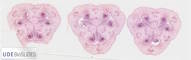 |
Plant |
Lilium sp. |
Lilly |
Ovary |
Cross section |
|
|
unknown |
unknown |
unknown |
Jungner/microthek |
2025-04-24 20:33:00 |
Olympus VS200 |
UPLXAPO 20x / 0.80 |
20x / 0.80 |
75 % |
|
Throughlight widefield |
iDS VS-264C |
Slide scanning microscope |
16.7 |
29,068 x
9,113 x
63 px³ |
7,939.1 x
2,488.8 x
36.6 µm³ |
✅
|
✅
|
✅
|
❌ |
| 443 |
Lilium sp. - ovary, older development stage, longitudinal section 20x |
 |
Plant |
Lilium sp. |
Lilly |
Ovary |
Longitudinal section |
|
|
unknown |
unknown |
unknown |
Jungner/microthek |
2025-04-25 14:23:18 |
Olympus VS200 |
UPLXAPO 20x / 0.80 |
20x / 0.80 |
75 % |
|
Throughlight widefield |
iDS VS-264C |
Slide scanning microscope |
59.7 |
32,722 x
28,951 x
63 px³ |
8,937.2 x
7,907.1 x
36.6 µm³ |
✅
|
✅
|
✅
|
❌ |
| 434 |
Lilium sp. - Pollen development, early prophase, cross section 20x |
 |
Plant |
Lilium sp. |
Lilly |
Pollen |
Cross section |
|
|
unknown |
unknown |
unknown |
Jungner/microthek |
2025-04-23 16:02:23 |
Olympus VS200 |
UPLXAPO 20x / 0.80 |
20x / 0.80 |
75 % |
|
Throughlight widefield |
iDS VS-264C |
Slide scanning microscope |
29.2 |
20,753 x
18,295 x
77 px³ |
5,668.0 x
4,996.7 x
44.8 µm³ |
✅
|
✅
|
✅
|
❌ |
| 435 |
Lilium sp. - Pollen development, late prophase, cross section 20x |
 |
Plant |
Lilium sp. |
Lilly |
Pollen |
Cross section |
|
|
unknown |
unknown |
unknown |
Jungner/microthek |
2025-04-23 16:27:44 |
Olympus VS200 |
UPLXAPO 20x / 0.80 |
20x / 0.80 |
75 % |
|
Throughlight widefield |
iDS VS-264C |
Slide scanning microscope |
2.8 |
8,365 x
5,229 x
63 px³ |
2,284.6 x
1,428.0 x
36.6 µm³ |
✅
|
✅
|
✅
|
❌ |
| 438 |
Lilium sp. - Pollen development, mature pollen grains, cross section 20x |
 |
Plant |
Lilium sp. |
Lilly |
Pollen |
Cross section |
|
|
unknown |
unknown |
unknown |
Jungner/microthek |
2025-04-24 15:50:31 |
Olympus VS200 |
UPLXAPO 20x / 0.80 |
20x / 0.80 |
75 % |
|
Throughlight widefield |
iDS VS-264C |
Slide scanning microscope |
18.6 |
19,219 x
18,295 x
53 px³ |
5,249.2 x
4,996.7 x
30.7 µm³ |
✅
|
✅
|
✅
|
❌ |
| 439 |
Lilium sp. - Pollen development, mature pollen grains, whole mount 40x |
 |
Plant |
Lilium sp. |
Lilly |
Pollen |
Whole mount |
|
|
unknown |
unknown |
unknown |
Jungner/microthek |
2025-04-24 16:21:16 |
Olympus VS200 |
UPLFLN 40x / 0.75 |
40x / 0.75 |
75 % |
|
Throughlight widefield |
iDS VS-264C |
Slide scanning microscope |
134.8 |
36,535 x
36,518 x
101 px³ |
4,999.0 x
4,996.7 x
67.0 µm³ |
✅
|
✅
|
✅
|
❌ |
| 437 |
Lilium sp. - Pollen development, mononuclear pollen grains, cross section 20x |
 |
Plant |
Lilium sp. |
Lilly |
Pollen |
Cross section |
|
|
unknown |
unknown |
unknown |
Jungner/microthek |
2025-04-24 15:27:03 |
Olympus VS200 |
UPLXAPO 20x / 0.80 |
20x / 0.80 |
75 % |
|
Throughlight widefield |
iDS VS-264C |
Slide scanning microscope |
24.1 |
20,902 x
18,295 x
63 px³ |
5,708.8 x
4,996.7 x
36.6 µm³ |
✅
|
✅
|
✅
|
❌ |
| 436 |
Lilium sp. - Pollen development, tetrad stage, cross section 20x |
 |
Plant |
Lilium sp. |
Lilly |
Pollen |
Cross section |
|
|
unknown |
unknown |
unknown |
Jungner/microthek |
2025-04-23 16:35:36 |
Olympus VS200 |
UPLXAPO 20x / 0.80 |
20x / 0.80 |
75 % |
|
Throughlight widefield |
iDS VS-264C |
Slide scanning microscope |
4.1 |
7,529 x
8,585 x
63 px³ |
2,056.2 x
2,344.7 x
36.6 µm³ |
✅
|
✅
|
✅
|
❌ |
| 440 |
Lilium sp. - Stigma, cross section 20x |
 |
Plant |
Lilium sp. |
Lilly |
Stigma |
Cross section |
|
|
unknown |
unknown |
unknown |
|
2025-04-24 19:37:20 |
Olympus VS200 |
UPLXAPO 20x / 0.80 |
20x / 0.80 |
75 % |
|
Throughlight widefield |
iDS VS-264C |
Slide scanning microscope |
12.7 |
13,723 x
10,876 x
85 px³ |
3,747.9 x
2,970.3 x
49.6 µm³ |
✅
|
✅
|
✅
|
❌ |
| 142 |
Liquidambar styraciflua - Branch cross section 20x 1 |
 |
Plant |
Liquidambar styraciflua |
American sweetgum |
Branch |
Cross section |
|
bark parenchyma, cambium/bast, cork, medullary parenchyma, primary xylem, sclerenchyma, wood |
Formol, acetic alcohol |
|
Euparal |
Rainer Teubner |
2021-10-28 14:13:49 |
Olympus VS200 |
UPLXAPO 20x / 0.80 |
20x / 0.80 |
50 % |
|
Throughlight widefield |
iDS VS-264C |
Slide scanning microscope |
18.1 |
14,269 x
14,944 x
85 px³ |
3,895.1 x
4,079.3 x
99.1 µm³ |
✅
|
✅
|
✅
|
✅
|
| 143 |
Liquidambar styraciflua - Branch cross section 20x 2 |
 |
Plant |
Liquidambar styraciflua |
American sweetgum |
Branch |
Cross section |
|
bark parenchyma, bast, cambium, cork, medullary parenchyma, primary xylem, sclerenchyma, wood |
Formol, acetic alcohol |
|
Euparal |
Rainer Teubner |
2021-10-28 14:34:13 |
Olympus VS200 |
UPLXAPO 20x / 0.80 |
20x / 0.80 |
50 % |
|
Throughlight widefield |
iDS VS-264C |
Slide scanning microscope |
22.8 |
16,163 x
16,566 x
85 px³ |
4,412.2 x
4,522.2 x
99.1 µm³ |
✅
|
✅
|
✅
|
✅
|
| 144 |
Liquidambar styraciflua - Branch cross section 20x 3 |
 |
Plant |
Liquidambar styraciflua |
American sweetgum |
Branch |
Cross section |
|
bark parenchyma, cambium/bast, cork, medullary parenchyma, primary xylem, sclerenchyma, wood |
Formol, acetic alcohol |
unstained |
Euparal |
Rainer Teubner |
2021-10-28 14:57:23 |
Olympus VS200 |
UPLXAPO 20x / 0.80 |
20x / 0.80 |
50 % |
|
Throughlight widefield |
iDS VS-264C |
Slide scanning microscope |
22.5 |
16,778 x
15,791 x
85 px³ |
4,580.0 x
4,310.4 x
99.1 µm³ |
✅
|
✅
|
✅
|
✅
|
| 140 |
Liquidambar styraciflua - Branch cross section 20x 4 |
 |
Plant |
Liquidambar styraciflua |
American sweetgum |
Branch |
Cross section |
|
bark parenchyma, bast, cambium, cork, medullary parenchyma, primary xylem, sclerenchyma, triomes, wood |
Formol, acetic alcohol |
|
Euparal |
Rainer Teubner |
2021-10-28 15:57:46 |
Olympus VS200 |
UPLXAPO 20x / 0.80 |
20x / 0.80 |
50 % |
|
Throughlight widefield |
iDS VS-264C |
Slide scanning microscope |
20.4 |
16,768 x
16,637 x
73 px³ |
4,577.2 x
4,541.5 x
85.0 µm³ |
✅
|
✅
|
✅
|
✅
|
| 138 |
Liquidambar styraciflua - Leaf cross section 20x 1 |
 |
Plant |
Liquidambar styraciflua |
American sweetgum |
Leaf |
Cross section |
|
bark parenchyma, collenchyma, crystal druse, epidermis, lower epidermis, medullary parenchyma, palisade parenchyma, phloem, sponge parenchyma, triomes, upper epidermis, xylem, xylem vessel or tracheae |
Formol, acetic alcohol |
|
Euparal |
Rainer Teubner |
2021-10-28 15:28:21 |
Olympus VS200 |
UPLXAPO 20x / 0.80 |
20x / 0.80 |
50 % |
|
Throughlight widefield |
iDS VS-264C |
Slide scanning microscope |
9.0 |
31,198 x
5,664 x
51 px³ |
8,516.3 x
1,545.9 x
59.0 µm³ |
✅
|
✅
|
✅
|
✅
|
| 139 |
Liquidambar styraciflua - Leaf cross section 20x 2 |
 |
Plant |
Liquidambar styraciflua |
American sweetgum |
Leaf |
Cross section |
|
bark parenchyma, collenchyma, crystal druse, epidermis & cuticle, lower epidermis, medullary parenchyma, palisade parenchyma, phloem, secretory duct?, sponge parenchyma, triomes, upper epidermis, xylem, xylem vessel or tracheae |
Formol, acetic alcohol |
|
Euparal |
Rainer Teubner |
2021-10-28 15:36:15 |
Olympus VS200 |
UPLXAPO 20x / 0.80 |
20x / 0.80 |
50 % |
|
Throughlight widefield |
iDS VS-264C |
Slide scanning microscope |
3.0 |
21,131 x
4,545 x
31 px³ |
5,768.3 x
1,240.6 x
35.4 µm³ |
✅
|
✅
|
✅
|
✅
|
| 141 |
Liquidambar styraciflua - Leaf stalk cross section 20x |
 |
Plant |
Liquidambar styraciflua |
American sweetgum |
Leaf stalk |
Cross section |
|
bark parenchyma, bast, collenchyma, crystal druse, epidermis & cuticle, medullary parenchyma, resin channel?, sclerenchyma, xylem/wood |
Formol, acetic alcohol, Chlorix |
|
Euparal |
Rainer Teubner |
2021-10-28 15:45:08 |
Olympus VS200 |
UPLXAPO 20x / 0.80 |
20x / 0.80 |
50 % |
|
Throughlight widefield |
iDS VS-264C |
Slide scanning microscope |
2.4 |
6,631 x
6,661 x
55 px³ |
1,810.0 x
1,818.2 x
63.7 µm³ |
✅
|
✅
|
✅
|
✅
|
| 382 |
Liquidambar styraciflua - Leaf stalk cross section 20x |
 |
Plant |
Liquidambar styraciflua |
American sweetgum |
Leaf stalk |
Cross section |
|
|
Formol, acetic alcohol |
W3Asim I: Acridine red, acriflavine, astra blue |
Euparal |
Jörg Weiß
www.mikroskopie-bonn.de |
2023-11-08 14:59:20 |
Olympus VS200 |
UPLXAPO 20x / 0.80 |
20x / 0.80 |
75 % |
|
Throughlight widefield |
iDS VS-264C |
Slide scanning microscope |
13.4 |
11,213 x
11,832 x
101 px³ |
3,062.5 x
3,231.4 x
59.0 µm³ |
✅
|
✅
|
✅
|
❌ |
| 329 |
Lithops sp. - Leaf cross section 20x |
 |
Plant |
Lithops sp. |
Living stone |
Leaf |
Cross section |
Wikipedia on Lithops sp..
Jungner/microthek micro preparation no. 1621 |
Hydrenchyme |
unknown |
unknown |
unknown |
Jungner/microthek |
2023-10-26 12:46:47 |
Olympus VS200 |
UPLXAPO 20x / 0.80 |
20x / 0.80 |
75 % |
|
Throughlight widefield |
iDS VS-264C |
Slide scanning microscope |
20.5 |
32,444 x
10,019 x
63 px³ |
8,861.1 x
2,736.3 x
36.6 µm³ |
✅
|
✅
|
✅
|
✅
|
| 339 |
Locusta migratoria - Meiosis 60x 1 |
 |
Animal |
Locusta migratoria |
Migratory locust |
Testes |
Squash technique |
|
Spermatogenesis |
Methanol : acetic acid (3:1) |
Orcein (2%) in acetic acid (50%) |
Entellan |
Christian Johannes |
2023-11-06 10:20:09 |
Olympus VS200 |
UPLXAPO OPH 60x / 1.42 |
60x / 1.42 |
75 % |
Oil |
Throughlight widefield |
iDS VS-264C |
Slide scanning microscope |
7.7 |
19,442 x
18,755 x
21 px³ |
1,767.9 x
1,705.5 x
5.6 µm³ |
✅
|
✅
|
✅
|
❌ |
| 340 |
Locusta migratoria - Meiosis 60x 2 |
 |
Animal |
Locusta migratoria |
Migratory locust |
Testes |
Squash technique |
|
Spermatogenesis |
Methanol : acetic acid (3:1) |
Orcein (2%) in acetic acid (50%) |
Entellan |
Christian Johannes |
2023-11-06 10:32:58 |
Olympus VS200 |
UPLXAPO OPH 60x / 1.42 |
60x / 1.42 |
75 % |
Oil |
Throughlight widefield |
iDS VS-264C |
Slide scanning microscope |
7.6 |
24,647 x
11,367 x
27 px³ |
2,241.3 x
1,033.6 x
7.3 µm³ |
✅
|
✅
|
✅
|
❌ |
| 341 |
Locusta migratoria - Meiosis 60x 3 |
 |
Animal |
Locusta migratoria |
Migratory locust |
Testes |
Squash technique |
|
Spermatogenesis |
Methanol : acetic acid (3:1) |
Orcein (2%) in acetic acid (50%) |
Entellan |
Christian Johannes |
2023-11-06 10:48:18 |
Olympus VS200 |
UPLXAPO OPH 60x / 1.42 |
60x / 1.42 |
75 % |
Oil |
Throughlight widefield |
iDS VS-264C |
Slide scanning microscope |
12.7 |
19,622 x
18,544 x
35 px³ |
1,784.3 x
1,686.3 x
9.5 µm³ |
✅
|
✅
|
✅
|
❌ |
| 223 |
Lumbricus terrestris - tip longitudinal section 40x |
 |
Animal |
Lumbricus terrestris |
Common Earthworm |
Tip |
Longitudinal section |
Wikipedia on Lumbricus terrestris
Due to its weak contrast, for this specimen it is beneficial to increase the value for "Gamma" to about 2.5 (see above unter "Image processing") |
|
Bouin, paraffin |
|
unknown |
|
2023-03-15 13:37:20 |
Olympus VS200 |
UPLXAPO O 40x / 1.40 |
40x / 1.40 |
75 % |
Oil |
Throughlight widefield |
iDS VS-264C |
Slide scanning microscope |
237.6 |
33,979 x
82,282 x
85 px³ |
4,655.8 x
11,274.5 x
24.4 µm³ |
✅
|
✅
|
✅
|
❌ |
| 460 |
Lupinus sp. - Root cross section 20x |
 |
Plant |
Lupinus sp. |
Lupin |
Root |
Cross section |
|
|
unknown |
unknown |
unknown |
Jungner/microthek |
2025-05-02 21:22:43 |
Olympus VS200 |
UPLXAPO 20x / 0.80 |
20x / 0.80 |
75 % |
|
Throughlight widefield |
iDS VS-264C |
Slide scanning microscope |
73.0 |
29,675 x
24,350 x
101 px³ |
8,105.0 x
6,650.5 x
59.0 µm³ |
✅
|
✅
|
✅
|
❌ |
| 592 |
Lycopodium sp. - Spores 40x |
 |
Plant |
Lycopodium sp. |
Ground pine |
Spores |
Whole mount |
|
|
unknown |
unknown |
unknown |
Jungner/microthek |
2025-06-25 15:12:52 |
Olympus VS200 |
UPLXAPO O 40x / 1.40 |
40x / 1.40 |
75 % |
Oil |
Throughlight widefield |
iDS VS-264C |
Slide scanning microscope |
55.9 |
24,690 x
22,429 x
101 px³ |
3,383.9 x
3,073.9 x
59.0 µm³ |
✅
|
✅
|
✅
|
❌ |
| 591 |
Lycopodium sp. - Sporophyll cross section 20x |
 |
Plant |
Lycopodium sp. |
Ground pine |
Sporophyll |
Cross section |
|
|
unknown |
unknown |
unknown |
Jungner/microthek |
2025-06-25 11:21:04 |
Olympus VS200 |
UPLXAPO 20x / 0.80 |
20x / 0.80 |
75 % |
|
Throughlight widefield |
iDS VS-264C |
Slide scanning microscope |
3.6 |
8,437 x
14,583 x
29 px³ |
2,304.1 x
3,982.5 x
16.5 µm³ |
✅
|
✅
|
✅
|
❌ |
| 590 |
Lycopodium sp. - Stem cross section 20x |
 |
Plant |
Lycopodium sp. |
Ground pine |
Stem |
Cross section |
|
|
unknown |
unknown |
unknown |
Jungner/microthek |
2025-06-25 10:39:39 |
Olympus VS200 |
UPLXAPO 20x / 0.80 |
20x / 0.80 |
75 % |
|
Throughlight widefield |
iDS VS-264C |
Slide scanning microscope |
11.8 |
13,585 x
11,017 x
79 px³ |
3,710.1 x
3,008.6 x
46.0 µm³ |
✅
|
✅
|
✅
|
❌ |
| 148 |
Malus domestica - Branch cross section 20x 1 |
 |
Plant |
Malus domestica |
Apple tree |
Branch |
Cross section |
|
bark parenchyma, bast, cambium, cork, epidermis, medullary parenchyma, primary xylem, sclerenchyma, wood |
Formol, acetic alcohol, sodium hypochlorite |
|
Euparal |
Rainer Teubner |
2021-11-05 11:07:40 |
Olympus VS200 |
UPLXAPO 20x / 0.80 |
20x / 0.80 |
50 % |
|
Throughlight widefield |
iDS VS-264C |
Slide scanning microscope |
16.4 |
13,896 x
13,886 x
85 px³ |
3,793.3 x
3,790.5 x
99.1 µm³ |
✅
|
✅
|
✅
|
✅
|
| 149 |
Malus domestica - Branch cross section 20x 2 |
 |
Plant |
Malus domestica |
Apple tree |
Branch |
Cross section |
|
bark parenchyma, bast, cambium, cork, epidermis & cuticle, medullary parenchyma, primary xylem, sclerenchyma, wood |
Formol, acetic alcohol, sodium hypochlorite |
|
Euparal |
Rainer Teubner |
2021-11-05 11:28:03 |
Olympus VS200 |
UPLXAPO 20x / 0.80 |
20x / 0.80 |
50 % |
|
Throughlight widefield |
iDS VS-264C |
Slide scanning microscope |
16.2 |
13,402 x
14,229 x
85 px³ |
3,658.5 x
3,884.0 x
99.1 µm³ |
✅
|
✅
|
✅
|
✅
|
| 150 |
Malus domestica - Branch cross section 20x 3 |
 |
Plant |
Malus domestica |
Apple tree |
Branch |
Cross section |
|
bark parenchyma, bast, cambium, cork, epidermis, medullary parenchyma, primary xylem, sclerenchyma, wood |
Formol, acetic alcohol, sodium hypochlorite |
Wacker |
Euparal |
Rainer Teubner |
2021-11-05 11:44:39 |
Olympus VS200 |
UPLXAPO 20x / 0.80 |
20x / 0.80 |
50 % |
|
Throughlight widefield |
iDS VS-264C |
Slide scanning microscope |
16.9 |
13,967 x
14,198 x
85 px³ |
3,812.5 x
3,875.8 x
99.1 µm³ |
✅
|
✅
|
✅
|
✅
|
| 151 |
Malus domestica - Branch cross section 20x 4 |
 |
Plant |
Malus domestica |
Apple tree |
Branch |
Cross section |
|
bark parenchyma, cambium/bast, cork, crystal druse, epidermis, medullary parenchyma, primary xylem, wood |
Formol, acetic alcohol, sodium hypochlorite |
unstained |
Euparal |
Rainer Teubner |
2021-11-05 12:59:13 |
Olympus VS200 |
UPLXAPO 20x / 0.80 |
20x / 0.80 |
50 % |
|
Throughlight widefield |
iDS VS-264C |
Slide scanning microscope |
18.6 |
14,118 x
15,468 x
85 px³ |
3,853.8 x
4,222.4 x
99.1 µm³ |
✅
|
✅
|
✅
|
✅
|
| 145 |
Malus domestica - Branch longitudinal section 20x 1 |
 |
Plant |
Malus domestica |
Apple tree |
Branch |
Longitudinal section |
|
bark parenchyma, bast, cork, crystal druse, medullary parenchyma, wood |
? |
|
Euparal |
Rainer Teubner |
2021-10-14 10:27:08 |
Olympus VS200 |
UPLXAPO 20x / 0.80 |
20x / 0.80 |
50 % |
|
Throughlight widefield |
iDS VS-264C |
Slide scanning microscope |
22.5 |
13,957 x
18,965 x
85 px³ |
3,809.8 x
5,176.9 x
99.1 µm³ |
✅
|
✅
|
✅
|
✅
|
| 146 |
Malus domestica - Branch longitudinal section 20x 2 |
 |
Plant |
Malus domestica |
Apple tree |
Branch |
Longitudinal section |
|
bark parenchyma, bast, cork, crystal druse, medullary parenchyma, wood |
? |
unstained |
Euparal |
Rainer Teubner |
2021-10-14 11:00:07 |
Olympus VS200 |
UPLXAPO 20x / 0.80 |
20x / 0.80 |
50 % |
|
Throughlight widefield |
iDS VS-264C |
Slide scanning microscope |
26.5 |
14,239 x
21,877 x
85 px³ |
3,886.8 x
5,971.8 x
99.1 µm³ |
✅
|
✅
|
✅
|
✅
|
| 546 |
Malus domestica - Fruit cross section 20x |
 |
Plant |
Malus domestica (a.k.a Pyrus malus) |
Apple |
Fruit |
Cross section |
|
Fleshy hypanthium, Individual follicle (fruit), Lenticel, Pericarp, Skin with wax layer, Vascular tissue |
unknown |
unknown |
unknown |
Jungner/microthek |
2025-05-22 16:41:11 |
Olympus VS200 |
UPLXAPO 20x / 0.80 |
20x / 0.80 |
75 % |
|
Throughlight widefield |
iDS VS-264C |
Slide scanning microscope |
76.4 |
27,367 x
28,194 x
99 px³ |
7,474.5 x
7,700.4 x
57.8 µm³ |
✅
|
✅
|
✅
|
✅
|
| 689 |
Malus domestica - fruiting shoot cross section 20x |
 |
Plant |
Malus domestica |
Apple tree |
Shoot |
Cross section |
|
|
unknown |
unknown |
unknown |
Jungner/microthek |
2025-08-13 10:47:47 |
Olympus VS200 |
UPLXAPO 20x / 0.80 |
20x / 0.80 |
75 % |
|
Throughlight widefield |
iDS VS-264C |
Slide scanning microscope |
20.0 |
13,985 x
14,144 x
101 px³ |
3,819.4 x
3,862.8 x
100.0 µm³ |
✅
|
✅
|
✅
|
❌ |
| 82 |
Malus domestica - Pedicel cross section 20x 1 |
 |
Plant |
Malus domestica |
Apple tree |
Pedicel |
Cross section |
|
bark parenchyma, cambium, collenchyma, epidermis & cuticle, medullary parenchyma, phloem/ bast, primary xylem, sclerenchyma, stone cell, xylem/wood |
Formol, acetic alcohol |
|
Euparal |
Rainer Teubner |
2021-07-01 14:26:42 |
Olympus VS200 |
UPLXAPO 20x / 0.80 |
20x / 0.80 |
40 % |
|
Throughlight widefield |
iDS VS-264C |
Slide scanning microscope |
6.8 |
9,966 x
11,175 x
61 px³ |
2,720.5 x
3,050.6 x
70.8 µm³ |
✅
|
✅
|
✅
|
✅
|
| 83 |
Malus domestica - Pedicel cross section 20x 2 |
 |
Plant |
Malus domestica |
Apple tree |
Pedicel |
Cross section |
|
bark parenchyma, cambium, collenchyma, epidermis & cuticle, medullary parenchyma without thickened cell wall, medullary parenchyma with thickened cell wall, phloem/bast, primary xylem, sclerenchyma, stone cell, triome, xylem/wood |
Formol, acetic alcohol |
|
Euparal |
Rainer Teubner |
2021-07-01 14:38:08 |
Olympus VS200 |
UPLXAPO 20x / 0.80 |
20x / 0.80 |
40 % |
|
Throughlight widefield |
iDS VS-264C |
Slide scanning microscope |
7.8 |
11,327 x
11,317 x
61 px³ |
3,091.8 x
3,089.1 x
70.8 µm³ |
✅
|
✅
|
✅
|
✅
|
| 84 |
Malus domestica - Pedicel cross section 20x 3 |
 |
Plant |
Malus domestica |
Apple tree |
Pedicel |
Cross section |
|
bark parenchyma, cambium, collenchyma, epidermis & cuticle, medullary parenchyma without thickened cell wall, medullary parenchyma with thikened cell walls, primary xylem, sclerenchyma, sec. phloem / bast, sec. xylem / wood, stone cell |
Formol, acetic alcohol |
|
Euparal |
Rainer Teubner |
2021-07-02 08:37:05 |
Olympus VS200 |
UPLXAPO 20x / 0.80 |
20x / 0.80 |
40 % |
|
Throughlight widefield |
iDS VS-264C |
Slide scanning microscope |
7.3 |
9,916 x
12,133 x
61 px³ |
2,706.7 x
3,311.9 x
70.8 µm³ |
✅
|
✅
|
✅
|
✅
|
| 147 |
Malus domestica - Pedicel cross section 20x 3 |
 |
Plant |
Malus domestica |
Apple tree |
Pedicel |
Cross section |
|
bark parenchyma, collenchyma, epidermis, medullary parenchyma, phloem, sclerenchyma, stone cell, xylem |
Formol, acetic alcohol |
unstained |
Euparal |
Rainer Teubner |
2021-11-05 10:32:05 |
Olympus VS200 |
UPLXAPO 20x / 0.80 |
20x / 0.80 |
50 % |
|
Throughlight widefield |
iDS VS-264C |
Slide scanning microscope |
16.9 |
14,370 x
15,297 x
77 px³ |
3,922.6 x
4,175.6 x
89.7 µm³ |
✅
|
✅
|
✅
|
✅
|
| 608 |
Malus domestica - Wood cross section 20x |
 |
Plant |
Malus domestica |
Apple |
Wood |
Cross section |
Wikipedia on Malus domestica (also known as Pyrus malus).
This sample was repaired after shrinking of the mounting medium due to aging, and thus has some minor damage.
Jungner/microthek micro preparation no. 745 |
|
unknown |
unstained |
unknown |
Jungner/microthek |
2025-07-03 20:28:00 |
Olympus VS200 |
UPLXAPO 20x / 0.80 |
20x / 0.80 |
75 % |
|
Throughlight widefield |
iDS VS-264C |
Slide scanning microscope |
74.3 |
28,189 x
26,087 x
101 px³ |
7,698.6 x
7,124.5 x
59.0 µm³ |
✅
|
✅
|
✅
|
❌ |
| 581 |
Marchantia sp. - Antheridial stalk longitudinal section 20x |
 |
Plant |
Marchantia sp. |
Marchantia |
Antheridial stalk |
Longitudinal section |
|
|
unknown |
unknown |
unknown |
Jungner/microthek |
2025-06-24 18:32:39 |
Olympus VS200 |
UPLXAPO 20x / 0.80 |
20x / 0.80 |
75 % |
|
Throughlight widefield |
iDS VS-264C |
Slide scanning microscope |
7.2 |
18,605 x
6,515 x
59 px³ |
5,081.0 x
1,779.1 x
34.2 µm³ |
✅
|
✅
|
✅
|
❌ |
| 582 |
Marchantia sp. - Archegonial stalk longitudinal section 20x |
 |
Plant |
Marchantia sp. |
Marchantia |
Archegonial stalk |
Longitudinal section |
|
|
unknown |
unknown |
unknown |
Jungner/microthek |
2025-06-24 18:48:38 |
Olympus VS200 |
UPLXAPO 20x / 0.80 |
20x / 0.80 |
75 % |
|
Throughlight widefield |
iDS VS-264C |
Slide scanning microscope |
11.8 |
18,884 x
7,331 x
85 px³ |
5,157.2 x
2,002.1 x
49.6 µm³ |
✅
|
✅
|
✅
|
❌ |
| 583 |
Marchantia sp. - Thallus cross section 20x |
 |
Plant |
Marchantia sp. |
Marchantia |
Thallus |
Cross section |
|
|
unknown |
unknown |
unknown |
Jungner/microthek |
2025-06-24 19:05:55 |
Olympus VS200 |
UPLXAPO 20x / 0.80 |
20x / 0.80 |
75 % |
|
Throughlight widefield |
iDS VS-264C |
Slide scanning microscope |
33.5 |
26,775 x
26,645 x
47 px³ |
7,312.3 x
7,276.8 x
27.1 µm³ |
✅
|
✅
|
✅
|
❌ |
| 606 |
Matricaria chamomilla - Flower cross section 20x |
 |
Plant |
Matricaria chamomilla |
German chamomile |
Flower |
Cross section |
|
Disc floret, Hollow receptacle, Ray floret |
unknown |
unknown |
unknown |
Jungner/microthek |
2025-07-02 12:41:34 |
Olympus VS200 |
UPLXAPO 20x / 0.80 |
20x / 0.80 |
75 % |
|
Throughlight widefield |
iDS VS-264C |
Slide scanning microscope |
57.9 |
31,801 x
23,029 x
79 px³ |
8,685.1 x
6,289.3 x
46.0 µm³ |
✅
|
✅
|
✅
|
✅
|
| 451 |
Medicago sativa - Root cross section 20x, Jungner 513 |
 |
Plant |
Medicago sativa |
Alfalfa |
Root |
Cross section |
|
|
unknown |
unknown |
unknown |
Jungner/microthek |
2025-04-30 22:05:57 |
Olympus VS200 |
UPLXAPO 20x / 0.80 |
20x / 0.80 |
75 % |
|
Throughlight widefield |
iDS VS-264C |
Slide scanning microscope |
26.5 |
18,154 x
16,413 x
89 px³ |
4,958.2 x
4,482.6 x
51.9 µm³ |
✅
|
✅
|
✅
|
❌ |
| 152 |
Mentha speciosa - Sprout cross section 20x 1 |
 |
Plant |
Mentha speciosa |
Mint |
Sprout |
Cross section |
|
cambium, collenchyma, endodermis?, epidermis & cuticle, medullary parenchyma, phloem, sclerenchyma, xylem, xylem vessel or tracheae |
Formol, acetic alcohol |
|
Euparal |
Rainer Teubner |
2021-11-02 14:13:41 |
Olympus VS200 |
UPLXAPO 20x / 0.80 |
20x / 0.80 |
50 % |
|
Throughlight widefield |
iDS VS-264C |
Slide scanning microscope |
9.0 |
10,349 x
10,762 x
81 px³ |
2,825.0 x
2,937.8 x
94.4 µm³ |
✅
|
✅
|
✅
|
✅
|
| 153 |
Mentha speciosa - Sprout cross section 20x 2 |
 |
Plant |
Mentha speciosa |
Mint |
Sprout |
Cross section |
|
cambium, collenchyma, endodermis?, epidermis & cuticle, medullary parenchyma, phloem, primary xylem, sclerenchyma, xylem, xylem vessel or tracheae |
Formol, acetic alcohol |
|
Euparal |
Rainer Teubner |
2021-11-02 14:31:38 |
Olympus VS200 |
UPLXAPO 20x / 0.80 |
20x / 0.80 |
50 % |
|
Throughlight widefield |
iDS VS-264C |
Slide scanning microscope |
10.8 |
11,468 x
11,085 x
85 px³ |
3,130.4 x
3,025.8 x
99.1 µm³ |
✅
|
✅
|
✅
|
✅
|
| 154 |
Mentha speciosa - Sprout cross section 20x 3 |
 |
Plant |
Mentha speciosa |
Mint |
Sprout |
Cross section |
|
assimilation parenchyma?, collenchyma, endodermis?, epidermis & cuticle, medullary parenchyma, phloem, triome, xylem, xylem vessel or tracheae |
Formol, acetic alcohol |
unstained |
Euparal |
Rainer Teubner |
2021-11-02 14:44:01 |
Olympus VS200 |
UPLXAPO 20x / 0.80 |
20x / 0.80 |
50 % |
|
Throughlight widefield |
iDS VS-264C |
Slide scanning microscope |
7.0 |
9,392 x
8,727 x
85 px³ |
2,563.7 x
2,382.1 x
99.1 µm³ |
✅
|
✅
|
✅
|
✅
|
| 155 |
Mentha spicata - Sprout cross section 20x 1 |
 |
Plant |
Mentha spicata |
Spearmint |
Sprout |
Cross section |
|
cambium, collenchyma, endodermis?, epidermis & cuticle, medullary parenchyma, phloem, primary xylem, sec. xylem / wood, triomes, xylem vessel or tracheae |
Formol, acetic alcohol |
|
Euparal |
Rainer Teubner |
2021-11-02 12:09:50 |
Olympus VS200 |
UPLXAPO 20x / 0.80 |
20x / 0.80 |
50 % |
|
Throughlight widefield |
iDS VS-264C |
Slide scanning microscope |
6.0 |
8,928 x
10,299 x
65 px³ |
2,437.2 x
2,811.2 x
75.5 µm³ |
✅
|
✅
|
✅
|
✅
|
| 156 |
Mentha spicata - Sprout cross section 20x 2 |
 |
Plant |
Mentha spicata |
Spearmint |
Sprout |
Cross section |
|
cambium, collenchyma, epidermis & cuticle, medullary parenchyma, parenchyma cells? endodermis?, phloem / Bast?, primary xylem, sec. xylem / wood, triomes, xylem vessel or tracheae |
Formol, acetic alcohol |
|
Euparal |
Rainer Teubner |
2021-11-02 13:45:10 |
Olympus VS200 |
UPLXAPO 20x / 0.80 |
20x / 0.80 |
50 % |
|
Throughlight widefield |
iDS VS-264C |
Slide scanning microscope |
5.9 |
9,352 x
8,626 x
73 px³ |
2,552.7 x
2,354.6 x
85.0 µm³ |
✅
|
✅
|
✅
|
✅
|
| 157 |
Mentha spicata - Sprout cross section 20x 3 |
 |
Plant |
Mentha spicata |
Spearmint |
Sprout |
Cross section |
|
collenchyma, endodermis, epidermis & cuticle, medullary parenchyma, phloem, triomes, xylem, xylem vessel or tracheae |
Formol, acetic alcohol |
unstained |
Euparal |
Rainer Teubner |
2021-11-02 13:59:01 |
Olympus VS200 |
UPLXAPO 20x / 0.80 |
20x / 0.80 |
50 % |
|
Throughlight widefield |
iDS VS-264C |
Slide scanning microscope |
5.5 |
8,132 x
8,414 x
81 px³ |
2,219.8 x
2,296.9 x
94.4 µm³ |
✅
|
✅
|
✅
|
✅
|
| 408 |
Mesothaenium sp. - 40x |
 |
Algae |
Mesotaenium sp. |
Mesotaenium |
Whole organism |
Whole mount |
|
|
unknown |
unknown |
unknown |
Jungner/microthek |
2025-03-31 18:16:41 |
Olympus VS200 |
UPLXAPO O 40x / 1.40 |
40x / 1.40 |
75 % |
Oil |
Throughlight widefield |
iDS VS-264C |
Slide scanning microscope |
82.2 |
26,896 x
30,253 x
101 px³ |
3,685.3 x
4,145.3 x
59.0 µm³ |
✅
|
✅
|
✅
|
❌ |
| 641 |
Micrasterias radiosa 20x |
 |
Protist |
Micrasterias radiosa |
Micrasterias radiosa |
Whole cell |
Whole organism |
|
|
not fixated |
not stained |
Water |
Michael Kloster / Eva Papendick |
2025-07-18 12:16:27 |
Olympus VS200 |
UPLXAPO 20x / 0.80 |
20x / 0.80 |
75 % |
|
Throughlight widefield |
iDS VS-264C |
Slide scanning microscope |
25.8 |
15,111 x
16,883 x
101 px³ |
4,126.8 x
4,610.9 x
59.0 µm³ |
✅
|
✅
|
✅
|
❌ |
| 197 |
Micrasterias rotata - Living sample 60x |
 |
Protist |
Micrasterias rotata |
Micrasterias rotata |
Whole cell |
Whole organism |
|
|
not fixated |
not stained |
Agarose 2% |
Nadine Graupner |
2022-02-22 23:10:02 |
Olympus VS200 |
UPLXAPO OPH 60x / 1.42 |
60x / 1.42 |
40 % |
Oil |
Throughlight widefield |
iDS VS-264C |
Slide scanning microscope |
184.9 |
55,054 x
55,053 x
61 px³ |
4,998.0 x
4,998.0 x
68.4 µm³ |
✅
|
✅
|
✅
|
❌ |
| 405 |
Microcystis sp. - 40x |
 |
Cyanobacterium |
Microcystis sp. |
Microcystis |
Whole organism |
Whole mount |
Microcystis sp. is a cyanobacterium that builds aggregates of many cells embedded in a gelatinous matrix. Wikipedia on Microcystis sp.
Jungner/microthek micro preparation no. 951 |
|
unknown |
unknown |
unknown |
Jungner/microthek |
2025-03-31 12:17:54 |
Olympus VS200 |
UPLXAPO O 40x / 1.40 |
40x / 1.40 |
75 % |
Oil |
Throughlight widefield |
iDS VS-264C |
Slide scanning microscope |
134.4 |
36,484 x
36,466 x
101 px³ |
4,999.0 x
4,996.7 x
29.0 µm³ |
✅
|
✅
|
✅
|
❌ |
| 586 |
Mnium sp. - Spore capsule cross section 20x |
 |
Plant |
Mnium sp. |
Mnium |
Spore capsule |
Cross section |
|
|
unknown |
unknown |
unknown |
Jungner/microthek |
2025-06-25 09:41:29 |
Olympus VS200 |
UPLXAPO 20x / 0.80 |
20x / 0.80 |
75 % |
|
Throughlight widefield |
iDS VS-264C |
Slide scanning microscope |
18.1 |
15,459 x
15,180 x
77 px³ |
4,222.0 x
4,145.7 x
44.8 µm³ |
✅
|
✅
|
✅
|
❌ |
| 587 |
Mnium sp. - Thallus cross section 20x |
 |
Plant |
Mnium sp. |
Mnium |
Thallus |
Cross section |
|
|
unknown |
unknown |
unknown |
Jungner/microthek |
2025-06-25 09:59:30 |
Olympus VS200 |
UPLXAPO 20x / 0.80 |
20x / 0.80 |
75 % |
|
Throughlight widefield |
iDS VS-264C |
Slide scanning microscope |
5.1 |
13,307 x
6,933 x
55 px³ |
3,634.1 x
1,893.3 x
31.9 µm³ |
✅
|
✅
|
✅
|
❌ |
| 356 |
Monocystis sp. - in earth worm cross section 40x |
 |
Protist |
Monocystis sp. |
Monocystis |
Whole cell |
Cross section |
Wikipedia on Monocystis sp.
Jungner/microthek micro preparation no. 128 |
Cyst with gregarines, Free oocysts, Parasite, Sperm cells of the earthworm |
unknown |
unknown |
unknown |
Jungner/microthek |
2023-12-05 13:02:26 |
Olympus VS200 |
UPLXAPO O 40x / 1.40 |
40x / 1.40 |
75 % |
Oil |
Throughlight widefield |
iDS VS-264C |
Slide scanning microscope |
125.0 |
39,699 x
31,166 x
101 px³ |
5,439.6 x
4,270.5 x
29.0 µm³ |
✅
|
✅
|
✅
|
✅
|
| 467 |
Monstera deliciosa - Aerial root cross section 20x |
 |
Plant |
Monstera deliciosa |
Split-leaf philodendron |
Root |
Cross section |
|
|
unknown |
unknown |
unknown |
Jungner/microthek |
2025-05-02 22:52:11 |
Olympus VS200 |
UPLXAPO 20x / 0.80 |
20x / 0.80 |
75 % |
|
Throughlight widefield |
iDS VS-264C |
Slide scanning microscope |
41.5 |
20,743 x
19,819 x
101 px³ |
5,665.3 x
5,412.9 x
100.0 µm³ |
✅
|
✅
|
✅
|
❌ |
| 651 |
Monstera deliciosa - Leaf stalk cross section 20x |
 |
Plant |
Monstera deliciosa |
Split-leaf philodendron |
Leaf stalk |
Cross section |
Wikipedia on Monstera deliciosa.
This sample has some damage (partly large bubbles) caused by shrinking of the mounting medium due to ageing.
Jungner/microthek micro preparation no. 1292 |
|
unknown |
unknown |
unknown |
Jungner/microthek |
2025-07-17 09:47:09 |
Olympus VS200 |
UPLXAPO 20x / 0.80 |
20x / 0.80 |
75 % |
|
Throughlight widefield |
iDS VS-264C |
Slide scanning microscope |
51.2 |
21,403 x
23,706 x
101 px³ |
5,845.4 x
6,474.3 x
59.0 µm³ |
✅
|
✅
|
✅
|
❌ |
| 572 |
Morchella sp. - Fruiting body cross section 20x |
 |
Fungus |
Morchella sp. |
Morel |
Fruiting body |
Cross section |
|
|
unknown |
unknown |
unknown |
Jungner/microthek |
2025-06-23 20:05:13 |
Olympus VS200 |
UPLXAPO 20x / 0.80 |
20x / 0.80 |
75 % |
|
Throughlight widefield |
iDS VS-264C |
Slide scanning microscope |
33.9 |
28,418 x
22,521 x
53 px³ |
7,761.2 x
6,150.6 x
30.7 µm³ |
✅
|
✅
|
✅
|
❌ |
| 548 |
Morus alba - Fruit longitudinal section 20x |
 |
Plant |
Morus alba |
White mulberry |
Fruit |
Longitudinal section |
|
Fleshy sepals, Individual fruit, Pericarp, Receptacle, Seed, Stem of the inflorescence, Trichomes, Vascular tissue |
unknown |
unknown |
unknown |
Jungner/microthek |
2025-05-23 10:13:29 |
Olympus VS200 |
UPLXAPO 20x / 0.80 |
20x / 0.80 |
75 % |
|
Throughlight widefield |
iDS VS-264C |
Slide scanning microscope |
23.1 |
27,763 x
17,727 x
47 px³ |
7,582.8 x
4,841.7 x
27.1 µm³ |
✅
|
✅
|
✅
|
✅
|
| 191 |
Mus musculus - Embryo HE 40x |
 |
Animal |
Mus musculus |
House mouse |
Embryo |
Sagittal thin section |
|
|
unknown |
|
DPX |
Manuela Wülling |
2021-12-06 13:07:41 |
Olympus VS200 |
UPLXAPO O 40x / 1.40 |
40x / 1.40 |
50 % |
Oil |
Throughlight widefield |
iDS VS-264C |
Slide scanning microscope |
253.4 |
69,736 x
49,783 x
73 px³ |
9,594.6 x
6,849.3 x
20.9 µm³ |
✅
|
✅
|
✅
|
❌ |
| 190 |
Mus musculus - Embryo Toluidine blue 40x |
 |
Animal |
Mus musculus |
House mouse |
Embryo |
Sagittal thin section |
|
|
unknown |
|
DPX |
Manuela Wülling |
2021-12-06 11:52:14 |
Olympus VS200 |
UPLXAPO O 40x / 1.40 |
40x / 1.40 |
50 % |
Oil |
Throughlight widefield |
iDS VS-264C |
Slide scanning microscope |
100.3 |
68,417 x
41,886 x
35 px³ |
9,413.1 x
5,762.8 x
9.9 µm³ |
✅
|
✅
|
✅
|
❌ |
| 37 |
Mus musculus - Forelimb 60x |
 |
Animal |
Mus musculus |
House mouse |
Forelimb |
Longitudinal section |
|
|
unknown |
SW-Stain |
unknown |
|
2021-07-13 09:00:43 |
Olympus VS200 |
UPLXAPO OPH 60x / 1.42 |
60x / 1.42 |
75 % |
Oil |
Throughlight widefield |
iDS VS-264C |
Slide scanning microscope |
204.6 |
76,598 x
43,783 x
61 px³ |
6,953.9 x
3,974.8 x
16.8 µm³ |
✅
|
✅
|
✅
|
❌ |
| 432 |
Mus musculus - Head longitudinal section quer 40x |
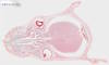 |
Animal |
Mus musculus |
House mouse |
Head |
Frontal thin section |
|
|
unknown |
|
unknown |
Karl Brügmann |
2023-11-30 14:25:09 |
Olympus VS200 |
UPLXAPO O 40x / 1.40 |
40x / 1.40 |
75 % |
Oil |
Throughlight widefield |
iDS VS-264C |
Slide scanning microscope |
1,020.9 |
152,583 x
91,652 x
73 px³ |
20,907.4 x
12,558.3 x
20.9 µm³ |
✅
|
✅
|
✅
|
❌ |
| 635 |
Musa sp. - Leaf stalk cross section 20x |
 |
Plant |
Musa sp. |
Banana plant |
Leaf stalk |
Cross section |
|
|
unknown |
unknown |
unknown |
Jungner/microthek |
2025-07-16 09:48:53 |
Olympus VS200 |
UPLXAPO 20x / 0.80 |
20x / 0.80 |
75 % |
|
Throughlight widefield |
iDS VS-264C |
Slide scanning microscope |
15.2 |
13,516 x
21,286 x
53 px³ |
3,691.1 x
5,813.3 x
30.7 µm³ |
✅
|
✅
|
✅
|
❌ |
| 72 |
Musa x paradisica - Pedicel cross section 20x |
 |
Plant |
Musa x paradisica |
Banana |
Pedicel |
Cross section |
|
bark parenchyma, epidermis, periderm, phloem, sclerenchyma, vascular bundle, xylem, xylem vessel or tracheae |
Formol, acetic alcohol |
|
Euparal |
Rainer Teubner |
2021-08-26 11:04:13 |
Olympus VS200 |
UPLXAPO 20x / 0.80 |
20x / 0.80 |
50 % |
|
Throughlight widefield |
iDS VS-264C |
Slide scanning microscope |
60.7 |
33,667 x
31,631 x
57 px³ |
9,190.2 x
8,634.5 x
99.1 µm³ |
✅
|
✅
|
✅
|
✅
|
| 709 |
Myriophyllum sp. - Stem cross section 20x |
 |
Plant |
Myriophyllum sp. |
Water milfoil |
Stem |
Cross section |
|
|
unknown |
unknown |
unknown |
Jungner/microthek |
2025-08-21 09:53:27 |
Olympus VS200 |
UPLXAPO 20x / 0.80 |
20x / 0.80 |
75 % |
|
Throughlight widefield |
iDS VS-264C |
Slide scanning microscope |
0.7 |
8,028 x
2,809 x
33 px³ |
2,192.4 x
767.1 x
18.9 µm³ |
✅
|
✅
|
✅
|
❌ |
| 328 |
Myriophyllum sp. Stem and leaves cross section 20x 1 |
 |
Plant |
Myriophyllum sp. |
Water milfoil |
Stem and Leaf |
Cross section |
|
Hydrophyte |
unknown |
unknown |
Eukitt |
Jungner/microthek |
2023-10-26 09:05:52 |
Olympus VS200 |
UPLXAPO 20x / 0.80 |
20x / 0.80 |
75 % |
|
Throughlight widefield |
iDS VS-264C |
Slide scanning microscope |
5.2 |
16,074 x
9,232 x
35 px³ |
4,390.1 x
2,521.5 x
20.1 µm³ |
✅
|
✅
|
✅
|
❌ |
| 327 |
Myriophyllum sp. Stem and leaves cross section 20x 2 |
 |
Plant |
Myriophyllum sp. |
Water milfoil |
Stem and Leaf |
Cross section |
|
Hydrophyte |
unknown |
unknown |
Eukitt |
Jungner/microthek |
2023-10-26 09:16:46 |
Olympus VS200 |
UPLXAPO 20x / 0.80 |
20x / 0.80 |
75 % |
|
Throughlight widefield |
iDS VS-264C |
Slide scanning microscope |
7.0 |
12,509 x
15,985 x
35 px³ |
3,416.3 x
4,365.6 x
20.1 µm³ |
✅
|
✅
|
✅
|
❌ |
| 537 |
Myristica sp. - Fruit cross section 20x |
 |
Plant |
Myristica sp. |
Nutmeg |
Fruit |
Cross section |
|
|
unknown |
unknown |
unknown |
Jungner/microthek |
2025-05-22 05:43:53 |
Olympus VS200 |
UPLXAPO 20x / 0.80 |
20x / 0.80 |
75 % |
|
Throughlight widefield |
iDS VS-264C |
Slide scanning microscope |
46.0 |
25,144 x
18,106 x
101 px³ |
6,867.5 x
4,945.0 x
70.0 µm³ |
✅
|
✅
|
✅
|
❌ |
| 163 |
Myrtus communis - Leaf cross section 20x 1 |
 |
Plant |
Myrtus communis |
Common myrtle |
Leaf |
Cross section |
|
lower epidermidis & cuticle, lower epidermis & cuticle, palisade parenchyma, sclernchyma, sponge parenchyma, upper epidermis & cuticle, vascular bundle |
Formol, acetic alcohol |
|
Euparal |
Rainer Teubner |
2021-09-24 11:19:41 |
Olympus VS200 |
UPLXAPO 20x / 0.80 |
20x / 0.80 |
50 % |
|
Throughlight widefield |
iDS VS-264C |
Slide scanning microscope |
13.9 |
34,100 x
6,661 x
61 px³ |
9,308.5 x
1,818.2 x
99.6 µm³ |
✅
|
✅
|
✅
|
✅
|
| 158 |
Myrtus communis - Leaf cross section 20x 2 |
 |
Plant |
Myrtus communis |
Common myrtle |
Leaf |
Cross section |
|
collenchyma, crystal druse, epidermis & cuticle, lower epidermis & cuticle, palisade parenchyma, parenchyma, phloem, ring of sklerenchyma, sponge parenchyma, upper epidermis & cuticle, vascular bundle, xylem |
Formol, acetic alcohol |
|
Euparal |
Rainer Teubner |
2021-09-24 11:29:19 |
Olympus VS200 |
UPLXAPO 20x / 0.80 |
20x / 0.80 |
50 % |
|
Throughlight widefield |
iDS VS-264C |
Slide scanning microscope |
8.4 |
35,208 x
3,920 x
61 px³ |
9,611.1 x
1,070.0 x
99.6 µm³ |
✅
|
✅
|
✅
|
✅
|
| 159 |
Myrtus communis - Leaf cross section 20x 3 |
 |
Plant |
Myrtus communis |
Common myrtle |
Leaf |
Cross section |
|
collenchyma, crystal druse, lower epidermis & cuticle, palisade parenchyma, parenchyma, phloem, ring of sclerenchyma, sponge Parenchym, upper epidermis & cuticle, vascular bundle, xylem |
Formol, acetic alcohol |
W3Asim I: Acridine red, acriflavine, astra blue |
Euparal |
Rainer Teubner |
2021-09-24 11:51:00 |
Olympus VS200 |
UPLXAPO 20x / 0.80 |
20x / 0.80 |
50 % |
|
Throughlight widefield |
iDS VS-264C |
Slide scanning microscope |
5.9 |
28,800 x
3,366 x
61 px³ |
7,861.6 x
918.7 x
78.0 µm³ |
✅
|
✅
|
✅
|
✅
|
| 160 |
Myrtus communis - Leaf cross section 20x 4 |
 |
Plant |
Myrtus communis |
Common myrtle |
Leaf |
Cross section |
|
collenchyma, crystal druse, epidermis & cuticle, lower epidermis & cuticle, palisade parenchyma, phloem, ring of sclerenchyma, sponge parenchyma, upper epidermis & cuticula, vascular bundle, xylem |
Formol, acetic alcohol |
unstained |
Euparal |
Rainer Teubner |
2021-09-24 14:53:08 |
Olympus VS200 |
UPLXAPO 20x / 0.80 |
20x / 0.80 |
50 % |
|
Throughlight widefield |
iDS VS-264C |
Slide scanning microscope |
19.5 |
31,551 x
10,138 x
61 px³ |
8,612.6 x
2,767.2 x
79.8 µm³ |
✅
|
✅
|
✅
|
✅
|
| 161 |
Myrtus communis - Leaf cross section 20x II 1 |
 |
Plant |
Myrtus communis |
Common myrtle |
Leaf |
Cross section |
|
collenchyma, crystal druse, lower epidermis & cuticle, lower epidermis & cuticle, palisade parenchyma, parenchyma, phloem, ring of sclerenchyma, sponge parenchyma, upper epidermis & cuticle, vascular bundle, xylem |
Formol, acetic alcohol |
|
Euparal |
Rainer Teubner |
2021-09-27 10:57:23 |
Olympus VS200 |
UPLXAPO 20x / 0.80 |
20x / 0.80 |
50 % |
|
Throughlight widefield |
iDS VS-264C |
Slide scanning microscope |
4.5 |
24,598 x
3,003 x
61 px³ |
6,714.6 x
819.7 x
70.8 µm³ |
✅
|
✅
|
✅
|
✅
|
| 162 |
Myrtus communis - Leaf cross section 20x II 2 |
 |
Plant |
Myrtus communis |
Common myrtle |
Leaf |
Cross section |
|
crystal druse, lower epidermis & cuticle, palisade parenchyma, parenchyma cells, phloem, ring of sclerenchyma, sponge parenchyma, upper epidermis & cuticle, vascular bundle, xylem |
Formol, acetic alcohol |
|
Euparal |
Rainer Teubner |
2021-09-27 11:05:27 |
Olympus VS200 |
UPLXAPO 20x / 0.80 |
20x / 0.80 |
50 % |
|
Throughlight widefield |
iDS VS-264C |
Slide scanning microscope |
3.8 |
22,945 x
2,731 x
61 px³ |
6,263.5 x
745.4 x
70.8 µm³ |
✅
|
✅
|
✅
|
✅
|
| 74 |
Myrtus communis - Leaf cross section 20x II 3 |
 |
Plant |
Myrtus communis |
Common myrtle |
Leaf |
Cross section |
|
crystal druse, longitudinal vascular bundle, lower epidermis & cuticle, palisade parenchyma, phloem, sclerenchyma, sponge parenchyma, upper epidermis & cuticle, vascular bundle, xylem |
Formol, acetic alcohol |
unstained |
Euparal |
Rainer Teubner |
2021-09-28 10:34:50 |
Olympus VS200 |
UPLXAPO 20x / 0.80 |
20x / 0.80 |
50 % |
|
Throughlight widefield |
iDS VS-264C |
Slide scanning microscope |
7.4 |
24,225 x
4,998 x
61 px³ |
6,612.8 x
1,364.4 x
70.8 µm³ |
✅
|
✅
|
✅
|
✅
|
| 466 |
Neottia nidus-avis - Root with mycorrhiza cross section 20x |
 |
Plant |
Neottia nidus-avis |
Bird's-nest orchid |
Root |
Cross section |
|
Fungal hyphae |
unknown |
unknown |
unknown |
Jungner/microthek |
2025-05-02 11:23:48 |
Olympus VS200 |
UPLXAPO 20x / 0.80 |
20x / 0.80 |
75 % |
|
Throughlight widefield |
iDS VS-264C |
Slide scanning microscope |
7.2 |
9,481 x
8,356 x
91 px³ |
2,589.2 x
2,282.1 x
53.1 µm³ |
✅
|
✅
|
✅
|
✅
|
| 76 |
Nerium oleander - Seed cross section 20x |
 |
Plant |
Nerium oleander |
Oleander, Nerium |
Seed |
Cross section |
|
epidermis & cuticle, hair, parenchyma cells, sclerenchyma, seed coat, vascular bundle |
not fixated |
|
Euparal |
Rainer Teubner |
2021-08-27 11:06:57 |
Olympus VS200 |
UPLXAPO 20x / 0.80 |
20x / 0.80 |
40 % |
|
Throughlight widefield |
iDS VS-264C |
Slide scanning microscope |
28.7 |
25,283 x
18,632 x
61 px³ |
6,901.6 x
5,086.1 x
70.8 µm³ |
✅
|
✅
|
✅
|
✅
|
| 487 |
Nerium sp. - Leaf cross section 20x |
 |
Plant |
Nerium sp. |
Oleander |
Leaf |
Cross section |
|
Calcium oxalate crystal druse, Cuticle, Epidermis, Epidermis and cuticle of the underside, Hypodermis, Midrib, Palisade parenchyma, Phloem, Sponge parenchyma, Stoma, Stomatal crypt, Trichome, Vascular bundle, Vascular tissue, Xylem |
unknown |
unknown |
unknown |
Jungner/microthek |
2025-05-08 18:20:49 |
Olympus VS200 |
UPLXAPO 20x / 0.80 |
20x / 0.80 |
75 % |
|
Throughlight widefield |
iDS VS-264C |
Slide scanning microscope |
25.5 |
35,981 x
12,887 x
55 px³ |
9,827.3 x
3,519.7 x
31.9 µm³ |
✅
|
✅
|
✅
|
✅
|
| 642 |
Netrium sp. 20x |
 |
Protist |
Netrium sp. |
Netrium |
Whole cell |
Whole organism |
|
|
not fixated |
not stained |
Water |
Michael Kloster / Eva Papendick |
2025-07-18 12:01:09 |
Olympus VS200 |
UPLXAPO 20x / 0.80 |
20x / 0.80 |
75 % |
|
Throughlight widefield |
iDS VS-264C |
Slide scanning microscope |
18.6 |
14,802 x
12,451 x
101 px³ |
4,042.4 x
3,400.4 x
59.0 µm³ |
✅
|
✅
|
✅
|
❌ |
| 489 |
Nicotiana tabacum - Leaf cross section 20x |
 |
Plant |
Nicotiana tabacum |
Cultivated tobacco |
Leaf |
Cross section |
|
|
unknown |
unknown |
unknown |
Jungner/microthek |
2025-05-08 18:55:09 |
Olympus VS200 |
UPLXAPO 20x / 0.80 |
20x / 0.80 |
75 % |
|
Throughlight widefield |
iDS VS-264C |
Slide scanning microscope |
55.1 |
50,571 x
11,722 x
93 px³ |
13,812.1 x
3,201.5 x
54.3 µm³ |
✅
|
✅
|
✅
|
❌ |
| 192 |
Nitzschia sp. - Culture living sample 60x |
 |
Protist |
Nitzschia sp. |
Nitzschia |
Whole cell |
Whole organism |
|
|
not fixated |
unstained |
Glycerine gelatine |
Michael Kloster |
2022-01-17 16:43:34 |
Olympus VS200 |
UPLXAPO OPH 60x / 1.42 |
60x / 1.42 |
50 % |
Oil |
Throughlight widefield |
iDS VS-264C |
Slide scanning microscope |
96.4 |
41,450 x
27,361 x
85 px³ |
3,763.0 x
2,483.9 x
23.5 µm³ |
✅
|
✅
|
✅
|
❌ |
| 557 |
Nostoc sp. - Whole 20x |
 |
Cynobacterium |
Nostoc sp. |
Witch's jelly |
Whole organism |
Whole mount |
Wikipedia on Nostoc sp. Forms colonies of unbranched cell cords in a gelatinous sheath.
Jungner/microthek micro preparation no. 650 |
|
unknown |
unknown |
unknown |
Jungner/microthek |
2025-05-30 11:29:08 |
Olympus VS200 |
UPLXAPO 20x / 0.80 |
20x / 0.80 |
75 % |
|
Throughlight widefield |
iDS VS-264C |
Slide scanning microscope |
12.4 |
14,270 x
8,625 x
101 px³ |
3,897.5 x
2,355.5 x
90.0 µm³ |
✅
|
✅
|
✅
|
❌ |
| 477 |
Nymphaea alba - Floating leaf cross section 20x |
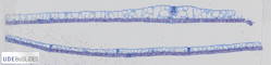 |
Plant |
Nymphaea alba |
White waterlily |
Leaf |
Cross section |
|
Astrosclereid, Epidermis, Epidermis of the leaf underside, Hydropote, Large intercellular space, Palisade parenchyma, Sclereid fragments, Sponge parenchyma, Stoma on leaf surface, Vascular bundle |
unknown |
unknown |
unknown |
Jungner/microthek |
2025-05-05 15:59:03 |
Olympus VS200 |
UPLXAPO 20x / 0.80 |
20x / 0.80 |
75 % |
|
Throughlight widefield |
iDS VS-264C |
Slide scanning microscope |
24.6 |
46,568 x
11,224 x
47 px³ |
12,718.7 x
3,065.5 x
27.1 µm³ |
✅
|
✅
|
✅
|
✅
|
| 503 |
Nymphaea sp. - Stalk cross section 20x |
 |
Plant |
Nymphaea sp. |
Waterlily |
Stalk |
Cross section |
|
Collenchyma, Epidermis, Intercellular with foreign bodies, Large intercellular, Parenchyma cells, Sclereid, Vascular bundle |
unknown |
unknown |
unknown |
Jungner/microthek |
2025-05-13 19:44:58 |
Olympus VS200 |
UPLXAPO 20x / 0.80 |
20x / 0.80 |
75 % |
|
Throughlight widefield |
iDS VS-264C |
Slide scanning microscope |
88.3 |
34,007 x
25,704 x
101 px³ |
9,288.1 x
7,020.4 x
59.0 µm³ |
✅
|
✅
|
✅
|
✅
|
| 610 |
Ochroma pyramidale - Wood cross section 20x |
 |
Plant |
Ochroma pyramidale (syn. lagopus) |
Balsa |
Wood |
Cross section |
Wikipedia on Ochroma pyramidale.
This sample was repaired after shrinking of the mounting medium due to aging, and thus has some minor damage.
Jungner/microthek micro preparation no. 747 |
|
unknown |
unstained |
unknown |
Jungner/microthek |
2025-07-03 22:15:31 |
Olympus VS200 |
UPLXAPO 20x / 0.80 |
20x / 0.80 |
75 % |
|
Throughlight widefield |
iDS VS-264C |
Slide scanning microscope |
70.7 |
33,146 x
33,846 x
63 px³ |
9,052.3 x
9,243.6 x
36.6 µm³ |
✅
|
✅
|
✅
|
❌ |
| 275 |
Oedogonium sp. - 20x |
 |
Algae |
Oedogonium sp. |
Oedogonium |
Whole organism |
Whole mount |
This un-branched filamentous green algae Oedogonium sp. is overgrown bei Cocconeis pediculus (a diatom). Wikipedia on Oedogonium sp. and Algenläuse.
Jungner/microthek micro preparation no. 953 |
|
unknown |
unknown |
unknown |
Jungner/microthek |
2023-09-19 16:46:34 |
Olympus VS200 |
UPLXAPO 20x / 0.80 |
20x / 0.80 |
75 % |
|
Throughlight widefield |
iDS VS-264C |
Slide scanning microscope |
24.0 |
15,366 x
18,405 x
85 px³ |
4,196.6 x
5,026.6 x
69.7 µm³ |
✅
|
✅
|
✅
|
❌ |
| 403 |
Oedogonium sp. 2 - 40x |
 |
Algae |
Oedogonium sp. |
Oedogonium |
Whole organism |
Whole mount |
|
|
unknown |
unknown |
unknown |
Jungner/microthek |
2025-03-31 15:20:16 |
Olympus VS200 |
UPLXAPO O 40x / 1.40 |
40x / 1.40 |
75 % |
Oil |
Throughlight widefield |
iDS VS-264C |
Slide scanning microscope |
92.9 |
28,584 x
32,179 x
101 px³ |
3,916.6 x
4,409.2 x
59.0 µm³ |
✅
|
✅
|
✅
|
❌ |
| 603 |
Ophioglossum sp. - Sporangia cross section 20x |
 |
Plant |
Ophioglossum sp. |
Adder's-tongue fern |
Sporangium |
Cross section |
|
|
unknown |
unknown |
unknown |
Jungner/microthek |
2025-07-02 10:38:09 |
Olympus VS200 |
UPLXAPO 20x / 0.80 |
20x / 0.80 |
75 % |
|
Throughlight widefield |
iDS VS-264C |
Slide scanning microscope |
16.4 |
26,406 x
7,292 x
85 px³ |
7,211.6 x
1,991.3 x
49.6 µm³ |
✅
|
✅
|
✅
|
❌ |
| 320 |
Opuntia sp. - Stem cross section 20x |
 |
Plant |
Opuntia sp. |
Prickly pear cactus |
Stem |
Thin section |
|
Xerophyte,CAM plant |
unknown |
unknown |
Eukitt |
Jungner/microthek |
2023-10-25 09:23:44 |
Olympus VS200 |
UPLXAPO 20x / 0.80 |
20x / 0.80 |
75 % |
|
Throughlight widefield |
iDS VS-264C |
Slide scanning microscope |
55.1 |
44,443 x
16,532 x
75 px³ |
12,138.4 x
4,515.3 x
43.7 µm³ |
✅
|
✅
|
✅
|
❌ |
| 383 |
Osmanthus x fortunei - Leaf cross section 20x |
 |
Plant |
Osmanthus x fortunei |
Osmanthus |
Leaf |
Cross section |
|
|
Formol, acetic alcohol |
W3Asim I: Acridine red, acriflavine, astra blue |
Euparal |
Jörg Weiß
www.mikroskopie-bonn.de |
2023-11-08 14:30:39 |
Olympus VS200 |
UPLXAPO 20x / 0.80 |
20x / 0.80 |
75 % |
|
Throughlight widefield |
iDS VS-264C |
Slide scanning microscope |
25.0 |
32,404 x
7,629 x
101 px³ |
8,850.2 x
2,083.5 x
59.0 µm³ |
✅
|
✅
|
✅
|
❌ |
| 28 |
Osmunda regalis - Petiole cross section 20x |
 |
Plant |
Osmunda regalis |
Royal fern |
Petiole |
Cross section |
|
bark parenchyma, chloroplasts, endodermis, epidermis & cuticle, medullary parenchyma, phloem, pit / pitting canal, ring of sclerenchyma, xylem |
Formol, acetic alcohol |
W3Asim I: Acridine red, acriflavine, astra blue |
Euparal |
Jörg Weiß
www.mikroskopie-bonn.de |
2021-07-21 16:22:52 |
Olympus VS200 |
UPLXAPO 20x / 0.80 |
20x / 0.80 |
75 % |
|
Throughlight widefield |
iDS VS-264C |
Slide scanning microscope |
12.8 |
14,753 x
14,209 x
61 px³ |
4,027.1 x
3,878.5 x
70.8 µm³ |
✅
|
✅
|
✅
|
✅
|
| 27 |
Osmunda regalis - Pinnule cross section 20x |
 |
Plant |
Osmunda regalis |
Royal fern |
Pinnule |
Cross section |
|
assimilation parenchyma, bark parenchyma, collenchyma, Concentric vascular bundle with inner xylem, endodermis, epidermis & cuticle, guard cells, phloem, sponge parenchyma, upper epidermis & cuticle, xylem |
Formol, acetic alcohol |
W3Asim I: Acridine red, acriflavine, astra blue |
Euparal |
Jörg Weiß
www.mikroskopie-bonn.de |
2021-07-21 11:50:12 |
Olympus VS200 |
UPLXAPO 20x / 0.80 |
20x / 0.80 |
75 % |
|
Throughlight widefield |
iDS VS-264C |
Slide scanning microscope |
7.8 |
32,649 x
3,920 x
61 px³ |
8,912.4 x
1,070.0 x
70.8 µm³ |
✅
|
✅
|
✅
|
✅
|
| 73 |
Papaver rhoeas - Peduncle cross section 20x |
 |
Plant |
Papaver rhoeas |
Common poppy |
Peduncle |
Cross section |
|
assimilation parenchyma, collenchyma, companion cells, epidermis, medullary parenchyma, phloem, ring of sclerenchyma, sclerenchyma, sive tubes, vascular bundle, xylem, xylem vessel or tracheae |
Formol, acetic alcohol |
|
Euparal |
Rainer Teubner |
2021-06-30 11:24:48 |
Olympus VS200 |
UPLXAPO 20x / 0.80 |
20x / 0.80 |
40 % |
|
Throughlight widefield |
iDS VS-264C |
Slide scanning microscope |
45.7 |
29,384 x
25,504 x
61 px³ |
8,021.2 x
6,962.1 x
70.8 µm³ |
✅
|
✅
|
✅
|
✅
|
| 164 |
Papaver rhoeas - Peduncle cross section 20x |
 |
Plant |
Papaver rhoeas |
Common poppy |
Peduncle |
Cross section |
|
assimilation parenchyma, collenchyma, epidermis & cuticle, medullary parenchyma, parenchyma cells (& phloem cells?), phloem, ring of sclerenchyma, sclerenchyma, vascular bundle, xylem, xylem parenchyma, xylem vessel or tracheae |
Formol, acetic alcohol |
|
Euparal |
Rainer Teubner |
2021-10-12 10:24:03 |
Olympus VS200 |
UPLXAPO 20x / 0.80 |
20x / 0.80 |
50 % |
|
Throughlight widefield |
iDS VS-264C |
Slide scanning microscope |
51.3 |
29,344 x
28,658 x
61 px³ |
8,010.2 x
7,823.1 x
99.6 µm³ |
✅
|
✅
|
✅
|
✅
|
| 178 |
Papaver rhoeas - Stalk cross section 20x 1 |
 |
Plant |
Papaver rhoeas |
Common poppy |
Stalk |
Cross section |
|
assimilation parenchyma, collenchyma, companion cell, epidermis & cuticle, guard cells, medullary parenchyma, phloem, phloem ???, ring of sclerenchyma, sclerenchyma, vascular bundle, xylem, xylem vessel or tracheae |
Formol, acetic alcohol |
|
Euparal |
Rainer Teubner |
2021-10-06 14:53:23 |
Olympus VS200 |
UPLXAPO 20x / 0.80 |
20x / 0.80 |
40 % |
|
Throughlight widefield |
iDS VS-264C |
Slide scanning microscope |
15.6 |
15,851 x
16,083 x
61 px³ |
4,326.9 x
4,390.2 x
70.8 µm³ |
✅
|
✅
|
✅
|
✅
|
| 179 |
Papaver rhoeas - Stalk cross section 20x 2 |
 |
Plant |
Papaver rhoeas |
Common poppy |
Stalk |
Cross section |
|
assimilation parenchyma, epidermis & cuticle, medullary parenchyma, phloem, phloem ?, ring of sclerenchyma, sclerenchyma, vascular bundle, xylem, xylem vessel or tracheae |
Formol, acetic alcohol |
|
Euparal |
Rainer Teubner |
2021-10-06 15:09:46 |
Olympus VS200 |
UPLXAPO 20x / 0.80 |
20x / 0.80 |
40 % |
|
Throughlight widefield |
iDS VS-264C |
Slide scanning microscope |
15.8 |
15,801 x
16,375 x
61 px³ |
4,313.2 x
4,469.9 x
70.8 µm³ |
✅
|
✅
|
✅
|
✅
|
| 180 |
Papaver rhoeas - Stalk cross section 20x 3 |
 |
Plant |
Papaver rhoeas |
Common poppy |
Stalk |
Cross section |
|
assmiliation parenchyma, epidermis & cuticle, medullary parenchyma, phloem, phloem ?, ring of collenchmya, ring of sclerenchyma, sclerenchyma, vascular bundle, xylem, xylem vessel or tracheae |
Formol, acetic alcohol |
HE according to Papani |
Euparal |
Rainer Teubner |
2021-10-06 15:32:22 |
Olympus VS200 |
UPLXAPO 20x / 0.80 |
20x / 0.80 |
40 % |
|
Throughlight widefield |
iDS VS-264C |
Slide scanning microscope |
15.2 |
15,821 x
15,760 x
61 px³ |
4,318.7 x
4,302.1 x
70.8 µm³ |
✅
|
✅
|
✅
|
✅
|
| 181 |
Papaver rhoeas - Stalk cross section 20x 4 |
 |
Plant |
Papaver rhoeas |
Common poppy |
Stalk |
Cross section |
|
assimilation parenchyma, epidermis & cuticle, ring of sclerenchyma |
Formol, acetic alcohol |
unstained |
Euparal |
Rainer Teubner |
2021-10-06 15:53:49 |
Olympus VS200 |
UPLXAPO 20x / 0.80 |
20x / 0.80 |
40 % |
|
Throughlight widefield |
iDS VS-264C |
Slide scanning microscope |
15.1 |
15,912 x
15,569 x
61 px³ |
4,343.4 x
4,249.9 x
70.8 µm³ |
✅
|
✅
|
✅
|
✅
|
| 510 |
Papaver sp. - Flower bud cross section 20x |
 |
Plant |
Papaver sp. |
Poppy |
Flower bud |
Cross section |
|
|
unknown |
unknown |
unknown |
Jungner/microthek |
2025-05-15 09:19:43 |
Olympus VS200 |
UPLXAPO 20x / 0.80 |
20x / 0.80 |
75 % |
|
Throughlight widefield |
iDS VS-264C |
Slide scanning microscope |
7.3 |
15,595 x
12,061 x
39 px³ |
4,259.3 x
3,294.0 x
22.4 µm³ |
✅
|
✅
|
✅
|
❌ |
| 60 |
Paramecium tetraurelia - Phase contrast 60x 1 |
 |
Protist |
Paramecium tetraurelia |
Paramecium |
Whole cell |
Whole organism |
|
|
not fixated |
unstained |
Agarose |
Nadine Graupner |
2021-09-28 10:19:59 |
Olympus VS200 |
UPLXAPO OPH 60x / 1.42 |
60x / 1.42 |
50 % |
Oil |
Phase contrast |
iDS VS-264C |
Slide scanning microscope |
0.8 |
3,879 x
3,515 x
61 px³ |
352.1 x
319.1 x
68.4 µm³ |
✅
|
✅
|
✅
|
❌ |
| 61 |
Paramecium tetraurelia - Phase contrast 60x 2 |
 |
Protist |
Paramecium tetraurelia |
Paramecium |
Whole cell |
Whole organism |
|
|
not fixated |
unstained |
Agarose |
Nadine Graupner |
2021-09-28 10:20:51 |
Olympus VS200 |
UPLXAPO OPH 60x / 1.42 |
60x / 1.42 |
50 % |
Oil |
Phase contrast |
iDS VS-264C |
Slide scanning microscope |
0.6 |
3,152 x
2,909 x
61 px³ |
286.1 x
264.1 x
68.4 µm³ |
✅
|
✅
|
✅
|
❌ |
| 62 |
Paramecium tetraurelia - Phase contrast 60x 3 |
 |
Protist |
Paramecium tetraurelia |
Paramecium |
Whole cell |
Whole organism |
|
|
not fixated |
unstained |
Agarose |
Nadine Graupner |
2021-09-28 10:22:47 |
Olympus VS200 |
UPLXAPO OPH 60x / 1.42 |
60x / 1.42 |
50 % |
Oil |
Phase contrast |
iDS VS-264C |
Slide scanning microscope |
0.2 |
1,879 x
1,667 x
61 px³ |
170.5 x
151.3 x
68.4 µm³ |
✅
|
✅
|
✅
|
❌ |
| 57 |
Paramecium tetraurelia - Widefield 60x 1 |
 |
Protist |
Paramecium tetraurelia |
Paramecium |
Whole cell |
Whole organism |
|
|
not fixated |
unstained |
Agarose |
Nadine Graupner |
2021-11-03 13:00:48 |
Olympus VS200 |
UPLXAPO OPH 60x / 1.42 |
60x / 1.42 |
40 % |
Oil |
Throughlight widefield |
iDS VS-264C |
Slide scanning microscope |
0.8 |
3,879 x
3,515 x
61 px³ |
352.1 x
319.1 x
68.4 µm³ |
✅
|
✅
|
✅
|
❌ |
| 58 |
Paramecium tetraurelia - Widefield 60x 2 |
 |
Protist |
Paramecium tetraurelia |
Paramecium |
Whole cell |
Whole organism |
|
|
not fixated |
unstained |
Agarose |
Nadine Graupner |
2021-11-03 10:27:54 |
Olympus VS200 |
UPLXAPO OPH 60x / 1.42 |
60x / 1.42 |
40 % |
Oil |
Throughlight widefield |
iDS VS-264C |
Slide scanning microscope |
0.6 |
3,152 x
2,909 x
61 px³ |
286.1 x
264.1 x
68.4 µm³ |
✅
|
✅
|
✅
|
❌ |
| 59 |
Paramecium tetraurelia - Widefield 60x 3 |
 |
Protist |
Paramecium tetraurelia |
Paramecium |
Whole cell |
Whole organism |
|
|
not fixated |
unstained |
Agarose |
Nadine Graupner |
2021-11-03 10:23:16 |
Olympus VS200 |
UPLXAPO OPH 60x / 1.42 |
60x / 1.42 |
40 % |
Oil |
Throughlight widefield |
iDS VS-264C |
Slide scanning microscope |
0.2 |
1,879 x
1,667 x
61 px³ |
170.5 x
151.3 x
68.4 µm³ |
✅
|
✅
|
✅
|
❌ |
| 38 |
Pelargonium sp. - Stalk cross section 20x |
 |
Plant |
Pelargonium sp. |
Geranium |
Stalk |
Cross section |
|
bark parenchyma, cambium, collenchyma, crystal druse, epidermis & cuticle, medullary parenchyma, phloem, sclerenchyma, xylem |
Formol, acetic alcohol |
|
Euparal |
Rainer Teubner |
2021-08-27 10:25:17 |
Olympus VS200 |
UPLXAPO 20x / 0.80 |
20x / 0.80 |
50 % |
|
Throughlight widefield |
iDS VS-264C |
Slide scanning microscope |
32.8 |
24,567 x
23,419 x
57 px³ |
6,706.3 x
6,392.7 x
99.1 µm³ |
✅
|
✅
|
✅
|
✅
|
| 570 |
Peziza sp. - Fruiting body cross section 20x |
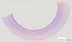 |
Fungus |
Peziza sp. |
Cup fungus |
Fruiting body |
Cross section |
|
|
unknown |
unknown |
unknown |
Jungner/microthek |
2025-06-23 17:03:56 |
Olympus VS200 |
UPLXAPO 20x / 0.80 |
20x / 0.80 |
75 % |
|
Throughlight widefield |
iDS VS-264C |
Slide scanning microscope |
47.1 |
28,147 x
16,575 x
101 px³ |
7,687.1 x
4,526.6 x
59.0 µm³ |
✅
|
✅
|
✅
|
❌ |
| 542 |
Phaseolus sp. - Fruit cross section 20x |
 |
Plant |
Phaseolus sp. |
Bean |
Fruit |
Cross section |
|
Dorsal suture, Endocarp, Exocarp, Funicle, Hilum region, Kotyledons, Mesocarp, Pericarp, Seed, Seed coat, Vascular bundle, Vascular tissue, Ventral suture |
unknown |
unknown |
unknown |
Jungner/microthek |
2025-05-22 12:39:11 |
Olympus VS200 |
UPLXAPO 20x / 0.80 |
20x / 0.80 |
75 % |
|
Throughlight widefield |
iDS VS-264C |
Slide scanning microscope |
78.4 |
43,259 x
34,179 x
53 px³ |
11,814.9 x
9,335.1 x
30.7 µm³ |
✅
|
✅
|
✅
|
✅
|
| 449 |
Phaseolus sp. - young root cross section 20x |
 |
Plant |
Phaseolus sp. |
Bean |
Root |
Cross section |
|
|
unknown |
unknown |
unknown |
Jungner/microthek |
2025-04-30 19:52:05 |
Olympus VS200 |
UPLXAPO 20x / 0.80 |
20x / 0.80 |
75 % |
|
Throughlight widefield |
iDS VS-264C |
Slide scanning microscope |
1.3 |
11,074 x
4,163 x
29 px³ |
3,024.4 x
1,137.0 x
16.5 µm³ |
✅
|
✅
|
✅
|
❌ |
| 326 |
Phoenix sp. - Leaflet cross section 20x |
 |
Plant |
Phoenix sp. |
Date palm |
Leaflet |
Cross section |
|
|
unknown |
unknown |
Eukitt |
Jungner/microthek |
2023-10-26 11:51:15 |
Olympus VS200 |
UPLXAPO 20x / 0.80 |
20x / 0.80 |
75 % |
|
Throughlight widefield |
iDS VS-264C |
Slide scanning microscope |
16.5 |
25,055 x
12,937 x
51 px³ |
6,843.0 x
3,533.3 x
29.5 µm³ |
✅
|
✅
|
✅
|
❌ |
| 655 |
Picea abies - Root cross section 20x |
 |
Plant |
Picea abies |
Norway spruce |
Root |
Cross section |
|
|
unknown |
unknown |
unknown |
Jungner/microthek |
2025-07-17 18:02:16 |
Olympus VS200 |
UPLXAPO 20x / 0.80 |
20x / 0.80 |
75 % |
|
Throughlight widefield |
iDS VS-264C |
Slide scanning microscope |
17.0 |
15,199 x
17,202 x
65 px³ |
4,150.8 x
4,698.0 x
37.8 µm³ |
✅
|
✅
|
✅
|
❌ |
| 654 |
Picea abies - Sprout tip cross section 20x |
 |
Plant |
Picea abies |
Norway spruce |
Sprout tip |
Cross section |
|
|
unknown |
unknown |
unknown |
Jungner/microthek |
2025-07-17 18:47:26 |
Olympus VS200 |
UPLXAPO 20x / 0.80 |
20x / 0.80 |
75 % |
|
Throughlight widefield |
iDS VS-264C |
Slide scanning microscope |
15.7 |
15,896 x
15,150 x
65 px³ |
4,341.2 x
4,137.6 x
37.8 µm³ |
✅
|
✅
|
✅
|
❌ |
| 617 |
Picea abies - Wood cross section 20x |
 |
Plant |
Picea abies |
Norway spruce |
Wood |
Cross section |
Wikipedia on Picea abies (also known as Picea excelsa).
This sample has some damages (cracks in the mounting medium) due to aging.
Jungner/microthek micro preparation no. 753 |
|
unknown |
unstained |
unknown |
Jungner/microthek |
2025-07-14 10:48:47 |
Olympus VS200 |
UPLXAPO 20x / 0.80 |
20x / 0.80 |
75 % |
|
Throughlight widefield |
iDS VS-264C |
Slide scanning microscope |
41.7 |
20,987 x
22,840 x
87 px³ |
5,731.8 x
6,237.6 x
50.7 µm³ |
✅
|
✅
|
✅
|
❌ |
| 656 |
Picea abies - young female strobile longitudinal section 20x |
 |
Plant |
Picea abies |
Norway spruce |
Strobile |
Longitudinal section |
|
|
unknown |
unknown |
unknown |
Jungner/microthek |
2025-07-17 16:41:34 |
Olympus VS200 |
UPLXAPO 20x / 0.80 |
20x / 0.80 |
75 % |
|
Throughlight widefield |
iDS VS-264C |
Slide scanning microscope |
23.2 |
16,334 x
33,049 x
43 px³ |
4,460.9 x
9,025.9 x
24.8 µm³ |
✅
|
✅
|
✅
|
❌ |
| 325 |
Picea sp. - Shoot bud cross section 20x |
 |
Plant |
Picea sp. |
Spruce |
Shoot bud |
Cross section |
|
Cell with crystal, Pith, Resin duct, Resin duct of the needle leaf, Vascular bundle, Vascular tissue, Vascular tissue of the needle leaf, Young needle leaf |
unknown |
Astra blue, fuchsine |
unknown |
Karl Brügmann |
2023-09-28 13:56:36 |
Olympus VS200 |
UPLXAPO 20x / 0.80 |
20x / 0.80 |
75 % |
|
Throughlight widefield |
iDS VS-264C |
Slide scanning microscope |
9.8 |
13,156 x
11,423 x
65 px³ |
3,593.1 x
3,119.9 x
37.8 µm³ |
✅
|
✅
|
✅
|
✅
|
| 511 |
Picea syn. excelsa - Sprout tip cross section 20x |
 |
Plant |
Picea excelsa (a.k.a. Picea abies) |
Norway spruce |
Sprout tip |
Cross section |
|
|
unknown |
unknown |
unknown |
Jungner/microthek |
2025-05-14 13:26:00 |
Olympus VS200 |
UPLXAPO 20x / 0.80 |
20x / 0.80 |
75 % |
|
Throughlight widefield |
iDS VS-264C |
Slide scanning microscope |
23.3 |
18,702 x
16,154 x
77 px³ |
5,107.8 x
4,411.9 x
44.8 µm³ |
✅
|
✅
|
✅
|
❌ |
| 490 |
Pinguicula sp. - Leaf cross section 20x |
 |
Plant |
Pinguicula sp. |
Butterwort |
Leaf |
Cross section |
|
Carnivorous plant, Cells with granula, Epidermis, Peduncular gland, Reservoir cell, Secretory cells, Sessile gland, Sessile gland on leaf underside, Stalk cell, Stomatal apparatus, Vascular tissue |
unknown |
unknown |
unknown |
Jungner/microthek |
2025-05-08 19:40:03 |
Olympus VS200 |
UPLXAPO 20x / 0.80 |
20x / 0.80 |
75 % |
|
Throughlight widefield |
iDS VS-264C |
Slide scanning microscope |
72.9 |
50,461 x
18,295 x
79 px³ |
13,782.2 x
4,996.7 x
46.0 µm³ |
✅
|
✅
|
✅
|
✅
|
| 661 |
Pinus mugo - one-year-old branch cross section 20x |
 |
Plant |
Pinus mugo |
Mountain pine |
Branch |
Cross section |
|
|
unknown |
unknown |
unknown |
Jungner/microthek |
2025-08-11 09:41:29 |
Olympus VS200 |
UPLXAPO 20x / 0.80 |
20x / 0.80 |
75 % |
|
Throughlight widefield |
iDS VS-264C |
Slide scanning microscope |
31.3 |
17,609 x
17,571 x
101 px³ |
4,809.0 x
4,798.6 x
91.0 µm³ |
✅
|
✅
|
✅
|
❌ |
| 660 |
Pinus mugo - Root cross section 20x |
 |
Plant |
Pinus mugo |
Mountain pine |
Root |
Cross section |
|
|
unknown |
unknown |
unknown |
Jungner/microthek |
2025-08-11 09:04:55 |
Olympus VS200 |
UPLXAPO 20x / 0.80 |
20x / 0.80 |
75 % |
|
Throughlight widefield |
iDS VS-264C |
Slide scanning microscope |
19.2 |
14,352 x
13,238 x
101 px³ |
3,919.6 x
3,615.3 x
89.0 µm³ |
✅
|
✅
|
✅
|
❌ |
| 521 |
Pinus sp. - male flower longitudinal section 20x |
 |
Plant |
Pinus sp. |
Pine |
Flower |
Longitudinal section |
|
|
unknown |
unknown |
unknown |
Jungner/microthek |
2025-05-15 20:05:27 |
Olympus VS200 |
UPLXAPO 20x / 0.80 |
20x / 0.80 |
75 % |
|
Throughlight widefield |
iDS VS-264C |
Slide scanning microscope |
19.3 |
22,177 x
13,794 x
63 px³ |
6,057.1 x
3,767.2 x
36.6 µm³ |
✅
|
✅
|
✅
|
❌ |
| 551 |
Pinus sp. - Embryo with endosperm cross section 20x |
 |
Plant |
Pinus sp. |
Pine |
Embryo |
Cross section |
|
|
unknown |
unknown |
unknown |
Jungner/microthek |
2025-05-30 10:02:11 |
Olympus VS200 |
UPLXAPO 20x / 0.80 |
20x / 0.80 |
75 % |
|
Throughlight widefield |
iDS VS-264C |
Slide scanning microscope |
16.4 |
18,454 x
15,018 x
59 px³ |
5,040.1 x
4,101.8 x
34.2 µm³ |
✅
|
✅
|
✅
|
❌ |
| 552 |
Pinus sp. - Embryo with Endosperm longitudinal section 20x |
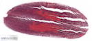 |
Plant |
Pinus sp. |
Pine |
Embryo |
Longitudinal section |
|
|
unknown |
unknown |
unknown |
Jungner/microthek |
2025-05-30 10:30:05 |
Olympus VS200 |
UPLXAPO 20x / 0.80 |
20x / 0.80 |
75 % |
|
Throughlight widefield |
iDS VS-264C |
Slide scanning microscope |
18.2 |
30,751 x
13,774 x
43 px³ |
8,398.8 x
3,761.8 x
24.8 µm³ |
✅
|
✅
|
✅
|
❌ |
| 418 |
Pinus sp. - female strobile 20x |
 |
Plant |
Pinus sp. |
Pine |
Strobile |
Longitudinal section |
|
|
unknown |
unknown |
unknown |
Bresser |
2025-04-08 10:45:23 |
Olympus VS200 |
UPLXAPO 20x / 0.80 |
20x / 0.80 |
75 % |
|
Throughlight widefield |
iDS VS-264C |
Slide scanning microscope |
44.2 |
19,510 x
22,446 x
101 px³ |
5,328.5 x
6,130.4 x
68.0 µm³ |
✅
|
✅
|
✅
|
❌ |
| 301 |
Pinus sp. - Lignified stem longitudinal section 20x |
 |
Plant |
Pinus sp. |
Pine |
Stem |
Longitudinal section |
|
Bordered pit, Fenestriform pit, Resin duct, Wood ray parenchyma |
unknown |
unknown |
Eukitt |
Jungner/microthek |
2023-10-16 09:53:21 |
Olympus VS200 |
UPLXAPO 20x / 0.80 |
20x / 0.80 |
75 % |
|
Throughlight widefield |
iDS VS-264C |
Slide scanning microscope |
79.7 |
37,572 x
21,004 x
101 px³ |
10,261.8 x
5,736.5 x
80.0 µm³ |
✅
|
✅
|
✅
|
✅
|
| 520 |
Pinus sp. - male flower cross section 20x |
 |
Plant |
Pinus sp. |
Pine |
Flower |
Cross section |
|
|
unknown |
unknown |
unknown |
Jungner/microthek |
2025-05-15 19:46:19 |
Olympus VS200 |
UPLXAPO 20x / 0.80 |
20x / 0.80 |
75 % |
|
Throughlight widefield |
iDS VS-264C |
Slide scanning microscope |
14.9 |
16,650 x
12,638 x
71 px³ |
4,547.5 x
3,451.7 x
41.3 µm³ |
✅
|
✅
|
✅
|
❌ |
| 417 |
Pinus sp. - male strobile 20x |
 |
Plant |
Pinus sp. |
Pine |
Strobile |
Longitudinal section |
|
|
unknown |
unknown |
unknown |
Bresser |
2025-04-08 09:36:49 |
Olympus VS200 |
UPLXAPO 20x / 0.80 |
20x / 0.80 |
75 % |
|
Throughlight widefield |
iDS VS-264C |
Slide scanning microscope |
58.8 |
19,769 x
29,456 x
101 px³ |
5,399.2 x
8,045.1 x
59.0 µm³ |
✅
|
✅
|
✅
|
❌ |
| 130 |
Pinus sp. - Needle cross section 20x 1 |
 |
Plant |
Pinus sp. |
Pine |
Needle |
Cross section |
|
assimilation parenchyma, closed collateral vascular bundle, endodermis, epidermis & cuticle, guard cells, hypodermis, resin channel, transfusion tissue |
Formol, acetic alcohol |
unstained |
Euparal |
Rainer Teubner |
2021-09-23 13:32:44 |
Olympus VS200 |
UPLXAPO 20x / 0.80 |
20x / 0.80 |
50 % |
|
Throughlight widefield |
iDS VS-264C |
Slide scanning microscope |
2.1 |
6,409 x
5,351 x
61 px³ |
1,749.5 x
1,460.6 x
99.6 µm³ |
✅
|
✅
|
✅
|
✅
|
| 131 |
Pinus sp. - Needle cross section 20x 2 |
 |
Plant |
Pinus sp. |
Pine |
Needle |
Cross section |
|
assimilation parenchyma, closed collateral vascular bundle, endodermis, epidermis & cuticle, guard cells, hypodermis, phloem, resin channel, transfusion tissue, xylem |
Formol, acetic alcohol |
|
Euparal |
Rainer Teubner |
2021-09-23 13:39:25 |
Olympus VS200 |
UPLXAPO 20x / 0.80 |
20x / 0.80 |
50 % |
|
Throughlight widefield |
iDS VS-264C |
Slide scanning microscope |
2.3 |
7,155 x
5,190 x
61 px³ |
1,953.0 x
1,416.6 x
70.8 µm³ |
✅
|
✅
|
✅
|
✅
|
| 132 |
Pinus sp. - Needle cross section 20x 3 |
 |
Plant |
Pinus sp. |
Pine |
Needle |
Cross section |
|
assimilation parenchyma, closed collateral vascular bundle, endodermis, epidermis & cuticle, guard cells, hypodermis, phloem, resin channel, transfusion tissue, xylem |
Formol, acetic alcohol |
Anthracene blue |
Euparal |
Rainer Teubner |
2021-09-23 13:46:12 |
Olympus VS200 |
UPLXAPO 20x / 0.80 |
20x / 0.80 |
50 % |
|
Throughlight widefield |
iDS VS-264C |
Slide scanning microscope |
1.7 |
6,460 x
4,293 x
61 px³ |
1,763.2 x
1,171.8 x
70.8 µm³ |
✅
|
✅
|
✅
|
✅
|
| 133 |
Pinus sp. - Needle cross section 20x 4 |
 |
Plant |
Pinus sp. |
Pine |
Needle |
Cross section |
|
assimilation parenchyma, closed collateral vascular bundle, endodermis, epidermis & cuticle, guard cells, hypodermis, phloem, resin channel, transfusion tissue, xylem |
Formol, acetic alcohol |
|
Euparal |
Rainer Teubner |
2021-09-23 14:42:29 |
Olympus VS200 |
UPLXAPO 20x / 0.80 |
20x / 0.80 |
50 % |
|
Throughlight widefield |
iDS VS-264C |
Slide scanning microscope |
2.0 |
6,490 x
4,928 x
61 px³ |
1,771.5 x
1,345.1 x
70.8 µm³ |
✅
|
✅
|
✅
|
✅
|
| 419 |
Pinus sp. - Needle cross section 20x 5 |
 |
Plant |
Pinus sp. |
Pine |
Strobile |
Cross section |
|
|
unknown |
unknown |
unknown |
Bresser |
2025-04-08 09:15:15 |
Olympus VS200 |
UPLXAPO 20x / 0.80 |
20x / 0.80 |
75 % |
|
Throughlight widefield |
iDS VS-264C |
Slide scanning microscope |
2.9 |
6,065 x
4,681 x
101 px³ |
1,656.5 x
1,278.3 x
59.0 µm³ |
✅
|
✅
|
✅
|
❌ |
| 420 |
Pinus sp. - Needle cross section 20x 6 |
 |
Plant |
Pinus sp. |
Pine |
Strobile |
Cross section |
|
|
unknown |
unknown |
unknown |
Bresser |
2025-04-08 09:22:58 |
Olympus VS200 |
UPLXAPO 20x / 0.80 |
20x / 0.80 |
75 % |
|
Throughlight widefield |
iDS VS-264C |
Slide scanning microscope |
2.0 |
5,000 x
5,438 x
73 px³ |
1,365.4 x
1,485.0 x
42.5 µm³ |
✅
|
✅
|
✅
|
❌ |
| 217 |
Pinus sp. - Needle fresh cross section 20x 1 |
 |
Plant |
Pinus sp. |
Pine |
Needle |
Cross section |
|
Central cylinder, Cuticle, Endodermis, Epidermis, Hypodermis, Large intercellular space, Mesophyll, Phloem, Resin duct, Sclerenchyma, Stoma, Stomatal complex, Vascular bundle, Xylem |
not fixated |
unstained |
Water |
Christiane Wittmann |
2020-10-05 13:33:13 |
Olympus VS200 |
UPLXAPO 20x / 0.80 |
20x / 0.80 |
75 % |
|
Throughlight widefield |
iDS VS-264C |
Slide scanning microscope |
1.7 |
6,258 x
4,545 x
61 px³ |
1,708.2 x
1,240.5 x
70.8 µm³ |
✅
|
✅
|
✅
|
✅
|
| 218 |
Pinus sp. - Needle fresh cross section 20x 2 |
 |
Plant |
Pinus sp. |
Pine |
Needle |
Cross section |
|
|
not fixated |
unstained |
Water |
Christiane Wittmann |
2020-10-05 13:38:05 |
Olympus VS200 |
UPLXAPO 20x / 0.80 |
20x / 0.80 |
75 % |
|
Throughlight widefield |
iDS VS-264C |
Slide scanning microscope |
1.6 |
6,490 x
3,971 x
61 px³ |
1,771.5 x
1,083.7 x
70.8 µm³ |
✅
|
✅
|
✅
|
❌ |
| 524 |
Pinus sp. - older female strobile longitudinal section 20x |
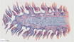 |
Plant |
Pinus sp. |
Pine |
Strobile |
Longitudinal section |
|
|
unknown |
unknown |
unknown |
Jungner/microthek |
2025-05-15 18:21:15 |
Olympus VS200 |
UPLXAPO 20x / 0.80 |
20x / 0.80 |
75 % |
|
Throughlight widefield |
iDS VS-264C |
Slide scanning microscope |
94.8 |
43,527 x
25,037 x
87 px³ |
11,888.2 x
6,838.2 x
50.7 µm³ |
✅
|
✅
|
✅
|
❌ |
| 523 |
Pinus sp. - young female strobile longitudinal section 20x |
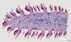 |
Plant |
Pinus sp. |
Pine |
Strobile |
Longitudinal section |
|
Bract scale, Central stem, Ovule, Pits, Resin ducts, Seed scale, Vascular tissue |
unknown |
unknown |
unknown |
Jungner/microthek |
2025-05-15 17:02:55 |
Olympus VS200 |
UPLXAPO 20x / 0.80 |
20x / 0.80 |
75 % |
|
Throughlight widefield |
iDS VS-264C |
Slide scanning microscope |
55.0 |
43,617 x
25,754 x
49 px³ |
11,912.7 x
7,034.0 x
28.3 µm³ |
✅
|
✅
|
✅
|
✅
|
| 454 |
Pinus sylvestris - junge Wurzel 20x, Jungner 516 |
 |
Plant |
Pinus sylvestris |
Baltic pine |
Root |
Cross section |
|
|
unknown |
unknown |
unknown |
Jungner/microthek |
2025-05-02 09:34:02 |
Olympus VS200 |
UPLXAPO 20x / 0.80 |
20x / 0.80 |
75 % |
|
Throughlight widefield |
iDS VS-264C |
Slide scanning microscope |
13.1 |
12,697 x
10,208 x
101 px³ |
3,467.7 x
2,788.0 x
59.0 µm³ |
✅
|
✅
|
✅
|
❌ |
| 7 |
Pinus sylvestris - Needle cross section 20x 1 |
 |
Plant |
Pinus sylvestris |
Baltic pine |
Needle leaf |
cross section |
|
assimilation parenchyma, closed collateral vascular bundle, endodermis, epidermis & cuticle, guard cells, hypodermis, phloem, resin channel, sclerenchyma, transfusion tissue, xylem |
Formol, acetic, alcohol |
|
Euparal |
Rainer Teubner |
2021-07-01 18:22:16 |
Olympus VS200 |
UPLXAPO 20x / 0.80 |
20x / 0.80 |
40 % |
|
Throughlight widefield |
iDS VS-264C |
Slide scanning microscope |
1.1 |
4,202 x
4,313 x
61 px³ |
1,147.1 x
1,177.3 x
70.8 µm³ |
✅
|
✅
|
✅
|
✅
|
| 85 |
Pinus sylvestris - Needle cross section 20x 2 |
 |
Plant |
Pinus sylvestris |
Baltic pine |
Needle leaf |
cross section |
|
assimilation parenchyma, closed collateral vascular bundle, endodermis, epidermis & cuticle, guard cells, hypodermis, phloem, resin channel, sclerenchyma, transfusion tissue, xylem |
Formol, acetic, alcohol |
|
Euparal |
Rainer Teubner |
2021-07-01 18:20:25 |
Olympus VS200 |
UPLXAPO 20x / 0.80 |
20x / 0.80 |
40 % |
|
Throughlight widefield |
iDS VS-264C |
Slide scanning microscope |
1.1 |
3,910 x
4,565 x
61 px³ |
1,067.3 x
1,246.1 x
70.8 µm³ |
✅
|
✅
|
✅
|
✅
|
| 86 |
Pinus sylvestris - Needle cross section 20x 3 |
 |
Plant |
Pinus sylvestris |
Baltic pine |
Needle leaf |
cross section |
|
assimilation parenchyma, closed collateral vascular bundle, endodermis, epidermis & cuticle, guard cells, hypodermis, phloem, resin channel, sclerenchyma, transfusion tissue, xylem |
Formol, acetic, alcohol |
|
Euparal |
Rainer Teubner |
2021-07-01 18:21:20 |
Olympus VS200 |
UPLXAPO 20x / 0.80 |
20x / 0.80 |
40 % |
|
Throughlight widefield |
iDS VS-264C |
Slide scanning microscope |
1.2 |
4,263 x
4,444 x
61 px³ |
1,163.6 x
1,213.1 x
70.8 µm³ |
✅
|
✅
|
✅
|
✅
|
| 87 |
Pinus sylvestris - Needle cross section 20x 4 |
 |
Plant |
Pinus sylvestris |
Baltic pine |
Needle leaf |
cross section |
|
assimilation parenchyma, closed collateral vascular bundle, endodermis, epidermis & cuticle, guard cells, phloem, resin channel, sclerenchyma, transfusion tissue, xylem |
Formol, acetic, alcohol |
|
Euparal |
Rainer Teubner |
2021-07-01 18:23:11 |
Olympus VS200 |
UPLXAPO 20x / 0.80 |
20x / 0.80 |
40 % |
|
Throughlight widefield |
iDS VS-264C |
Slide scanning microscope |
1.0 |
3,648 x
4,374 x
61 px³ |
995.8 x
1,193.8 x
70.8 µm³ |
✅
|
✅
|
✅
|
✅
|
| 494 |
Pinus sylvestris - Needle cross section 5 20x |
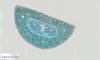 |
Plant |
Pinus sylvestris |
Baltic pine |
Needle leaf |
Cross section |
|
|
unknown |
unknown |
unknown |
Jungner/microthek |
2025-05-09 08:29:29 |
Olympus VS200 |
UPLXAPO 20x / 0.80 |
20x / 0.80 |
75 % |
|
Throughlight widefield |
iDS VS-264C |
Slide scanning microscope |
4.8 |
8,914 x
5,329 x
101 px³ |
2,434.4 x
1,455.2 x
59.0 µm³ |
✅
|
✅
|
✅
|
❌ |
| 297 |
Pinus sylvestris - stem cross section 20x 1/4 |
 |
Plant |
Pinus sylvestris |
Baltic pine |
Stem |
cross section |
|
Annual ring boundary, Cell with two bordered pits, Early wood, Fenestriform pit, Late wood, Resin ducts, Wood ray parenchyma |
unknown |
unknown |
Eukitt |
Jungner/microthek |
2023-10-12 10:05:20 |
Olympus VS200 |
UPLXAPO 20x / 0.80 |
20x / 0.80 |
75 % |
|
Throughlight widefield |
iDS VS-264C |
Slide scanning microscope |
25.6 |
15,565 x
16,303 x
101 px³ |
4,251.1 x
4,452.7 x
100.0 µm³ |
✅
|
✅
|
✅
|
✅
|
| 298 |
Pinus sylvestris - stem cross section 20x 2/4 |
 |
Plant |
Pinus sylvestris |
Baltic pine |
Stem |
cross section |
|
|
unknown |
unknown |
Eukitt |
Jungner/microthek |
2023-10-12 10:29:57 |
Olympus VS200 |
UPLXAPO 20x / 0.80 |
20x / 0.80 |
75 % |
|
Throughlight widefield |
iDS VS-264C |
Slide scanning microscope |
30.3 |
18,045 x
16,602 x
101 px³ |
4,928.3 x
4,534.3 x
100.0 µm³ |
✅
|
✅
|
✅
|
❌ |
| 299 |
Pinus sylvestris - stem cross section 20x 3/4 |
 |
Plant |
Pinus sylvestris |
Baltic pine |
Stem |
cross section |
|
|
unknown |
unknown |
Eukitt |
Jungner/microthek |
2023-10-12 10:57:00 |
Olympus VS200 |
UPLXAPO 20x / 0.80 |
20x / 0.80 |
75 % |
|
Throughlight widefield |
iDS VS-264C |
Slide scanning microscope |
31.8 |
17,955 x
17,558 x
101 px³ |
4,903.9 x
4,795.4 x
100.0 µm³ |
✅
|
✅
|
✅
|
❌ |
| 300 |
Pinus sylvestris - stem cross section 20x 4/4 |
 |
Plant |
Pinus sylvestris |
Baltic pine |
Stem |
cross section |
|
|
unknown |
unknown |
Eukitt |
Jungner/microthek |
2023-10-12 11:38:17 |
Olympus VS200 |
UPLXAPO 20x / 0.80 |
20x / 0.80 |
75 % |
|
Throughlight widefield |
iDS VS-264C |
Slide scanning microscope |
28.4 |
16,123 x
17,429 x
101 px³ |
4,403.4 x
4,760.1 x
100.0 µm³ |
✅
|
✅
|
✅
|
❌ |
| 618 |
Pinus sylvestris - Wood cross section 20x |
 |
Plant |
Pinus sylvestris |
Baltic pine |
Wood |
Cross section |
Wikipedia on Pinus sylvestris.
This sample was repaired after shrinking of the mounting medium due to aging, and thus has some minor damage.
Jungner/microthek micro preparation no. 754 |
Annual ring boundary, Bordered pit, Early wood, Fenestriform pit, Late wood, Resin duct, Wood ray parenchyma with fenestriform pits |
unknown |
unstained |
unknown |
Jungner/microthek |
2025-07-14 12:15:02 |
Olympus VS200 |
UPLXAPO 20x / 0.80 |
20x / 0.80 |
75 % |
|
Throughlight widefield |
iDS VS-264C |
Slide scanning microscope |
92.7 |
29,292 x
31,326 x
101 px³ |
7,999.9 x
8,555.3 x
59.0 µm³ |
✅
|
✅
|
✅
|
✅
|
| 465 |
Pisum sativum - Root with tubercles 40x |
 |
Plant |
Pisum sativum |
Pea |
Root |
Cross section |
|
Cells with rhizobia |
unknown |
unknown |
unknown |
Jungner/microthek |
2025-05-05 08:42:39 |
Olympus VS200 |
UPLXAPO O 40x / 1.40 |
40x / 1.40 |
75 % |
Oil |
Throughlight widefield |
iDS VS-264C |
Slide scanning microscope |
46.7 |
22,986 x
20,109 x
101 px³ |
3,149.5 x
2,755.4 x
33.0 µm³ |
✅
|
✅
|
✅
|
✅
|
| 492 |
Pisum sativum, Leaf tendril cross section 20x |
 |
Plant |
Pisum sativum |
Pea |
Leaf tendril |
Cross section |
|
|
unknown |
unknown |
unknown |
Jungner/microthek |
2025-05-08 21:20:26 |
Olympus VS200 |
UPLXAPO 20x / 0.80 |
20x / 0.80 |
75 % |
|
Throughlight widefield |
iDS VS-264C |
Slide scanning microscope |
9.2 |
20,725 x
8,715 x
51 px³ |
5,660.3 x
2,380.0 x
29.5 µm³ |
✅
|
✅
|
✅
|
❌ |
| 491 |
Plantago sp. - Leaf stalk cross section 20x |
 |
Plant |
Plantago sp. |
Plantain |
Leaf stalk |
Cross section |
Wikipedia on Plantago sp.
This sample has some minor damage caused by shrinking of the mounting medium due to ageing.
Jungner/microthek micro preparation no. 549 |
|
unknown |
unknown |
unknown |
Jungner/microthek |
2025-05-08 20:37:50 |
Olympus VS200 |
UPLXAPO 20x / 0.80 |
20x / 0.80 |
75 % |
|
Throughlight widefield |
iDS VS-264C |
Slide scanning microscope |
40.2 |
32,476 x
25,266 x
49 px³ |
8,869.9 x
6,900.7 x
28.3 µm³ |
✅
|
✅
|
✅
|
❌ |
| 728 |
Plasmodium falciparum Malaria tropica in a blood smear |
 |
Animal |
Homo sapiens |
Human |
Blood |
Smear |
Wikipedia on malaria.
The plasmodia are located inside the erythrocytes and look somewhat like a signet ring. |
|
Methanol |
|
Eukitt |
Anna M. Kehl |
2026-03-24 11:14:08 |
Olympus VS200 |
UPLXAPO OPH 60x / 1.42 |
60x / 1.42 |
75 % |
Oil |
Throughlight widefield |
iDS VS-264C |
Slide scanning microscope |
14.0 |
21,983 x
22,013 x
29 px³ |
1,999.4 x
2,002.1 x
7.8 µm³ |
✅
|
✅
|
✅
|
❌ |
| 348 |
Plasmodium sp. - Blood smear 40x |
 |
Protist |
Plasmodium sp. |
Cause of malaria |
Whole cell |
Blood smear |
|
Parasite |
unknown |
unknown |
unknown |
Jungner/microthek |
2023-12-05 12:25:18 |
Olympus VS200 |
UPLXAPO O 40x / 1.40 |
40x / 1.40 |
75 % |
Oil |
Throughlight widefield |
iDS VS-264C |
Slide scanning microscope |
25.3 |
22,370 x
19,871 x
57 px³ |
3,065.2 x
2,722.7 x
16.2 µm³ |
✅
|
✅
|
✅
|
❌ |
| 355 |
Plasmodium sp. - Blood smear 40x 2 |
 |
Protist |
Plasmodium sp. |
Cause of malaria |
Whole cell |
Blood smear |
|
Parasite |
unknown |
unknown |
unknown |
Jungner/microthek |
2023-12-06 16:55:40 |
Olympus VS200 |
UPLXAPO O 40x / 1.40 |
40x / 1.40 |
75 % |
Oil |
Throughlight widefield |
iDS VS-264C |
Slide scanning microscope |
38.6 |
29,417 x
25,727 x
51 px³ |
4,030.7 x
3,525.2 x
14.5 µm³ |
✅
|
✅
|
✅
|
❌ |
| 625 |
Platanus orientalis - Wood cross section 20x |
 |
Plant |
Platanus orientalis |
Oriental plane |
Wood |
Cross section |
Wikipedia on Platanus orientalis.
This sample was repaired after shrinking of the mounting medium due to aging, and thus has some minor damage.
Jungner/microthek micro preparation no. 761 |
|
unknown |
unstained |
unknown |
Jungner/microthek |
2025-07-14 18:08:01 |
Olympus VS200 |
UPLXAPO 20x / 0.80 |
20x / 0.80 |
75 % |
|
Throughlight widefield |
iDS VS-264C |
Slide scanning microscope |
69.5 |
28,697 x
23,995 x
101 px³ |
7,837.3 x
6,553.2 x
100.0 µm³ |
✅
|
✅
|
✅
|
❌ |
| 647 |
Platycerium sp. - Leaf cross section 20x |
 |
Plant |
Platycerium sp. |
Staghorn fern |
Leaf |
Cross section |
Wikipedia on Platycerium sp.
This sample has some damage (partly large bubbles) caused by shrinking of the mounting medium due to ageing.
Jungner/microthek micro preparation no. 1285 |
|
unknown |
unknown |
unknown |
Jungner/microthek |
2025-07-16 20:20:42 |
Olympus VS200 |
UPLXAPO 20x / 0.80 |
20x / 0.80 |
75 % |
|
Throughlight widefield |
iDS VS-264C |
Slide scanning microscope |
90.9 |
31,353 x
39,723 x
73 px³ |
8,562.7 x
10,848.5 x
42.5 µm³ |
✅
|
✅
|
✅
|
❌ |
| 584 |
Polytrichum sp. - Stalk cross section 20x |
 |
Plant |
Polytrichum sp. |
Hair moss |
Stalk |
Cross section |
|
|
unknown |
unknown |
unknown |
Jungner/microthek |
2025-06-24 19:36:32 |
Olympus VS200 |
UPLXAPO 20x / 0.80 |
20x / 0.80 |
75 % |
|
Throughlight widefield |
iDS VS-264C |
Slide scanning microscope |
0.5 |
4,433 x
3,457 x
35 px³ |
1,210.5 x
943.9 x
20.1 µm³ |
✅
|
✅
|
✅
|
❌ |
| 585 |
Polytrichum sp. - Thalli cross section 20x |
 |
Plant |
Polytrichum sp. |
Hair moss |
Thallus |
Cross section |
|
|
unknown |
unknown |
unknown |
Jungner/microthek |
2025-06-25 09:33:48 |
Olympus VS200 |
UPLXAPO 20x / 0.80 |
20x / 0.80 |
75 % |
|
Throughlight widefield |
iDS VS-264C |
Slide scanning microscope |
1.7 |
7,022 x
4,463 x
53 px³ |
1,917.6 x
1,218.7 x
30.7 µm³ |
✅
|
✅
|
✅
|
❌ |
| 624 |
Populus alba - Wood cross section 20x |
 |
Plant |
Populus alba |
Silver poplar |
Wood |
unknown |
Wikipedia on Populus alba.
This sample was repaired after shrinking of the mounting medium due to aging, and thus has some minor damage.
Jungner/microthek micro preparation no. 760 |
|
unknown |
unknown |
unknown |
Jungner/microthek |
2025-07-14 17:30:38 |
Olympus VS200 |
UPLXAPO 20x / 0.80 |
20x / 0.80 |
75 % |
|
Throughlight widefield |
iDS VS-264C |
Slide scanning microscope |
35.8 |
15,806 x
22,441 x
101 px³ |
4,316.7 x
6,128.8 x
59.0 µm³ |
✅
|
✅
|
✅
|
❌ |
| 673 |
Populus tremula - Seed fibers 20x |
 |
Plant |
Populus tremula |
Aspen |
Seed fibers |
Whole mount |
Wikipedia on Populus tremula.
This sample has some damage (partly large bubbles) caused by shrinking of the mounting medium due to ageing.
Jungner/microthek micro preparation no. 1246 |
|
unknown |
unknown |
unknown |
Jungner/microthek |
2025-08-13 17:03:19 |
Olympus VS200 |
UPLXAPO 20x / 0.80 |
20x / 0.80 |
75 % |
|
Throughlight widefield |
iDS VS-264C |
Slide scanning microscope |
69.8 |
34,863 x
19,822 x
101 px³ |
9,521.2 x
5,413.4 x
59.0 µm³ |
✅
|
✅
|
✅
|
❌ |
| 165 |
Prunus avium - Pedicel cross section 20x 1 |
 |
Plant |
Prunus avium |
Wild cherry |
Pedicel |
Cross section |
|
bark parenchyma, epidermis & cuticle, guard cells, medullary parenchyma, open collateral vascular bundle, phloem, sclerenchyma, sponge parenchyma, xylem |
Formol, acetic alcohol |
|
Euparal |
Rainer Teubner |
2021-10-19 16:20:30 |
Olympus VS200 |
UPLXAPO 20x / 0.80 |
20x / 0.80 |
50 % |
|
Throughlight widefield |
iDS VS-264C |
Slide scanning microscope |
2.0 |
5,381 x
5,553 x
67 px³ |
1,468.9 x
1,515.7 x
77.9 µm³ |
✅
|
✅
|
✅
|
✅
|
| 166 |
Prunus avium - Pedicel cross section 20x 2 |
 |
Plant |
Prunus avium |
Wild cherry |
Pedicel |
Cross section |
|
bark parenchyma, epidermis & cuticle, guard cells, medullary parenchyma, open collateral vascular bundle, phloem, sclerenchyma, sponge parenchyma, xylem |
Formol, acetic alcohol |
|
Euparal |
Rainer Teubner |
2021-10-19 16:27:21 |
Olympus VS200 |
UPLXAPO 20x / 0.80 |
20x / 0.80 |
50 % |
|
Throughlight widefield |
iDS VS-264C |
Slide scanning microscope |
2.5 |
6,117 x
6,107 x
67 px³ |
1,669.7 x
1,666.9 x
77.9 µm³ |
✅
|
✅
|
✅
|
✅
|
| 167 |
Prunus avium - Pedicel cross section 20x 3 |
 |
Plant |
Prunus avium |
Wild cherry |
Pedicel |
Cross section |
|
bark parenchyma, epidermis & cuticle, guard cells, medullary parenchyma, open collateral vascular bundle, phloem, sclerenchyma, sponge parenchyma, xylem |
Formol, acetic alcohol |
unstained |
Euparal |
Rainer Teubner |
2021-10-19 17:01:35 |
Olympus VS200 |
UPLXAPO 20x / 0.80 |
20x / 0.80 |
50 % |
|
Throughlight widefield |
iDS VS-264C |
Slide scanning microscope |
3.5 |
7,568 x
7,749 x
59 px³ |
2,065.8 x
2,115.3 x
68.4 µm³ |
✅
|
✅
|
✅
|
✅
|
| 168 |
Prunus avium - Pedicel cross section 20x 4 |
 |
Plant |
Prunus avium |
Wild cherry |
Pedicel |
Cross section |
|
bark parenchyma, epidermis & cuticle, guard cells, medullary parenchyma, open collateral vascular bundle, phloem, sclerenchyma, sponge parenchyma, xylem |
Formol, acetic alcohol |
|
Euparal |
Rainer Teubner |
2021-10-27 10:10:06 |
Olympus VS200 |
UPLXAPO 20x / 0.80 |
20x / 0.80 |
50 % |
|
Throughlight widefield |
iDS VS-264C |
Slide scanning microscope |
2.5 |
5,422 x
5,966 x
77 px³ |
1,479.9 x
1,628.4 x
89.7 µm³ |
✅
|
✅
|
✅
|
✅
|
| 169 |
Prunus avium - Pedicel cross section 20x 5 |
 |
Plant |
Prunus avium |
Wild cherry |
Pedicel |
Cross section |
|
bark parenchyma, epidermis & cuticle, guard cells, medullary parenchyma, open collateral vascular bundle, pholem, sclerenchyma, sponge parenchyma, xylem |
Formol, acetic alcohol |
|
Euparal |
Rainer Teubner |
2021-10-27 10:27:30 |
Olympus VS200 |
UPLXAPO 20x / 0.80 |
20x / 0.80 |
50 % |
|
Throughlight widefield |
iDS VS-264C |
Slide scanning microscope |
3.7 |
7,830 x
7,669 x
61 px³ |
2,137.3 x
2,093.3 x
70.8 µm³ |
✅
|
✅
|
✅
|
✅
|
| 170 |
Prunus avium - Pedicel cross section 20x 6 |
 |
Plant |
Prunus avium |
Wild cherry |
Pedicel |
Cross section |
|
bark parenchyma, epidermis & cuticle, medullary parenchyma, open collateral vascular bundle, phloem, sclerenchyma, sponge parenchyma, xylem |
Formol, acetic alcohol |
unstained |
Euparal |
Rainer Teubner |
2021-10-27 12:35:49 |
Olympus VS200 |
UPLXAPO 20x / 0.80 |
20x / 0.80 |
50 % |
|
Throughlight widefield |
iDS VS-264C |
Slide scanning microscope |
4.2 |
7,195 x
7,618 x
77 px³ |
1,964.0 x
2,079.5 x
89.7 µm³ |
✅
|
✅
|
✅
|
✅
|
| 619 |
Prunus avium - Wood cross section 20x |
 |
Plant |
Prunus avium |
Wild cherry |
Wood |
Cross section |
|
|
unknown |
unstained |
unknown |
Jungner/microthek |
2025-07-14 13:27:56 |
Olympus VS200 |
UPLXAPO 20x / 0.80 |
20x / 0.80 |
75 % |
|
Throughlight widefield |
iDS VS-264C |
Slide scanning microscope |
21.2 |
18,655 x
18,079 x
63 px³ |
5,094.6 x
4,937.3 x
36.6 µm³ |
✅
|
✅
|
✅
|
❌ |
| 540 |
Prunus domestica - young fruit cross section 20x |
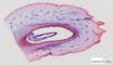 |
Plant |
Prunus domestica |
Plum |
Fruit |
Cross section |
|
Endocarp, Exocarp, Mesocarp, Seed, Trichome, Vascular bundles |
unknown |
unknown |
unknown |
Jungner/microthek |
2025-05-22 09:04:12 |
Olympus VS200 |
UPLXAPO 20x / 0.80 |
20x / 0.80 |
75 % |
|
Throughlight widefield |
iDS VS-264C |
Slide scanning microscope |
70.7 |
34,886 x
20,058 x
101 px³ |
9,528.1 x
5,478.1 x
59.0 µm³ |
✅
|
✅
|
✅
|
✅
|
| 531 |
Prunus dulcis - Fruit cross section 20x |
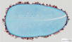 |
Plant |
Prunus dulcis |
Almond |
Fruit |
Cross section |
|
|
unknown |
unknown |
unknown |
Jungner/microthek |
2025-05-21 22:10:14 |
Olympus VS200 |
UPLXAPO 20x / 0.80 |
20x / 0.80 |
75 % |
|
Throughlight widefield |
iDS VS-264C |
Slide scanning microscope |
81.0 |
36,965 x
21,701 x
101 px³ |
10,095.8 x
5,927.0 x
59.0 µm³ |
✅
|
✅
|
✅
|
❌ |
| 171 |
Prunus laurocerasus - Branch cross section 20x |
 |
Plant |
Prunus avium |
Cherry laurel |
Pedicel |
Cross section |
|
bark parenchyma with assimilation parenchyma, bast, cambium, cuticle, epidermis, medullary parenchyma, medullary ray, primary xylem, sclerenchyma, wood, xylem vessel or tracheae |
Formol, acetic alcohol |
|
Euparal |
Rainer Teubner |
2021-10-14 13:09:05 |
Olympus VS200 |
UPLXAPO 20x / 0.80 |
20x / 0.80 |
50 % |
|
Throughlight widefield |
iDS VS-264C |
Slide scanning microscope |
11.4 |
14,521 x
15,448 x
51 px³ |
3,963.8 x
4,216.9 x
59.0 µm³ |
✅
|
✅
|
✅
|
✅
|
| 384 |
Prunus laurocerasus - Shade leaf old cross section 20x |
 |
Plant |
Prunus laurocerasus |
Cherry laurel |
Shade leaf |
Cross section |
|
|
Formol, acetic alcohol |
W3Asim I: Acridine red, acriflavine, astra blue |
Euparal |
Jörg Weiß
www.mikroskopie-bonn.de |
2023-11-08 10:51:18 |
Olympus VS200 |
UPLXAPO 20x / 0.80 |
20x / 0.80 |
75 % |
|
Throughlight widefield |
iDS VS-264C |
Slide scanning microscope |
43.2 |
36,297 x
11,772 x
101 px³ |
9,913.6 x
3,215.1 x
73.0 µm³ |
✅
|
✅
|
✅
|
❌ |
| 387 |
Prunus laurocerasus - Sprout one-year-old cross section 20x |
 |
Plant |
Prunus laurocerasus |
Cherry laurel |
Sprout |
Cross section |
|
|
Formol, acetic alcohol |
W3Asim I: Acridine red, acriflavine, astra blue |
Euparal |
Jörg Weiß
www.mikroskopie-bonn.de |
2023-11-08 15:57:19 |
Olympus VS200 |
UPLXAPO 20x / 0.80 |
20x / 0.80 |
75 % |
|
Throughlight widefield |
iDS VS-264C |
Slide scanning microscope |
22.0 |
15,386 x
14,172 x
101 px³ |
4,202.1 x
3,870.6 x
74.0 µm³ |
✅
|
✅
|
✅
|
❌ |
| 388 |
Prunus laurocerasus - Sprout two-year-old cross section 20x |
 |
Plant |
Prunus laurocerasus |
Cherry laurel |
Sprout |
Cross section |
|
|
Formol, acetic alcohol |
W3Asim I: Acridine red, acriflavine, astra blue |
Euparal |
Jörg Weiß
www.mikroskopie-bonn.de |
2023-11-08 12:45:33 |
Olympus VS200 |
UPLXAPO 20x / 0.80 |
20x / 0.80 |
75 % |
|
Throughlight widefield |
iDS VS-264C |
Slide scanning microscope |
35.4 |
18,881 x
18,554 x
101 px³ |
5,156.7 x
5,067.4 x
78.0 µm³ |
✅
|
✅
|
✅
|
❌ |
| 385 |
Prunus laurocerasus - Sun leaf one-year-old cross section 20x |
 |
Plant |
Prunus laurocerasus |
Cherry laurel |
Sun leaf |
Cross section |
|
|
Formol, acetic alcohol |
W3Asim I: Acridine red, acriflavine, astra blue |
Euparal |
Jörg Weiß
www.mikroskopie-bonn.de |
2023-11-08 11:25:03 |
Olympus VS200 |
UPLXAPO 20x / 0.80 |
20x / 0.80 |
75 % |
|
Throughlight widefield |
iDS VS-264C |
Slide scanning microscope |
57.0 |
38,767 x
14,560 x
101 px³ |
10,588.1 x
3,976.7 x
59.0 µm³ |
✅
|
✅
|
✅
|
❌ |
| 677 |
Prunus spinosa - Leaf cross section 20x |
 |
Plant |
Prunus spinosa |
Blackthorn |
Leaf |
Cross section |
|
|
unknown |
unknown |
unknown |
Jungner/microthek |
2025-08-13 20:33:43 |
Olympus VS200 |
UPLXAPO 20x / 0.80 |
20x / 0.80 |
75 % |
|
Throughlight widefield |
iDS VS-264C |
Slide scanning microscope |
56.3 |
20,708 x
31,993 x
85 px³ |
5,655.5 x
8,737.6 x
49.6 µm³ |
✅
|
✅
|
✅
|
❌ |
| 589 |
Psilotum sp. - Stem cross section 20x |
 |
Plant |
Psilotum sp. |
Whisk fern |
Stem |
Cross section |
|
|
unknown |
unknown |
unknown |
Jungner/microthek |
2025-06-25 10:28:47 |
Olympus VS200 |
UPLXAPO 20x / 0.80 |
20x / 0.80 |
75 % |
|
Throughlight widefield |
iDS VS-264C |
Slide scanning microscope |
4.3 |
9,701 x
6,066 x
73 px³ |
2,649.3 x
1,656.7 x
42.5 µm³ |
✅
|
✅
|
✅
|
❌ |
| 549 |
Pyrus communis - Fruit pulp cross section 20x |
 |
Plant |
Pyrus communis |
Common pear |
Fruit |
Cross section |
|
|
unknown |
unknown |
unknown |
Jungner/microthek |
2025-05-30 08:41:22 |
Olympus VS200 |
UPLXAPO 20x / 0.80 |
20x / 0.80 |
75 % |
|
Throughlight widefield |
iDS VS-264C |
Slide scanning microscope |
56.8 |
25,901 x
24,619 x
89 px³ |
7,074.2 x
6,723.9 x
51.9 µm³ |
✅
|
✅
|
✅
|
❌ |
| 612 |
Pyrus communis - Wood cross section 20x |
 |
Plant |
Pyrus communis |
Common pear |
Wood |
Cross section |
|
|
unknown |
unstained |
unknown |
Jungner/microthek |
2025-07-04 09:36:23 |
Olympus VS200 |
UPLXAPO 20x / 0.80 |
20x / 0.80 |
75 % |
|
Throughlight widefield |
iDS VS-264C |
Slide scanning microscope |
45.0 |
22,808 x
19,523 x
101 px³ |
6,228.9 x
5,331.8 x
59.0 µm³ |
✅
|
✅
|
✅
|
❌ |
| 294 |
Pyrus communis - young fruit cross section |
 |
Plant |
Pyrus communis |
Pear tree |
Fruit |
Cross section |
Wikipedia on Pyrus communis.
This sample has some minor damage caused by shrinking of the mounting medium due to ageing, mostly outside of the specimen.
Jungner/microthek micro preparation no. 1240 |
Cell lumen, Cells with crystals, Lignified cell wall, Sclerenchyma, Stone cell nest |
unknown |
unknown |
Eukitt |
Jungner/microthek |
2023-10-10 14:06:44 |
Olympus VS200 |
UPLXAPO 20x / 0.80 |
20x / 0.80 |
75 % |
|
Throughlight widefield |
iDS VS-264C |
Slide scanning microscope |
136.7 |
40,947 x
39,288 x
85 px³ |
11,183.6 x
10,730.5 x
49.6 µm³ |
✅
|
✅
|
✅
|
✅
|
| 691 |
Pyrus communis - young fruit cross section 20x |
 |
Plant |
Pyrus communis |
Common pear |
Fruit |
Cross section |
Wikipedia on Pyrus communis.
This sample has some minor damage caused by shrinking of the mounting medium due to ageing.
Jungner/microthek micro preparation no. 1240 |
|
unknown |
unknown |
unknown |
Jungner/microthek |
2025-08-13 11:18:20 |
Olympus VS200 |
UPLXAPO 20x / 0.80 |
20x / 0.80 |
75 % |
|
Throughlight widefield |
iDS VS-264C |
Slide scanning microscope |
122.2 |
41,075 x
39,673 x
75 px³ |
11,217.9 x
10,834.9 x
43.7 µm³ |
✅
|
✅
|
✅
|
❌ |
| 106 |
Pyrus sp. - Pedicel cross section 20x 1 |
 |
Plant |
Pyrus sp. |
Pear tree |
Pedicel |
Cross section |
|
bark parenchyma, collenchyma, cork, medullary parenchyma, phloem, stone cell, vascular bundle, xylem |
Formol, acetic alcohol |
|
Euparal |
Rainer Teubner |
2021-10-19 13:00:38 |
Olympus VS200 |
UPLXAPO 20x / 0.80 |
20x / 0.80 |
50 % |
|
Throughlight widefield |
iDS VS-264C |
Slide scanning microscope |
40.0 |
21,242 x
22,149 x
85 px³ |
5,798.6 x
6,046.1 x
99.1 µm³ |
✅
|
✅
|
✅
|
✅
|
| 107 |
Pyrus sp. - Pedicel cross section 20x 2 |
 |
Plant |
Pyrus sp. |
Pear tree |
Pedicel |
Cross section |
|
bark parenchyma, collenchyma, cork, medullary parenchyma, phloem, sclerenchyma, stone cell, vascular bundle, xylem |
Formol, acetic alcohol |
|
Euparal |
Rainer Teubner |
2021-10-19 15:24:54 |
Olympus VS200 |
UPLXAPO 20x / 0.80 |
20x / 0.80 |
50 % |
|
Throughlight widefield |
iDS VS-264C |
Slide scanning microscope |
42.2 |
21,867 x
22,723 x
85 px³ |
5,969.1 x
6,202.9 x
99.1 µm³ |
✅
|
✅
|
✅
|
✅
|
| 292 |
Pyrus sp. - Stone cells 40x |
 |
Plant |
Pyrus sp. |
Pear tree |
Fruit flesh |
Thin section |
|
|
unknown |
unknown |
Eukitt |
Jungner/microthek |
2023-10-10 13:10:13 |
Olympus VS200 |
UPLXAPO O 40x / 1.40 |
40x / 1.40 |
75 % |
Oil |
Throughlight widefield |
iDS VS-264C |
Slide scanning microscope |
1.2 |
3,792 x
3,732 x
85 px³ |
519.5 x
511.4 x
35.3 µm³ |
✅
|
✅
|
✅
|
❌ |
| 293 |
Pyrus sp. - Stone cells in fruit flesh cross section 20x |
 |
Plant |
Pyrus sp. |
Pear tree |
Fruit |
Cross section |
|
Sclerenchyma |
unknown |
unknown |
Eukitt |
Jungner/microthek |
2023-10-10 13:19:59 |
Olympus VS200 |
UPLXAPO 20x / 0.80 |
20x / 0.80 |
75 % |
|
Throughlight widefield |
iDS VS-264C |
Slide scanning microscope |
47.5 |
33,011 x
16,921 x
85 px³ |
9,016.0 x
4,621.3 x
49.6 µm³ |
✅
|
✅
|
✅
|
❌ |
| 615 |
Quercus robur - Wood cross section 20x |
 |
Plant |
Quercus robur |
Pedunculate oak |
Wood |
Cross section |
Wikipedia on Quercus robur.
This sample was repaired after shrinking of the mounting medium due to aging, and thus has some minor damage.
Jungner/microthek micro preparation no. 751 |
|
unknown |
unstained |
unknown |
Jungner/microthek |
2025-07-14 08:38:15 |
Olympus VS200 |
UPLXAPO 20x / 0.80 |
20x / 0.80 |
75 % |
|
Throughlight widefield |
iDS VS-264C |
Slide scanning microscope |
24.7 |
17,571 x
15,100 x
93 px³ |
4,798.7 x
4,124.0 x
54.3 µm³ |
✅
|
✅
|
✅
|
❌ |
| 555 |
Quercus sp. - Bark cross section 20x |
 |
Plant |
Quercus sp. |
Oak |
Bark |
Cross section |
|
Cells with crystals, Lenticel, Phellem, Phelloderm, Phellogen |
unknown |
unknown |
unknown |
Jungner/microthek |
2025-05-20 09:10:03 |
Olympus VS200 |
UPLXAPO 20x / 0.80 |
20x / 0.80 |
75 % |
|
Throughlight widefield |
iDS VS-264C |
Slide scanning microscope |
45.8 |
20,275 x
22,388 x
101 px³ |
5,537.5 x
6,114.6 x
73.0 µm³ |
✅
|
✅
|
✅
|
✅
|
| 679 |
Quercus sp. - Root with mycorrhiza cross section 20x |
 |
Plant |
Quercus sp. |
Oak |
Root |
Cross section |
Wikipedia on Quercus sp.
This sample has some damage (partly large bubbles) caused by shrinking of the mounting medium due to ageing.
Jungner/microthek micro preparation no. 1227 |
|
unknown |
unknown |
unknown |
Jungner/microthek |
2025-08-11 14:13:28 |
Olympus VS200 |
UPLXAPO 20x / 0.80 |
20x / 0.80 |
75 % |
|
Throughlight widefield |
iDS VS-264C |
Slide scanning microscope |
3.0 |
6,335 x
9,543 x
49 px³ |
1,729.9 x
2,606.0 x
28.3 µm³ |
✅
|
✅
|
✅
|
❌ |
| 324 |
Quercus sp. - Stem cross section 20x |
 |
Plant |
Quercus sp. |
Oak |
Stem |
Cross section |
|
Cork |
unknown |
unknown |
Eukitt |
Jungner/microthek |
2023-10-26 10:24:30 |
Olympus VS200 |
UPLXAPO 20x / 0.80 |
20x / 0.80 |
75 % |
|
Throughlight widefield |
iDS VS-264C |
Slide scanning microscope |
51.0 |
22,625 x
22,308 x
101 px³ |
6,179.4 x
6,092.9 x
70.0 µm³ |
✅
|
✅
|
✅
|
❌ |
| 452 |
Quercus sp. - woody root cross section 20x |
 |
Plant |
Quercus sp. |
Oak |
Root |
Cross section |
|
|
unknown |
unknown |
unknown |
Jungner/microthek |
2025-05-01 07:43:33 |
Olympus VS200 |
UPLXAPO 20x / 0.80 |
20x / 0.80 |
75 % |
|
Throughlight widefield |
iDS VS-264C |
Slide scanning microscope |
56.6 |
23,740 x
23,623 x
101 px³ |
6,484.0 x
6,451.9 x
70.0 µm³ |
✅
|
✅
|
✅
|
❌ |
| 671 |
Quercus sp. - young fruit cross section 20x |
 |
Plant |
Quercus sp. |
Oak |
Fruit |
Cross section |
Wikipedia on Quercus sp.
This sample has some damage (partly large bubbles) caused by shrinking of the mounting medium due to ageing.
Jungner/microthek micro preparation no. 1242 |
|
unknown |
unknown |
unknown |
Jungner/microthek |
2025-08-13 13:42:50 |
Olympus VS200 |
UPLXAPO 20x / 0.80 |
20x / 0.80 |
75 % |
|
Throughlight widefield |
iDS VS-264C |
Slide scanning microscope |
72.0 |
36,651 x
19,463 x
101 px³ |
10,009.8 x
5,315.5 x
59.0 µm³ |
✅
|
✅
|
✅
|
❌ |
| 682 |
Quercus suber - Bark cross section 20x |
 |
Plant |
Quercus suber |
Cork oak |
Bark |
Cross section |
Wikipedia on Quercus suber.
This sample has some damage (partly large bubbles) caused by shrinking of the mounting medium due to ageing.
Jungner/microthek micro preparation no. 1232 |
|
unknown |
unknown |
unknown |
Jungner/microthek |
2025-08-11 15:59:10 |
Olympus VS200 |
UPLXAPO 20x / 0.80 |
20x / 0.80 |
75 % |
|
Throughlight widefield |
iDS VS-264C |
Slide scanning microscope |
35.0 |
18,933 x
18,308 x
101 px³ |
5,170.8 x
4,999.9 x
100.0 µm³ |
✅
|
✅
|
✅
|
❌ |
| 456 |
Ranunculus ficaria - Root cross section 20x |
 |
Plant |
Ranunculus ficaria |
Lesser celandine |
Root |
Cross section |
|
|
unknown |
unknown |
unknown |
Jungner/microthek |
2025-05-02 09:55:40 |
Olympus VS200 |
UPLXAPO 20x / 0.80 |
20x / 0.80 |
75 % |
|
Throughlight widefield |
iDS VS-264C |
Slide scanning microscope |
19.2 |
13,802 x
13,794 x
101 px³ |
3,769.6 x
3,767.2 x
100.0 µm³ |
✅
|
✅
|
✅
|
❌ |
| 543 |
Ranunculus sp. - Fruit cross section 20x |
 |
Plant |
Ranunculus sp. |
Buttercup |
Fruit |
Cross section |
|
Endocarp, Exocarp, Funicle, Mesocarp, Pericarp, Placenta, Receptacle, Seed |
unknown |
unknown |
unknown |
Jungner/microthek |
2025-05-22 13:36:00 |
Olympus VS200 |
UPLXAPO 20x / 0.80 |
20x / 0.80 |
75 % |
|
Throughlight widefield |
iDS VS-264C |
Slide scanning microscope |
112.6 |
36,449 x
30,594 x
101 px³ |
9,955.2 x
8,355.9 x
100.0 µm³ |
✅
|
✅
|
✅
|
✅
|
| 504 |
Ranunculus sp. - Stalk cross section 20x |
 |
Plant |
Ranunculus sp. |
Buttercup |
Stalk |
Cross section |
|
|
unknown |
unknown |
unknown |
Jungner/microthek |
2025-05-13 21:29:28 |
Olympus VS200 |
UPLXAPO 20x / 0.80 |
20x / 0.80 |
75 % |
|
Throughlight widefield |
iDS VS-264C |
Slide scanning microscope |
29.1 |
17,588 x
16,403 x
101 px³ |
4,803.5 x
4,479.9 x
59.0 µm³ |
✅
|
✅
|
✅
|
❌ |
| 457 |
Raphanus sp. - Root cross section 20x |
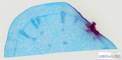 |
Plant |
Raphanus sp. |
Radish |
Root |
Cross section |
|
|
unknown |
unknown |
unknown |
Jungner/microthek |
2025-05-02 17:27:33 |
Olympus VS200 |
UPLXAPO 20x / 0.80 |
20x / 0.80 |
75 % |
|
Throughlight widefield |
iDS VS-264C |
Slide scanning microscope |
109.8 |
47,072 x
23,095 x
101 px³ |
12,856.4 x
6,307.8 x
90.0 µm³ |
✅
|
✅
|
✅
|
❌ |
| 88 |
Rhododendron spec. - Branch cross section 20x 1 |
 |
Plant |
Rhododendron spec. |
Rhododendron |
Branch |
Cross section |
|
bark parenchyma, bast, cambium, cork, medullary parenchyma, primary xylem, resin channel, stone cell, wood, xylem vessel or tracheae |
Formol, acetic alcohol |
|
Euparal |
Rainer Teubner |
2021-07-01 18:06:27 |
Olympus VS200 |
UPLXAPO 20x / 0.80 |
20x / 0.80 |
40 % |
|
Throughlight widefield |
iDS VS-264C |
Slide scanning microscope |
4.6 |
9,170 x
8,213 x
61 px³ |
2,503.2 x
2,241.8 x
70.8 µm³ |
✅
|
✅
|
✅
|
✅
|
| 89 |
Rhododendron spec. - Branch cross section 20x 2 |
 |
Plant |
Rhododendron spec. |
Rhododendron |
Branch |
Cross section |
|
bark parenchyma, bast, cambium, cork, medullary parenchyma, primary xylem, resin channel, stone cell, wood |
Formol, acetic alcohol |
|
Euparal |
Rainer Teubner |
2021-07-01 18:12:43 |
Olympus VS200 |
UPLXAPO 20x / 0.80 |
20x / 0.80 |
40 % |
|
Throughlight widefield |
iDS VS-264C |
Slide scanning microscope |
3.3 |
7,185 x
7,528 x
61 px³ |
1,961.3 x
2,054.8 x
70.8 µm³ |
✅
|
✅
|
✅
|
✅
|
| 545 |
Ribes grossularia - Berry cross section 20x |
 |
Plant |
Ribes grossularia |
Gooseberry |
Fruit |
Cross section |
|
Aril, Connection point of funicle, Embryo, Endocarp, Endosperm, Exocarp, Gelatinous sheath, Mesocarp, Oblong cells of the cellulose-pectinous layer?, Outer layer of the endoderm, Pericarp, Placenta, Sclerotesta & Cellulose-pectinous layer, Trichome Fragment, Vascular bundle, Vascular tissue |
unknown |
unknown |
unknown |
Jungner/microthek |
2025-05-22 15:21:27 |
Olympus VS200 |
UPLXAPO 20x / 0.80 |
20x / 0.80 |
75 % |
|
Throughlight widefield |
iDS VS-264C |
Slide scanning microscope |
71.3 |
39,029 x
25,734 x
71 px³ |
10,659.8 x
7,028.6 x
41.3 µm³ |
✅
|
✅
|
✅
|
✅
|
| 678 |
Ribes rubrum - young shoot cross section 20x |
 |
Plant |
Ribes rubrum |
Redcurrant |
Shoot |
Cross section |
Wikipedia on Ribes rubrum.
This sample has some damage (partly large bubbles) caused by shrinking of the mounting medium due to ageing.
Jungner/microthek micro preparation no. 1252 |
|
unknown |
unknown |
unknown |
Jungner/microthek |
2025-08-14 10:38:59 |
Olympus VS200 |
UPLXAPO 20x / 0.80 |
20x / 0.80 |
75 % |
|
Throughlight widefield |
iDS VS-264C |
Slide scanning microscope |
41.6 |
20,706 x
19,881 x
101 px³ |
5,655.0 x
5,429.7 x
100.0 µm³ |
✅
|
✅
|
✅
|
❌ |
| 692 |
Ribes uva-crispa - young fruit cross section 20x |
 |
Plant |
Ribes uva-crispa |
Gooseberry |
Fruit |
Cross section |
|
|
unknown |
unknown |
unknown |
Jungner/microthek |
2025-08-15 09:07:12 |
Olympus VS200 |
UPLXAPO 20x / 0.80 |
20x / 0.80 |
75 % |
|
Throughlight widefield |
iDS VS-264C |
Slide scanning microscope |
84.9 |
32,711 x
30,519 x
85 px³ |
8,933.6 x
8,335.0 x
49.6 µm³ |
✅
|
✅
|
✅
|
❌ |
| 580 |
Riccia sp. - Thallus cross section 20x |
 |
Plant |
Riccia sp. |
Riccia |
Thallus |
Cross section |
|
|
unknown |
unknown |
unknown |
Jungner/microthek |
2025-06-24 18:19:19 |
Olympus VS200 |
UPLXAPO 20x / 0.80 |
20x / 0.80 |
75 % |
|
Throughlight widefield |
iDS VS-264C |
Slide scanning microscope |
5.4 |
13,257 x
9,015 x
45 px³ |
3,620.5 x
2,461.9 x
26.0 µm³ |
✅
|
✅
|
✅
|
❌ |
| 526 |
Ricinus communis - Aleurone grains in seed 20x |
 |
Plant |
Ricinus communis |
Castor bean |
Seed |
unknown |
|
|
unknown |
unknown |
unknown |
Jungner/microthek |
2025-05-16 10:14:09 |
Olympus VS200 |
UPLXAPO 20x / 0.80 |
20x / 0.80 |
75 % |
|
Throughlight widefield |
iDS VS-264C |
Slide scanning microscope |
15.9 |
11,602 x
13,594 x
101 px³ |
3,168.6 x
3,712.8 x
100.0 µm³ |
✅
|
✅
|
✅
|
❌ |
| 637 |
Ricinus communis - Seed cross section 20x |
 |
Plant |
Ricinus communis |
Castor bean |
Seed |
Cross section |
|
Cotyledon, Endosperm with aleurone globules, Leaf primordium of the embryo, Seed coat |
unknown |
unknown |
unknown |
Jungner/microthek |
2025-07-16 11:44:52 |
Olympus VS200 |
UPLXAPO 20x / 0.80 |
20x / 0.80 |
75 % |
|
Throughlight widefield |
iDS VS-264C |
Slide scanning microscope |
55.5 |
23,547 x
24,802 x
95 px³ |
6,430.9 x
6,773.5 x
55.5 µm³ |
✅
|
✅
|
✅
|
✅
|
| 681 |
Robinia pseudoacacia - Heartwood longitudinal section 20x |
 |
Plant |
Robinia pseudoacacia |
Black locust |
Heartwood |
Longitudinal section |
Wikipedia on Robinia pseudoacacia.
This sample has some damage (partly large and many small bubbles) caused by shrinking of the mounting medium due to ageing.
Jungner/microthek micro preparation no. 1231 |
|
unknown |
unknown |
unknown |
Jungner/microthek |
2025-08-11 15:01:05 |
Olympus VS200 |
UPLXAPO 20x / 0.80 |
20x / 0.80 |
75 % |
|
Throughlight widefield |
iDS VS-264C |
Slide scanning microscope |
78.5 |
30,247 x
25,688 x
101 px³ |
8,260.8 x
7,015.6 x
93.0 µm³ |
✅
|
✅
|
✅
|
❌ |
| 685 |
Robinia pseudoacacia - Leaflet joint cross section 20x |
 |
Plant |
Robinia pseudoacacia |
Black locust |
Leaflet joint |
Cross section |
|
|
unknown |
unknown |
unknown |
Jungner/microthek |
2025-08-11 20:53:01 |
Olympus VS200 |
UPLXAPO 20x / 0.80 |
20x / 0.80 |
75 % |
|
Throughlight widefield |
iDS VS-264C |
Slide scanning microscope |
4.1 |
15,099 x
7,680 x
35 px³ |
4,123.6 x
2,097.3 x
20.1 µm³ |
✅
|
✅
|
✅
|
❌ |
| 626 |
Robinia pseudoacacia - Wood cross section 20x |
 |
Plant |
Robinia pseudoacacia |
Black locust |
Wood |
Cross section |
Wikipedia on Robinia pseudoacacia.
This sample was repaired after shrinking of the mounting medium due to aging, and thus has some minor damage.
Jungner/microthek micro preparation no. 762 |
|
unknown |
unstained |
unknown |
Jungner/microthek |
2025-07-14 19:12:04 |
Olympus VS200 |
UPLXAPO 20x / 0.80 |
20x / 0.80 |
75 % |
|
Throughlight widefield |
iDS VS-264C |
Slide scanning microscope |
77.0 |
25,757 x
29,583 x
101 px³ |
7,034.3 x
8,079.3 x
100.0 µm³ |
✅
|
✅
|
✅
|
❌ |
| 700 |
Rosa canina - Fruit cross section 20x |
 |
Plant |
Rosa canina |
Dog rose |
Fruit |
Cross section |
Wikipedia on Rosa canina.
This sample has some damage (partly large bubbles) caused by shrinking of the mounting medium due to ageing.
Jungner/microthek micro preparation no. 1264 |
|
unknown |
unknown |
unknown |
Jungner/microthek |
2025-08-19 19:32:00 |
Olympus VS200 |
UPLXAPO 20x / 0.80 |
20x / 0.80 |
75 % |
|
Throughlight widefield |
iDS VS-264C |
Slide scanning microscope |
145.1 |
43,902 x
32,730 x
101 px³ |
11,989.9 x
8,938.9 x
69.0 µm³ |
✅
|
✅
|
✅
|
❌ |
| 173 |
Rosa sp. - Branch cross section 20x |
 |
Plant |
Rosa sp. |
Rose |
Branch |
Cross section |
|
bark parenchyma, cambium/bast, collenchyma, epidermis & cuticle, medullary parenchyma, medullary ray, primary xylem, sklerenchyma, wood, xylem vessel or tracheae |
Formol, acetic alcohol |
|
Euparal |
Rainer Teubner |
2021-11-05 13:53:46 |
Olympus VS200 |
UPLXAPO 20x / 0.80 |
20x / 0.80 |
50 % |
|
Throughlight widefield |
iDS VS-264C |
Slide scanning microscope |
115.2 |
38,675 x
38,675 x
77 px³ |
10,557.3 x
10,557.3 x
89.7 µm³ |
✅
|
✅
|
✅
|
✅
|
| 172 |
Rosa sp. - Branch cross section 20x 2 |
 |
Plant |
Rosa sp. |
Rose |
Branch |
Cross section |
|
bark parenchyma, cambium/bast, cork, epidermis, medullary parenchyma, primary xylem, wood, xylem vessel or tracheae |
Formol, acetic alcohol |
|
Euparal |
Rainer Teubner |
2021-11-05 15:16:21 |
Olympus VS200 |
UPLXAPO 20x / 0.80 |
20x / 0.80 |
50 % |
|
Throughlight widefield |
iDS VS-264C |
Slide scanning microscope |
18.0 |
15,257 x
13,886 x
85 px³ |
4,164.6 x
3,790.5 x
99.1 µm³ |
✅
|
✅
|
✅
|
✅
|
| 472 |
Rosa sp. - Leaf cross section 20x |
 |
Plant |
Rosa sp. |
Rose |
Leaf |
Cross section |
|
|
unknown |
unknown |
unknown |
Jungner/microthek |
2025-05-05 13:40:21 |
Olympus VS200 |
UPLXAPO 20x / 0.80 |
20x / 0.80 |
75 % |
|
Throughlight widefield |
iDS VS-264C |
Slide scanning microscope |
43.0 |
24,009 x
17,717 x
101 px³ |
6,557.4 x
4,838.9 x
59.0 µm³ |
✅
|
✅
|
✅
|
❌ |
| 696 |
Rubus fruticosus - Shoot with prickle cross section 20x |
 |
Plant |
Rubus fruticosus |
Blackberry |
Shoot |
Cross section |
Wikipedia on Rubus fruticosus.
This sample has some damage (partly large bubbles) caused by shrinking of the mounting medium due to ageing.
Jungner/microthek micro preparation no. 1257 |
Prickle |
unknown |
unknown |
unknown |
Jungner/microthek |
2025-08-15 11:21:18 |
Olympus VS200 |
UPLXAPO 20x / 0.80 |
20x / 0.80 |
75 % |
|
Throughlight widefield |
iDS VS-264C |
Slide scanning microscope |
34.0 |
17,210 x
19,583 x
101 px³ |
4,700.2 x
5,348.1 x
100.0 µm³ |
✅
|
✅
|
✅
|
✅
|
| 697 |
Rubus idaeus - Fruit cross section 20x |
 |
Plant |
Rubus idaeus |
Raspberry |
Fruit |
Cross section |
Wikipedia on Rubus idaeus.
This sample has some damage (partly large bubbles) caused by shrinking of the mounting medium due to ageing.
Jungner/microthek micro preparation no. 1258 |
Drupelet, Endocarp, Exocarp, Exocarp with trichomes, Mesocarp, Receptacle / Torus, Seed |
unknown |
unknown |
unknown |
Jungner/microthek |
2025-08-19 14:39:03 |
Olympus VS200 |
UPLXAPO 20x / 0.80 |
20x / 0.80 |
75 % |
|
Throughlight widefield |
iDS VS-264C |
Slide scanning microscope |
54.7 |
26,433 x
25,529 x
81 px³ |
7,219.0 x
6,972.1 x
47.2 µm³ |
✅
|
✅
|
✅
|
✅
|
| 566 |
Saccharina latissima - thallus cross section 20x |
 |
Algae |
Saccharina latissima |
Sugar kelp |
Thallus |
Cross section |
|
|
unknown |
unknown |
unknown |
Jungner/microthek |
2025-06-23 13:34:56 |
Olympus VS200 |
UPLXAPO 20x / 0.80 |
20x / 0.80 |
75 % |
|
Throughlight widefield |
iDS VS-264C |
Slide scanning microscope |
29.4 |
44,225 x
12,561 x
53 px³ |
12,078.1 x
3,430.3 x
30.7 µm³ |
✅
|
✅
|
✅
|
❌ |
| 323 |
Sagittaria sp. - Leaf cross section 20x |
 |
Plant |
Sagittaria sp. |
Swamp potato |
Leaf |
Cross section |
|
Hydrophyte |
unknown |
unknown |
Eukitt |
Jungner/microthek |
2023-10-26 09:37:20 |
Olympus VS200 |
UPLXAPO 20x / 0.80 |
20x / 0.80 |
75 % |
|
Throughlight widefield |
iDS VS-264C |
Slide scanning microscope |
25.2 |
48,190 x
11,144 x
47 px³ |
13,161.8 x
3,043.7 x
27.1 µm³ |
✅
|
✅
|
✅
|
❌ |
| 632 |
Salix alba - Wood cross section 20x |
 |
Plant |
Salix alba |
White willow |
Wood |
unknown |
Wikipedia on Salix alba.
This sample was repaired after shrinking of the mounting medium due to aging, and thus has some minor damage.
Jungner/microthek micro preparation no. 768 |
|
unknown |
unknown |
unknown |
Jungner/microthek |
2025-07-15 18:42:30 |
Olympus VS200 |
UPLXAPO 20x / 0.80 |
20x / 0.80 |
75 % |
|
Throughlight widefield |
iDS VS-264C |
Slide scanning microscope |
105.5 |
32,143 x
32,511 x
101 px³ |
8,778.6 x
8,879.0 x
59.0 µm³ |
✅
|
✅
|
✅
|
❌ |
| 675 |
Salix sp. - female flower cross section 20x |
 |
Plant |
Salix sp. |
Willow |
Flower |
Cross section |
|
|
unknown |
unknown |
unknown |
Jungner/microthek |
2025-08-13 19:10:05 |
Olympus VS200 |
UPLXAPO 20x / 0.80 |
20x / 0.80 |
75 % |
|
Throughlight widefield |
iDS VS-264C |
Slide scanning microscope |
39.0 |
18,694 x
20,668 x
101 px³ |
5,105.5 x
5,644.6 x
59.0 µm³ |
✅
|
✅
|
✅
|
❌ |
| 674 |
Salix sp. - male flower cross section 20x |
 |
Plant |
Salix sp. |
Willow |
Flower |
Cross section |
Wikipedia on Salix sp.
This sample has some damage (partly large bubbles) caused by shrinking of the mounting medium due to ageing.
Jungner/microthek micro preparation no. 1247 |
|
unknown |
unknown |
unknown |
Jungner/microthek |
2025-08-13 18:13:18 |
Olympus VS200 |
UPLXAPO 20x / 0.80 |
20x / 0.80 |
75 % |
|
Throughlight widefield |
iDS VS-264C |
Slide scanning microscope |
71.4 |
35,490 x
24,852 x
81 px³ |
9,692.6 x
6,787.1 x
47.2 µm³ |
✅
|
✅
|
✅
|
❌ |
| 668 |
Salix sp. - Root cross section 20x |
 |
Plant |
Salix sp. |
Willow |
Root |
Cross section |
Wikipedia on Salix sp.
This sample has some minor damage caused by shrinking of the mounting medium due to ageing.
Jungner/microthek micro preparation no. 1226 |
|
unknown |
unknown |
unknown |
Jungner/microthek |
2025-08-11 13:51:47 |
Olympus VS200 |
UPLXAPO 20x / 0.80 |
20x / 0.80 |
75 % |
|
Throughlight widefield |
iDS VS-264C |
Slide scanning microscope |
9.4 |
18,087 x
7,790 x
67 px³ |
4,939.6 x
2,127.3 x
38.9 µm³ |
✅
|
✅
|
✅
|
❌ |
| 604 |
Salvia officinalis - Leaf cross section 20x |
 |
Plant |
Salvia officinalis |
Common sage |
Leaf |
Cross section |
|
|
unknown |
unknown |
unknown |
Jungner/microthek |
2025-07-02 11:10:23 |
Olympus VS200 |
UPLXAPO 20x / 0.80 |
20x / 0.80 |
75 % |
|
Throughlight widefield |
iDS VS-264C |
Slide scanning microscope |
28.1 |
36,572 x
13,985 x
55 px³ |
9,988.0 x
3,819.3 x
31.9 µm³ |
✅
|
✅
|
✅
|
❌ |
| 377 |
Sambucus nigra - Stem cross section 20x |
 |
Plant |
Sambucus nigra |
Black elderberry |
Stem |
Cross section |
Wikipedia on Sambucus nigra
Jungner/microthek micro preparation no. 567 |
|
unknown |
unknown |
unknown |
Jungner/microthek |
2024-01-09 12:36:55 |
Olympus VS200 |
UPLXAPO 20x / 0.80 |
20x / 0.80 |
75 % |
|
Throughlight widefield |
iDS VS-264C |
Slide scanning microscope |
22.5 |
15,545 x
14,311 x
101 px³ |
4,245.6 x
3,908.7 x
100.0 µm³ |
✅
|
✅
|
✅
|
❌ |
| 713 |
Sambucus sp. - Flower buds cross section 20x |
 |
Plant |
Sambucus sp. |
Elder |
Flower bud |
Cross section |
Wikipedia on Sambucus sp.
This sample has some minor damage (partly large bubbles) caused by shrinking of the mounting medium due to ageing.
Jungner/microthek micro preparation no. 1261 |
|
unknown |
unknown |
unknown |
Jungner/microthek |
2025-08-20 09:45:10 |
Olympus VS200 |
UPLXAPO 20x / 0.80 |
20x / 0.80 |
75 % |
|
Throughlight widefield |
iDS VS-264C |
Slide scanning microscope |
6.7 |
11,276 x
11,236 x
53 px³ |
3,079.4 x
3,068.5 x
30.7 µm³ |
✅
|
✅
|
✅
|
❌ |
| 529 |
Sambucus sp. - Medulla 20x |
 |
Plant |
Sambucus sp. |
Elder |
Medulla |
unknown |
|
|
unknown |
unknown |
unknown |
Jungner/mikrothek |
2025-05-16 16:01:08 |
Olympus VS200 |
UPLXAPO 20x / 0.80 |
20x / 0.80 |
75 % |
|
Throughlight widefield |
iDS VS-264C |
Slide scanning microscope |
21.9 |
18,622 x
11,662 x
101 px³ |
5,086.0 x
3,185.2 x
72.0 µm³ |
✅
|
✅
|
✅
|
❌ |
| 573 |
Scleroderma citrinum - Fruiting body cross section 20x, Jungner 666 |
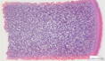 |
Fungus |
Scleroderma citrinum |
Common earthball |
Fruiting body |
Cross section |
|
|
unknown |
unknown |
unknown |
Jungner/microthek |
2025-06-23 20:41:39 |
Olympus VS200 |
UPLXAPO 20x / 0.80 |
20x / 0.80 |
75 % |
|
Throughlight widefield |
iDS VS-264C |
Slide scanning microscope |
54.5 |
37,358 x
21,784 x
67 px³ |
10,202.9 x
5,949.3 x
38.9 µm³ |
✅
|
✅
|
✅
|
❌ |
| 321 |
Sedum sp. - Leaf cross section 20x |
 |
Plant |
Sedum sp. |
Stonecrop |
Leaf |
Thin section |
|
CAM plant |
unknown |
unknown |
Eukitt |
Jungner/microthek |
2023-10-25 08:55:36 |
Olympus VS200 |
UPLXAPO 20x / 0.80 |
20x / 0.80 |
75 % |
|
Throughlight widefield |
iDS VS-264C |
Slide scanning microscope |
15.8 |
23,780 x
12,051 x
55 px³ |
6,494.9 x
3,291.2 x
31.9 µm³ |
✅
|
✅
|
✅
|
❌ |
| 525 |
Simple plant cells 20x |
 |
Plant |
? |
unknown |
unknown |
unknown |
|
|
unknown |
unknown |
unknown |
Jungner/microthek |
2025-05-16 08:32:20 |
Olympus VS200 |
UPLXAPO 20x / 0.80 |
20x / 0.80 |
75 % |
|
Throughlight widefield |
iDS VS-264C |
Slide scanning microscope |
31.4 |
16,680 x
18,653 x
101 px³ |
4,555.6 x
5,094.6 x
69.0 µm³ |
✅
|
✅
|
✅
|
❌ |
| 445 |
Sinapis sp. - Root tip cross section 20x |
 |
Plant |
Sinapis sp. |
Mustard |
Root |
Cross section |
|
|
unknown |
unknown |
unknown |
Jungner/microthek |
2025-04-30 16:26:56 |
Olympus VS200 |
UPLXAPO 20x / 0.80 |
20x / 0.80 |
75 % |
|
Throughlight widefield |
iDS VS-264C |
Slide scanning microscope |
10.6 |
10,417 x
10,069 x
101 px³ |
2,845.1 x
2,750.0 x
100.0 µm³ |
✅
|
✅
|
✅
|
❌ |
| 706 |
Solanum dulcamara - Stem cross section 20x |
 |
Plant |
Solanum dulcamara |
Bittersweet nightshade |
Stem |
Cross section |
|
|
unknown |
unknown |
unknown |
Jungner/microthek |
2025-08-21 06:51:16 |
Olympus VS200 |
UPLXAPO 20x / 0.80 |
20x / 0.80 |
75 % |
|
Throughlight widefield |
iDS VS-264C |
Slide scanning microscope |
66.8 |
24,810 x
32,441 x
83 px³ |
6,775.6 x
8,860.0 x
48.4 µm³ |
✅
|
✅
|
✅
|
❌ |
| 544 |
Solanum lycopersicum - Fruit cross section 20x |
 |
Plant |
Solanum lycopersicum |
Tomato |
Fruit |
Cross section |
|
Central vascular tissue, Endocarp, Exocarp, Locule, Mesocarp, Outer pericarp, Placenta, Radial/internal pericarp, Seed, Vascular bundle to seed |
unknown |
unknown |
unknown |
Jungner/microthek |
2025-05-22 14:50:48 |
Olympus VS200 |
UPLXAPO 20x / 0.80 |
20x / 0.80 |
75 % |
|
Throughlight widefield |
iDS VS-264C |
Slide scanning microscope |
26.3 |
22,039 x
19,530 x
61 px³ |
6,019.3 x
5,334.0 x
35.4 µm³ |
✅
|
✅
|
✅
|
✅
|
| 431 |
Solanum lycopersicum - Fruit cross section 20x |
 |
Plant |
Solanum lycopersicum |
Tomato |
Fruit |
Cross section |
|
|
unknown |
unknown |
unknown |
Bresser |
2025-04-08 15:35:06 |
Olympus VS200 |
UPLXAPO 20x / 0.80 |
20x / 0.80 |
75 % |
|
Throughlight widefield |
iDS VS-264C |
Slide scanning microscope |
116.0 |
40,871 x
35,042 x
81 px³ |
11,162.9 x
9,570.8 x
47.2 µm³ |
✅
|
✅
|
✅
|
❌ |
| 512 |
Solanum tuberosum - Flower cross section 20x |
 |
Plant |
Solanum tuberosum |
Potato |
Flower |
Cross section |
|
Anther, Calyx with trichome bases, Connective, Fragments of trichomes, Petals, Pistil, Pollen grains inside anther, Stamen, Vascular tissue |
unknown |
unknown |
unknown |
Jungner/microthek |
2025-05-15 08:30:56 |
Olympus VS200 |
UPLXAPO 20x / 0.80 |
20x / 0.80 |
75 % |
|
Throughlight widefield |
iDS VS-264C |
Slide scanning microscope |
23.7 |
19,011 x
16,632 x
75 px³ |
5,192.2 x
4,542.4 x
43.7 µm³ |
✅
|
✅
|
✅
|
✅
|
| 334 |
Solanum tuberosum - Leaf cross section 20x |
 |
Plant |
Solanum tuberosum |
Potato |
Leaf |
Cross section |
|
C3 plant,dicotyledonous |
unknown |
unknown |
unknown |
Jungner/microthek |
2023-10-26 13:49:17 |
Olympus VS200 |
UPLXAPO 20x / 0.80 |
20x / 0.80 |
75 % |
|
Throughlight widefield |
iDS VS-264C |
Slide scanning microscope |
49.0 |
49,565 x
12,828 x
77 px³ |
13,537.4 x
3,503.4 x
44.8 µm³ |
✅
|
✅
|
✅
|
❌ |
| 553 |
Solanum tuberosum - Tuber 20x |
 |
Plant |
Solanum tuberosum |
Potato |
Tuber |
unknown |
|
|
unknown |
unknown |
unknown |
Jungner/microthek |
2025-05-16 10:36:30 |
Olympus VS200 |
UPLXAPO 20x / 0.80 |
20x / 0.80 |
75 % |
|
Throughlight widefield |
iDS VS-264C |
Slide scanning microscope |
69.2 |
29,785 x
28,672 x
81 px³ |
8,134.9 x
7,831.0 x
47.2 µm³ |
✅
|
✅
|
✅
|
❌ |
| 506 |
Solanum tuberosum - Tuber cross section 20x |
 |
Plant |
Solanum tuberosum |
Potato |
Tuber |
Cross section |
|
|
unknown |
unknown |
unknown |
Jungner/microthek |
2025-05-14 08:32:06 |
Olympus VS200 |
UPLXAPO 20x / 0.80 |
20x / 0.80 |
75 % |
|
Throughlight widefield |
iDS VS-264C |
Slide scanning microscope |
54.5 |
42,733 x
21,621 x
59 px³ |
11,671.5 x
5,905.2 x
34.2 µm³ |
✅
|
✅
|
✅
|
❌ |
| 182 |
Solidago canadensis - Stalk cross section 20x |
 |
Plant |
Solidago canadensis |
Canadian goldenrod |
Stalk |
Cross section |
|
cork, crystal druse, sclerenchyma, wood |
Formol, acetic alcohol |
|
Euparal |
Rainer Teubner |
2021-11-03 10:20:43 |
Olympus VS200 |
UPLXAPO 20x / 0.80 |
20x / 0.80 |
50 % |
|
Throughlight widefield |
iDS VS-264C |
Slide scanning microscope |
74.9 |
30,593 x
28,820 x
85 px³ |
8,351.3 x
7,867.1 x
99.1 µm³ |
✅
|
✅
|
✅
|
✅
|
| 670 |
Sorbus aucuparia - Fruit cross section 20x |
 |
Plant |
Sorbus aucuparia |
Rowan |
Fruit |
Cross section |
Wikipedia on Sorbus aucuparia.
This sample has some tears and shifts of tissue due to its preperation.
Jungner/microthek micro preparation no. 1244 |
|
unknown |
unknown |
unknown |
Jungner/microthek |
2025-08-13 15:52:39 |
Olympus VS200 |
UPLXAPO 20x / 0.80 |
20x / 0.80 |
75 % |
|
Throughlight widefield |
iDS VS-264C |
Slide scanning microscope |
53.4 |
31,444 x
25,360 x
67 px³ |
8,587.5 x
6,925.9 x
38.9 µm³ |
✅
|
✅
|
✅
|
❌ |
| 614 |
Sorbus aucuparia - Wood cross section 20x |
 |
Plant |
Sorbus aucuparia |
Rowan |
Wood |
Cross section |
Wikipedia on Sorbus aucuparia.
This sample was repaired after shrinking of the mounting medium due to aging, and thus has some minor damage.
Jungner/microthek micro preparation no. 750 |
|
unknown |
unstained |
unknown |
Jungner/microthek |
2025-07-04 10:16:35 |
Olympus VS200 |
UPLXAPO 20x / 0.80 |
20x / 0.80 |
75 % |
|
Throughlight widefield |
iDS VS-264C |
Slide scanning microscope |
92.4 |
29,882 x
30,619 x
101 px³ |
8,161.0 x
8,362.2 x
67.0 µm³ |
✅
|
✅
|
✅
|
❌ |
| 588 |
Sphagnum sp. - Thallus cross section 20x |
 |
Plant |
Sphagnum sp. |
Sphagnum moss |
Thallus |
Cross section |
|
|
unknown |
unknown |
unknown |
Jungner/microthek |
2025-06-25 10:08:18 |
Olympus VS200 |
UPLXAPO 20x / 0.80 |
20x / 0.80 |
75 % |
|
Throughlight widefield |
iDS VS-264C |
Slide scanning microscope |
2.3 |
7,670 x
5,140 x
59 px³ |
2,094.5 x
1,403.7 x
34.2 µm³ |
✅
|
✅
|
✅
|
❌ |
| 274 |
Spirogyra sp. - 20x |
 |
Algae |
Spirogyra sp. |
Water silk |
Whole organism |
Whole mount |
|
|
unknown |
unknown |
Eukitt |
Jungner/microthek |
2023-09-19 15:47:06 |
Olympus VS200 |
UPLXAPO 20x / 0.80 |
20x / 0.80 |
75 % |
|
Throughlight widefield |
iDS VS-264C |
Slide scanning microscope |
27.0 |
19,498 x
16,313 x
85 px³ |
5,325.4 x
4,455.4 x
70.6 µm³ |
✅
|
✅
|
✅
|
❌ |
| 563 |
Spirogyra sp. - Whole 20x |
 |
Algae |
Spirogyra sp. |
Water silk |
Whole organism |
Whole mount |
|
|
unknown |
unknown |
unknown |
Jungner/microthek |
2025-06-23 10:06:54 |
Olympus VS200 |
UPLXAPO 20x / 0.80 |
20x / 0.80 |
75 % |
|
Throughlight widefield |
iDS VS-264C |
Slide scanning microscope |
38.8 |
22,848 x
16,814 x
101 px³ |
6,239.9 x
4,591.9 x
100.0 µm³ |
✅
|
✅
|
✅
|
❌ |
| 643 |
Staurastrum sp. 20x |
 |
Protist |
Staurastrum sp. |
Staurastrum |
Whole cell |
Whole organism |
|
|
not fixated |
not stained |
Water |
Michael Kloster / Eva Papendick |
2025-07-18 13:43:13 |
Olympus VS200 |
UPLXAPO 20x / 0.80 |
20x / 0.80 |
75 % |
|
Throughlight widefield |
iDS VS-264C |
Slide scanning microscope |
10.2 |
13,168 x
7,700 x
101 px³ |
3,596.3 x
2,102.8 x
59.0 µm³ |
✅
|
✅
|
✅
|
❌ |
| 561 |
Stigonema sp. - Whole 20x |
 |
Cyanobacterium |
Stigonema sp. |
Stigonema |
Whole organism |
Whole mount |
|
|
unknown |
unknown |
unknown |
Jungner/microthek |
2025-06-02 09:46:36 |
Olympus VS200 |
UPLXAPO 20x / 0.80 |
20x / 0.80 |
75 % |
|
Throughlight widefield |
iDS VS-264C |
Slide scanning microscope |
19.0 |
17,855 x
10,557 x
101 px³ |
4,876.6 x
2,883.2 x
59.0 µm³ |
✅
|
✅
|
✅
|
❌ |
| 704 |
Syringa sp. - Root cross section 1 20x |
 |
Plant |
Syringa sp. |
Lilac |
Root |
Cross section |
|
|
unknown |
unknown |
unknown |
Jungner/microthek |
2025-08-20 13:21:43 |
Olympus VS200 |
UPLXAPO 20x / 0.80 |
20x / 0.80 |
75 % |
|
Throughlight widefield |
iDS VS-264C |
Slide scanning microscope |
33.8 |
17,161 x
19,473 x
101 px³ |
4,686.6 x
5,318.2 x
100.0 µm³ |
✅
|
✅
|
✅
|
❌ |
| 705 |
Syringa sp. - Root cross section 2 20x |
 |
Plant |
Syringa sp. |
Lilac |
Root |
Cross section |
|
|
unknown |
unknown |
unknown |
Jungner/microthek |
2025-08-20 13:56:57 |
Olympus VS200 |
UPLXAPO 20x / 0.80 |
20x / 0.80 |
75 % |
|
Throughlight widefield |
iDS VS-264C |
Slide scanning microscope |
14.0 |
13,416 x
10,359 x
101 px³ |
3,663.9 x
2,829.1 x
100.0 µm³ |
✅
|
✅
|
✅
|
❌ |
| 468 |
Syringa vulgaris - Leaf cross section 20x |
 |
Plant |
Syringa vulgaris |
Lilac |
Leaf |
Cross section |
|
|
unknown |
unknown |
unknown |
Jungner/microthek |
2025-05-05 09:44:44 |
Olympus VS200 |
UPLXAPO 20x / 0.80 |
20x / 0.80 |
75 % |
|
Throughlight widefield |
iDS VS-264C |
Slide scanning microscope |
48.1 |
51,224 x
14,023 x
67 px³ |
13,990.6 x
3,829.8 x
38.9 µm³ |
✅
|
✅
|
✅
|
❌ |
| 352 |
Taenia saginata - Body segment cross section 40x |
 |
Animal |
Taenia saginata |
Beef tapeworm |
Body segment |
Cross section |
|
Parasite |
unknown |
unknown |
unknown |
Jungner/microthek |
2023-12-05 16:05:31 |
Olympus VS200 |
UPLXAPO O 40x / 1.40 |
40x / 1.40 |
75 % |
Oil |
Throughlight widefield |
iDS VS-264C |
Slide scanning microscope |
36.1 |
27,928 x
18,740 x
69 px³ |
3,826.8 x
2,567.7 x
19.7 µm³ |
✅
|
✅
|
✅
|
❌ |
| 371 |
Taenia sp. - Proglottis cross section quer 40x |
 |
Animal |
Taenia sp. |
Tapeworm |
Proglottis |
Cross section |
|
Parasite |
unknown |
unknown |
unknown |
Jungner/microthek |
2023-12-08 11:45:16 |
Olympus VS200 |
UPLXAPO O 40x / 1.40 |
40x / 1.40 |
75 % |
Oil |
Throughlight widefield |
iDS VS-264C |
Slide scanning microscope |
24.3 |
34,858 x
6,909 x
101 px³ |
4,776.3 x
946.6 x
29.0 µm³ |
✅
|
✅
|
✅
|
❌ |
| 174 |
Tanacetum vulgare - Stalk cross secion 20x |
 |
Plant |
Tanacetum vulgare |
Tansy |
Stalk |
Cross section |
|
assimilation parenchyma, epidermis & cuticle, medullary parenchyma, phloem / bast, sclerenchyma, wood / xylem, xylem vessel or tracheae |
Formol, acetic alcohol |
|
Euparal |
Rainer Teubner |
2021-10-27 12:58:52 |
Olympus VS200 |
UPLXAPO 20x / 0.80 |
20x / 0.80 |
50 % |
|
Throughlight widefield |
iDS VS-264C |
Slide scanning microscope |
14.9 |
13,453 x
13,009 x
85 px³ |
3,672.3 x
3,551.2 x
99.1 µm³ |
✅
|
✅
|
✅
|
✅
|
| 459 |
Taraxacum officinale - Tap root cross section 20x |
 |
Plant |
Taraxacum officinale |
Dandelion |
Root |
Cross section |
|
|
unknown |
unknown |
unknown |
Jungner/microthek |
2025-05-02 20:32:07 |
Olympus VS200 |
UPLXAPO 20x / 0.80 |
20x / 0.80 |
75 % |
|
Throughlight widefield |
iDS VS-264C |
Slide scanning microscope |
52.7 |
23,033 x
22,657 x
101 px³ |
6,290.9 x
6,188.1 x
100.0 µm³ |
✅
|
✅
|
✅
|
❌ |
| 513 |
Taraxacum sp. - Flower bud cross section 20x |
 |
Plant |
Taraxacum sp. |
Dandelion |
Flower bud |
Cross section |
|
Free-standing stamen filament , Individual ray floret, Leaf of the involucre, Pappi fragments, Pollen grains, Ruptured anther, Stamen tube, Stigmata of pistil, Style with collecting hairs |
unknown |
unknown |
unknown |
Jungner/microthek |
2025-05-14 22:29:10 |
Olympus VS200 |
UPLXAPO 20x / 0.80 |
20x / 0.80 |
75 % |
|
Throughlight widefield |
iDS VS-264C |
Slide scanning microscope |
21.8 |
24,607 x
14,999 x
59 px³ |
6,720.6 x
4,096.4 x
34.2 µm³ |
✅
|
✅
|
✅
|
✅
|
| 514 |
Taraxacum sp. - Flower bud longitudinal section 20x |
 |
Plant |
Taraxacum sp. |
Dandelion |
Flower bud |
Longitudinal section |
|
Base of filament of stamen, Fused petals, Involucre, Ovary with base of style, Pappus, Pollen grains, Ray floret, Receptacle, Stamen tube, Style with collecting hairs |
unknown |
unknown |
unknown |
Jungner/mikrothek |
2025-05-14 19:23:55 |
Olympus VS200 |
UPLXAPO 20x / 0.80 |
20x / 0.80 |
75 % |
|
Throughlight widefield |
iDS VS-264C |
Slide scanning microscope |
53.8 |
42,661 x
24,728 x
51 px³ |
11,651.6 x
6,753.8 x
29.5 µm³ |
✅
|
✅
|
✅
|
✅
|
| 664 |
Taxus baccata - Fruit cross section 20x |
 |
Plant |
Taxus baccata |
European yew |
Fruit |
Cross section |
|
|
unknown |
unknown |
unknown |
Jungner/microthek |
2025-08-11 10:58:04 |
Olympus VS200 |
UPLXAPO 20x / 0.80 |
20x / 0.80 |
75 % |
|
Throughlight widefield |
iDS VS-264C |
Slide scanning microscope |
1.0 |
4,960 x
5,220 x
39 px³ |
1,354.6 x
1,425.4 x
22.4 µm³ |
✅
|
✅
|
✅
|
❌ |
| 662 |
Taxus baccata - Long shoot cross section 20x |
 |
Plant |
Taxus baccata |
European yew |
Long shoot |
Cross section |
|
|
unknown |
unknown |
unknown |
Jungner/microthek |
2025-08-11 10:11:53 |
Olympus VS200 |
UPLXAPO 20x / 0.80 |
20x / 0.80 |
75 % |
|
Throughlight widefield |
iDS VS-264C |
Slide scanning microscope |
1.3 |
5,697 x
5,190 x
45 px³ |
1,555.9 x
1,417.3 x
26.0 µm³ |
✅
|
✅
|
✅
|
❌ |
| 663 |
Taxus baccata - Needle leaves cross section 20x |
 |
Plant |
Taxus baccata |
European yew |
Needle leaf |
Cross section |
|
|
unknown |
unknown |
unknown |
Jungner/microthek |
2025-08-11 10:50:14 |
Olympus VS200 |
UPLXAPO 20x / 0.80 |
20x / 0.80 |
75 % |
|
Throughlight widefield |
iDS VS-264C |
Slide scanning microscope |
3.9 |
5,011 x
7,620 x
101 px³ |
1,368.3 x
2,081.0 x
64.0 µm³ |
✅
|
✅
|
✅
|
❌ |
| 453 |
Taxus baccata - Root cross section 20x |
 |
Plant |
Taxus baccata |
European yew |
Root |
Cross section |
|
|
unknown |
unknown |
unknown |
Jungner/microthek |
2025-05-01 08:45:12 |
Olympus VS200 |
UPLXAPO 20x / 0.80 |
20x / 0.80 |
75 % |
|
Throughlight widefield |
iDS VS-264C |
Slide scanning microscope |
12.1 |
12,707 x
9,392 x
101 px³ |
3,470.4 x
2,565.0 x
100.0 µm³ |
✅
|
✅
|
✅
|
❌ |
| 463 |
Taxus baccata - Root cross section 20x 2, Jungner 515 |
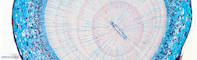 |
Plant |
Taxus baccata |
European yew |
Root |
Cross section |
|
|
unknown |
unknown |
unknown |
Jungner/microthek |
2025-05-01 09:00:57 |
Olympus VS200 |
UPLXAPO 20x / 0.80 |
20x / 0.80 |
75 % |
|
Throughlight widefield |
iDS VS-264C |
Slide scanning microscope |
10.9 |
18,821 x
5,737 x
101 px³ |
5,140.4 x
1,566.7 x
100.0 µm³ |
✅
|
✅
|
✅
|
❌ |
| 631 |
Tectona grandis - Wood cross section 20x |
 |
Plant |
Tectona grandis |
Teak |
Wood |
Cross section |
|
|
unknown |
unstained |
unknown |
Jungner/microthek |
2025-07-15 17:05:35 |
Olympus VS200 |
UPLXAPO 20x / 0.80 |
20x / 0.80 |
75 % |
|
Throughlight widefield |
iDS VS-264C |
Slide scanning microscope |
23.5 |
17,479 x
17,013 x
79 px³ |
4,773.7 x
4,646.3 x
46.0 µm³ |
✅
|
✅
|
✅
|
❌ |
| 666 |
Thuja sp. - One-year-old branch cross section 20x |
 |
Plant |
Thuja sp. |
Arborvitae |
Branch |
Cross section |
Wikipedia on Thuja sp.
This sample has some damage (partly large bubbles) caused by shrinking of the mounting medium due to ageing.
Jungner/microthek micro preparation no. 1224 |
|
unknown |
unknown |
unknown |
Jungner/microthek |
2025-08-11 12:13:11 |
Olympus VS200 |
UPLXAPO 20x / 0.80 |
20x / 0.80 |
75 % |
|
Throughlight widefield |
iDS VS-264C |
Slide scanning microscope |
44.5 |
21,125 x
20,858 x
101 px³ |
5,769.2 x
5,696.3 x
100.0 µm³ |
✅
|
✅
|
✅
|
❌ |
| 665 |
Thuja sp. - Short shoot cross section 20x |
 |
Plant |
Thuja sp. |
Arborvitae |
Short shoot |
Cross section |
|
|
unknown |
unknown |
unknown |
Jungner/microthek |
2025-08-11 12:54:57 |
Olympus VS200 |
UPLXAPO 20x / 0.80 |
20x / 0.80 |
75 % |
|
Throughlight widefield |
iDS VS-264C |
Slide scanning microscope |
4.6 |
7,888 x
10,997 x
53 px³ |
2,154.3 x
3,003.2 x
30.7 µm³ |
✅
|
✅
|
✅
|
❌ |
| 621 |
Tilia platyphyllos - Wood cross section 20x |
 |
Plant |
Tilia platyphyllos |
Large-leaved lime |
Wood |
Cross section |
|
|
unknown |
unstained |
unknown |
Jungner/microthek |
2025-07-14 15:00:48 |
Olympus VS200 |
UPLXAPO 20x / 0.80 |
20x / 0.80 |
75 % |
|
Throughlight widefield |
iDS VS-264C |
Slide scanning microscope |
44.8 |
27,611 x
20,519 x
79 px³ |
7,540.8 x
5,603.8 x
46.0 µm³ |
✅
|
✅
|
✅
|
❌ |
| 183 |
Tilia sp. - Branch cross section 20x 1 |
 |
Plant |
Tilia sp. |
Linden |
Branch |
Cross section |
|
bark parenchyma, bast, bast ray, cambium, epidermis, medullary parenchyma, periderm, primary xylem, sclerenchyma, secretory duct, wood |
Formol, acetic alcohol |
|
Euparal |
Rainer Teubner |
2021-11-11 11:59:26 |
Olympus VS200 |
UPLXAPO 20x / 0.80 |
20x / 0.80 |
50 % |
|
Throughlight widefield |
iDS VS-264C |
Slide scanning microscope |
136.8 |
44,922 x
44,136 x
69 px³ |
12,262.8 x
12,048.2 x
80.2 µm³ |
✅
|
✅
|
✅
|
✅
|
| 184 |
Tilia sp. - Branch cross section 20x 2 |
 |
Plant |
Tilia sp. |
Linden |
Branch |
Cross section |
|
bark parenchyma, bast, bast ray, cambium, epidermis, medullary parenchyma, periderm, primary xylem, sclerenchyma, secretory duct, wood |
Formol, acetic alcohol |
|
Euparal |
Rainer Teubner |
2021-11-11 13:51:46 |
Olympus VS200 |
UPLXAPO 20x / 0.80 |
20x / 0.80 |
50 % |
|
Throughlight widefield |
iDS VS-264C |
Slide scanning microscope |
158.6 |
45,446 x
41,053 x
85 px³ |
12,405.8 x
11,206.5 x
99.1 µm³ |
✅
|
✅
|
✅
|
✅
|
| 185 |
Tilia sp. - Branch cross section 20x 3 |
 |
Plant |
Tilia sp. |
Linden |
Branch |
Cross section |
|
bark parenchyma, bark parenchyma? bast ray?, bast, cambium, epidermis & cuticle, medullary parenchyma, periderm, protoxylem, secretory duct?, wood |
Formol, acetic alcohol |
Anthracene blue |
Euparal |
Rainer Teubner |
2021-11-15 12:29:17 |
Olympus VS200 |
UPLXAPO 20x / 0.80 |
20x / 0.80 |
50 % |
|
Throughlight widefield |
iDS VS-264C |
Slide scanning microscope |
140.7 |
42,302 x
45,547 x
73 px³ |
11,547.6 x
12,433.3 x
85.0 µm³ |
✅
|
✅
|
✅
|
✅
|
| 186 |
Tilia sp. - Branch cross section 20x 4 |
 |
Plant |
Tilia sp. |
Linden |
Branch |
Cross section |
|
bark parenchyma, bast, cambium, epidermis, medullary parenchyma, periderm, protoxylem, wood |
Formol, acetic alcohol |
|
Euparal |
Rainer Teubner |
2021-11-15 15:10:10 |
Olympus VS200 |
UPLXAPO 20x / 0.80 |
20x / 0.80 |
50 % |
|
Throughlight widefield |
iDS VS-264C |
Slide scanning microscope |
14.0 |
12,687 x
13,019 x
85 px³ |
3,463.2 x
3,553.9 x
99.1 µm³ |
✅
|
✅
|
✅
|
✅
|
| 187 |
Tilia sp. - Branch cross section 20x 5 |
 |
Plant |
Tilia sp. |
Linden |
Branch |
Cross section |
|
bark parenchyma, bast, bast ray?, cambium, epidermis, medullary parenchyma, periderm, protoxylem, wood |
Formol, acetic alcohol |
|
Euparal |
Rainer Teubner |
2021-11-15 16:21:00 |
Olympus VS200 |
UPLXAPO 20x / 0.80 |
20x / 0.80 |
50 % |
|
Throughlight widefield |
iDS VS-264C |
Slide scanning microscope |
14.5 |
13,785 x
12,364 x
85 px³ |
3,763.0 x
3,375.1 x
99.1 µm³ |
✅
|
✅
|
✅
|
✅
|
| 188 |
Tilia sp. - Branch cross section 20x 6 |
 |
Plant |
Tilia sp. |
Linden |
Branch |
Cross section |
|
bark parenchyma, bark parenchyma? Baststrahl?, bast, cambium, cork, medullary parenchyma, periderm, protoxylem, wood |
Formol, acetic alcohol |
unstained |
Euparal |
Rainer Teubner |
2021-11-15 16:40:14 |
Olympus VS200 |
UPLXAPO 20x / 0.80 |
20x / 0.80 |
50 % |
|
Throughlight widefield |
iDS VS-264C |
Slide scanning microscope |
14.0 |
13,594 x
12,103 x
85 px³ |
3,710.8 x
3,303.6 x
99.1 µm³ |
✅
|
✅
|
✅
|
✅
|
| 667 |
Tilia sp. - Root cross section 20x |
 |
Plant |
Tilia sp. |
Linden |
Root |
Cross section |
Wikipedia on Tilia sp.
This sample has some damage (partly large bubbles) caused by shrinking of the mounting medium due to ageing.
Jungner/microthek micro preparation no. 1225 |
|
unknown |
unknown |
unknown |
Jungner/microthek |
2025-08-11 13:08:05 |
Olympus VS200 |
UPLXAPO 20x / 0.80 |
20x / 0.80 |
75 % |
|
Throughlight widefield |
iDS VS-264C |
Slide scanning microscope |
32.8 |
16,862 x
19,244 x
101 px³ |
4,605.0 x
5,255.6 x
100.0 µm³ |
✅
|
✅
|
✅
|
❌ |
| 501 |
Tilia sp. - Stem cross section 20x |
 |
Plant |
Tilia sp. |
Linden |
Stem |
Cross section |
|
|
unknown |
unknown |
unknown |
Jungner/microthek |
2025-05-13 18:17:20 |
Olympus VS200 |
UPLXAPO 20x / 0.80 |
20x / 0.80 |
75 % |
|
Throughlight widefield |
iDS VS-264C |
Slide scanning microscope |
20.9 |
23,304 x
8,864 x
101 px³ |
6,364.9 x
2,420.8 x
100.0 µm³ |
✅
|
✅
|
✅
|
❌ |
| 502 |
Tilia sp. - Stem longitudinal section 20x |
 |
Plant |
Tilia sp. |
Linden |
Stem |
Longitudinal section |
|
|
unknown |
unknown |
unknown |
Jungner/microthek |
2025-05-13 18:43:01 |
Olympus VS200 |
UPLXAPO 20x / 0.80 |
20x / 0.80 |
75 % |
|
Throughlight widefield |
iDS VS-264C |
Slide scanning microscope |
65.8 |
26,977 x
24,141 x
101 px³ |
7,367.9 x
6,593.4 x
85.0 µm³ |
✅
|
✅
|
✅
|
❌ |
| 558 |
Tolypothrix sp. - Whole 20x |
 |
Cyanobacterium |
Tolyphotrix sp. |
Tolyphotrix |
Whole organism |
Whole mount |
|
|
unknown |
unknown |
unknown |
Jungner/microthek |
2025-05-30 11:50:20 |
Olympus VS200 |
UPLXAPO 20x / 0.80 |
20x / 0.80 |
75 % |
|
Throughlight widefield |
iDS VS-264C |
Slide scanning microscope |
10.1 |
9,869 x
10,139 x
101 px³ |
2,695.3 x
2,769.0 x
73.0 µm³ |
✅
|
✅
|
✅
|
❌ |
| 354 |
Toxoplasma gondii - Smear 40x |
 |
Protist |
Toxoplasma gondii |
Cause of toxoplasmosis |
Whole cell |
Smear |
|
Parasite |
unknown |
unknown |
unknown |
Jungner/microthek |
2023-12-06 13:19:09 |
Olympus VS200 |
UPLXAPO O 40x / 1.40 |
40x / 1.40 |
75 % |
Oil |
Throughlight widefield |
iDS VS-264C |
Slide scanning microscope |
48.9 |
28,742 x
27,871 x
61 px³ |
3,938.3 x
3,818.9 x
17.4 µm³ |
✅
|
✅
|
✅
|
❌ |
| 177 |
Tradescantia spathacea - lower epidermis before plasmolysis 20x |
 |
Plant |
Tradescantia spathacea |
Oyster plant |
Leaf |
Surface cut |
|
cell wall, central gap, chloroplasts, epidermis cells, guard cells, secondary cells, vacuole |
not fixated |
unstained |
Water |
Christiane Wittmann |
2021-10-14 14:31:51 |
Olympus VS200 |
UPLXAPO 20x / 0.80 |
20x / 0.80 |
50 % |
|
Throughlight widefield |
iDS VS-264C |
Slide scanning microscope |
27.0 |
13,060 x
24,295 x
85 px³ |
3,565.0 x
6,632.0 x
99.1 µm³ |
✅
|
✅
|
✅
|
✅
|
| 175 |
Tradescantia spathacea - lower epidermis plasmolysis 20x 1 |
 |
Plant |
Tradescantia spathacea |
Oyster plant |
Leaf |
Surface cut |
|
cell wall, central gap, chloroplasts, epidermis cells, guard cells, secondary cells, vacuole |
not fixated |
unstained |
Potassium nitrate solution (1M) |
Christiane Wittmann |
2021-10-14 13:27:24 |
Olympus VS200 |
UPLXAPO 20x / 0.80 |
20x / 0.80 |
50 % |
|
Throughlight widefield |
iDS VS-264C |
Slide scanning microscope |
40.3 |
23,489 x
20,164 x
85 px³ |
6,412.0 x
5,504.2 x
99.1 µm³ |
✅
|
✅
|
✅
|
✅
|
| 176 |
Tradescantia spathacea - lower epidermis plasmolysis 20x 2 |
 |
Plant |
Tradescantia spathacea |
Oyster plant |
Leaf |
Surface cut |
|
cell wall, central gap, chloroplasts, epidermis cells, guard cells, secondary cells, vacuole |
not fixated |
unstained |
Potassium nitrate solution (1M) |
Christiane Wittmann |
2021-10-14 14:55:36 |
Olympus VS200 |
UPLXAPO 20x / 0.80 |
20x / 0.80 |
50 % |
|
Throughlight widefield |
iDS VS-264C |
Slide scanning microscope |
11.6 |
9,724 x
13,997 x
85 px³ |
2,654.5 x
3,820.8 x
99.1 µm³ |
✅
|
✅
|
✅
|
✅
|
| 373 |
Trichinella sp. - within muscle tissue 40x |
 |
Animal |
Trichinella sp. |
Trichina worm |
Whole animal |
Thin section |
|
Parasite |
unknown |
unknown |
unknown |
Jungner/microthek |
2023-12-05 16:57:31 |
Olympus VS200 |
UPLXAPO O 40x / 1.40 |
40x / 1.40 |
75 % |
Oil |
Throughlight widefield |
iDS VS-264C |
Slide scanning microscope |
192.4 |
67,686 x
55,722 x
51 px³ |
9,274.5 x
7,635.1 x
18.5 µm³ |
✅
|
✅
|
✅
|
❌ |
| 535 |
Triticum sp. - Seed kernel cross section 20x |
 |
Plant |
Triticum sp. |
Wheat |
Seed |
Cross section |
|
Cells with aleurone, Embryo, Pigment strand, Seed coat, Starchy endosperm |
unknown |
unknown |
unknown |
Jungner/mikrothek |
2025-05-21 18:54:17 |
Olympus VS200 |
UPLXAPO 20x / 0.80 |
20x / 0.80 |
75 % |
|
Throughlight widefield |
iDS VS-264C |
Slide scanning microscope |
16.2 |
15,485 x
11,244 x
93 px³ |
4,229.2 x
3,070.9 x
54.3 µm³ |
✅
|
✅
|
✅
|
✅
|
| 527 |
Triticum sp. - starch grains 20x |
 |
Plant |
Triticum sp. |
Wheat |
Starch grain |
Stray preparation |
|
|
unknown |
unknown |
unknown |
Jungner/microthek |
2025-05-16 11:34:03 |
Olympus VS200 |
UPLXAPO 20x / 0.80 |
20x / 0.80 |
75 % |
|
Throughlight widefield |
iDS VS-264C |
Slide scanning microscope |
22.4 |
18,303 x
18,295 x
67 px³ |
4,999.0 x
4,996.7 x
38.9 µm³ |
✅
|
✅
|
✅
|
❌ |
| 189 |
Tropaeolum majus - Sprout cross section 20x |
 |
Plant |
Tropaeolum majus |
Garden nasturtium |
Sprout |
Cross section |
|
cambium, collenchyma, epidermis & cuticle, medullary parenchyma, phloem, xylem, xylem vessel or tracheae |
Formol, acetic alcohol |
|
Euparal |
Rainer Teubner |
2021-11-03 13:09:58 |
Olympus VS200 |
UPLXAPO 20x / 0.80 |
20x / 0.80 |
50 % |
|
Throughlight widefield |
iDS VS-264C |
Slide scanning microscope |
31.5 |
19,297 x
19,217 x
85 px³ |
5,267.7 x
5,245.6 x
99.1 µm³ |
✅
|
✅
|
✅
|
✅
|
| 345 |
Trypanosoma brucei gambiense - Blood smear 40x |
 |
Protist |
Trypanosoma brucei gambiense |
Cause of the African trypanosomiasis |
Whole cell |
Blood smear |
|
Group of pathogens, Parasite, Trypanosoma |
unknown |
unknown |
unknown |
Jungner/microthek |
2023-12-05 10:17:45 |
Olympus VS200 |
UPLXAPO O 40x / 1.40 |
40x / 1.40 |
75 % |
Oil |
Throughlight widefield |
iDS VS-264C |
Slide scanning microscope |
21.4 |
23,303 x
12,963 x
71 px³ |
3,193.0 x
1,776.2 x
20.3 µm³ |
✅
|
✅
|
✅
|
✅
|
| 353 |
Trypanosoma brucei - Blood smear 40x |
 |
Protist |
Trypanosoma brucei gambiense |
Cause of animal trypanosomiasis |
Whole cell |
Blood smear |
|
Cluster of Trypanosoma, Parasite |
unknown |
unknown |
unknown |
Jungner/microthek |
2023-12-06 15:08:57 |
Olympus VS200 |
UPLXAPO O 40x / 1.40 |
40x / 1.40 |
75 % |
Oil |
Throughlight widefield |
iDS VS-264C |
Slide scanning microscope |
17.7 |
15,463 x
19,434 x
59 px³ |
2,118.7 x
2,662.9 x
16.8 µm³ |
✅
|
✅
|
✅
|
✅
|
| 344 |
Trypanosoma equiperdum - Blood smear 40x |
 |
Protist |
Trypanosoma equiperdum |
Cause of the covering sickness |
Whole cell |
Blood smear |
|
Group of pathogens, Parasite, Trypanosoma |
unknown |
unknown |
unknown |
Jungner/microthek |
2023-12-05 11:17:16 |
Olympus VS200 |
UPLXAPO O 40x / 1.40 |
40x / 1.40 |
75 % |
Oil |
Throughlight widefield |
iDS VS-264C |
Slide scanning microscope |
77.1 |
29,715 x
25,687 x
101 px³ |
4,071.6 x
3,519.7 x
29.0 µm³ |
✅
|
✅
|
✅
|
✅
|
| 571 |
Tuber rufum - Fruiting body cross section 20x |
 |
Fungus |
Tuber rufum |
Red truffle |
Fruiting body |
Cross section |
|
|
unknown |
unknown |
unknown |
Jungner/microthek |
2025-06-23 19:18:44 |
Olympus VS200 |
UPLXAPO 20x / 0.80 |
20x / 0.80 |
75 % |
|
Throughlight widefield |
iDS VS-264C |
Slide scanning microscope |
54.9 |
32,890 x
23,517 x
71 px³ |
8,982.6 x
6,422.6 x
41.3 µm³ |
✅
|
✅
|
✅
|
❌ |
| 474 |
Tulipa sp. or Iris sp., Leaf epidermis 20x, Jungner 535 |
 |
Plant |
Tulipa sp. / Iris sp. |
Tulip or iris |
Leaf epidermis |
Peel |
|
Epidermal cell, Singular guard cell, Stoma, Stomatal apparatus |
unknown |
unknown |
unknown |
Jungner/microthek |
2025-05-05 13:21:00 |
Olympus VS200 |
UPLXAPO 20x / 0.80 |
20x / 0.80 |
75 % |
|
Throughlight widefield |
iDS VS-264C |
Slide scanning microscope |
14.9 |
12,528 x
11,772 x
101 px³ |
3,421.5 x
3,215.1 x
75.0 µm³ |
✅
|
✅
|
✅
|
✅
|
| 629 |
Ulmus glabra - Wood cross section 20x |
 |
Plant |
Ulmus glabra |
Scots elm |
Wood |
Cross section |
|
|
unknown |
unstained |
unknown |
Jungner/microthek |
2025-07-15 15:49:08 |
Olympus VS200 |
UPLXAPO 20x / 0.80 |
20x / 0.80 |
75 % |
|
Throughlight widefield |
iDS VS-264C |
Slide scanning microscope |
36.7 |
25,241 x
16,694 x
87 px³ |
6,893.4 x
4,559.2 x
50.7 µm³ |
✅
|
✅
|
✅
|
❌ |
| 708 |
Utricularia sp. - Leaf cross section 20x |
 |
Plant |
Utricularia sp. |
Bladderwort |
Leaf |
Cross section |
|
|
unknown |
unknown |
unknown |
|
2025-08-21 09:07:44 |
Olympus VS200 |
UPLXAPO 20x / 0.80 |
20x / 0.80 |
75 % |
|
Throughlight widefield |
iDS VS-264C |
Slide scanning microscope |
54.4 |
32,084 x
25,310 x
67 px³ |
8,762.2 x
6,912.3 x
38.9 µm³ |
✅
|
✅
|
✅
|
❌ |
| 715 |
Vaccinium myrtillus - Fruit cross section 20x |
 |
Plant |
Vaccinium myrtillus |
European blueberry |
Fruit |
Cross section |
Wikipedia on Vaccinium myrtillus.
This sample has some minor damage caused by shrinking of the mounting medium due to ageing.
Jungner/microthek micro preparation no. 1263 |
Columella, Endocarp, Exocarp, Internal/radial pericarp, Loculus, Mesocarp, Outer pericarp, Placenta, Seed, Vascular tissue |
unknown |
unknown |
unknown |
Jungner/microthek |
2025-08-20 10:13:31 |
Olympus VS200 |
UPLXAPO 20x / 0.80 |
20x / 0.80 |
75 % |
|
Throughlight widefield |
iDS VS-264C |
Slide scanning microscope |
21.4 |
21,652 x
21,067 x
47 px³ |
5,913.4 x
5,753.4 x
27.1 µm³ |
✅
|
✅
|
✅
|
✅
|
| 714 |
Vaccinium vitis-idaea - Leaf cross section 20x |
 |
Plant |
Vaccinium vitis-idaea |
Lingonberry |
Leaf |
Cross section |
|
|
unknown |
unknown |
unknown |
Jungner/microthek |
2025-08-20 09:56:59 |
Olympus VS200 |
UPLXAPO 20x / 0.80 |
20x / 0.80 |
75 % |
|
Throughlight widefield |
iDS VS-264C |
Slide scanning microscope |
7.7 |
15,538 x
12,660 x
39 px³ |
4,243.4 x
3,457.5 x
22.4 µm³ |
✅
|
✅
|
✅
|
❌ |
| 6 |
Vanilla planifolia - Aerial root cross section 20x |
 |
Plant |
Vanilla planifolia |
Flat-leaved vanilla |
Root |
Cross section |
|
bark parenchyma, endodermis, exodermis, medullary parenchyma, passage cells, phloem, rhizodermis, root hairs, tracheae, xylem |
Formol, acetic alcohol |
|
Euparal |
Rainer Teubner |
2021-10-12 15:35:44 |
Olympus VS200 |
UPLXAPO 20x / 0.80 |
20x / 0.80 |
50 % |
|
Throughlight widefield |
iDS VS-264C |
Slide scanning microscope |
15.9 |
15,629 x
16,677 x
61 px³ |
4,266.4 x
4,552.5 x
70.8 µm³ |
✅
|
✅
|
✅
|
✅
|
| 479 |
Verbascum sp. - Leaf cross section 20x |
 |
Plant |
Verbascum sp. |
Mullein |
Leaf |
Cross section |
|
|
unknown |
unknown |
unknown |
Jungner/microthek |
2025-05-06 10:35:12 |
Olympus VS200 |
UPLXAPO 20x / 0.80 |
20x / 0.80 |
75 % |
|
Throughlight widefield |
iDS VS-264C |
Slide scanning microscope |
17.6 |
38,438 x
8,017 x
57 px³ |
10,498.4 x
2,189.6 x
33.0 µm³ |
✅
|
✅
|
✅
|
❌ |
| 532 |
Verbascum thapsus - Leaf trichome 20x |
 |
Plant |
Verbascum thapsus |
Great mullein |
Leaf trichome |
Whole mount |
|
|
unknown |
unknown |
unknown |
Jungner/mikrothek |
2025-05-20 11:21:01 |
Olympus VS200 |
UPLXAPO 20x / 0.80 |
20x / 0.80 |
75 % |
|
Throughlight widefield |
iDS VS-264C |
Slide scanning microscope |
4.2 |
6,513 x
6,394 x
101 px³ |
1,778.7 x
1,746.3 x
100.0 µm³ |
✅
|
✅
|
✅
|
❌ |
| 703 |
Viburnum lantana - Leaf cross section 20x |
 |
Plant |
Viburnum lantana |
Wayfarer |
Leaf |
Cross section |
|
Trichomes of the leaf underside |
unknown |
unknown |
unknown |
Jungner/microthek |
2025-08-20 12:13:58 |
Olympus VS200 |
UPLXAPO 20x / 0.80 |
20x / 0.80 |
75 % |
|
Throughlight widefield |
iDS VS-264C |
Slide scanning microscope |
100.6 |
31,692 x
41,207 x
77 px³ |
8,655.2 x
11,253.9 x
44.8 µm³ |
✅
|
✅
|
✅
|
✅
|
| 33 |
Vigna unguiculata subsp. sesquipedalis - Sprout cross section 20x |
 unguiculata - Spross quer_Thumbnail.jpg) |
Plant |
Vigna (subsp. sesquipedalis) unguiculata |
Asparagus bean |
Sprout |
Cross section |
|
bark parenchyma, cambium, collenchyma, cuticula, medullary parenchyma, medullary ray, phloem, primary xylem, sclerenchyma, starch granules, xylem, xylem parenchyma, xylem vessel or tracheae |
Formol, acetic alcohol |
|
Euparal |
Jörg Weiß
www.mikroskopie-bonn.de |
2021-07-27 10:28:30 |
Olympus VS200 |
UPLXAPO 20x / 0.80 |
20x / 0.80 |
50 % |
|
Throughlight widefield |
iDS VS-264C |
Slide scanning microscope |
40.7 |
24,588 x
27,147 x
61 px³ |
6,711.8 x
7,410.5 x
70.8 µm³ |
✅
|
✅
|
✅
|
✅
|
| 605 |
Viscum album - Leaf cross section 20x |
 |
Plant |
Viscum album |
European mistletoe |
Leaf |
Cross section |
|
|
unknown |
unknown |
unknown |
Jungner/microthek |
2025-07-02 11:43:39 |
Olympus VS200 |
UPLXAPO 20x / 0.80 |
20x / 0.80 |
75 % |
|
Throughlight widefield |
iDS VS-264C |
Slide scanning microscope |
39.5 |
52,616 x
15,320 x
49 px³ |
14,370.0 x
4,183.8 x
28.3 µm³ |
✅
|
✅
|
✅
|
❌ |
| 32 |
Viscum album - Sprout cross section 20x |
 |
Plant |
Viscum album |
Mistletoe |
Sprout |
Cross section |
|
bark parenchyma, bast, cambium, epidermis & cuticle, medullary parenchyma, medullary ray, sclerenchyma, xylem / wood |
Formol, acetic alcohol |
|
Euparal |
Rainer Teubner |
2021-09-02 15:48:21 |
Olympus VS200 |
UPLXAPO 20x / 0.80 |
20x / 0.80 |
50 % |
|
Throughlight widefield |
iDS VS-264C |
Slide scanning microscope |
9.1 |
11,609 x
12,878 x
61 px³ |
3,168.9 x
3,515.4 x
87.0 µm³ |
✅
|
✅
|
✅
|
✅
|
| 564 |
Volvox sp. - Whole 20x |
 |
Algae |
Volvox sp. |
Globe algae |
Whole organism |
Whole mount |
|
|
unknown |
unknown |
unknown |
Jungner/microthek |
2025-06-23 11:03:00 |
Olympus VS200 |
UPLXAPO 20x / 0.80 |
20x / 0.80 |
75 % |
|
Throughlight widefield |
iDS VS-264C |
Slide scanning microscope |
43.8 |
34,481 x
30,977 x
41 px³ |
9,416.9 x
8,460.1 x
23.6 µm³ |
✅
|
✅
|
✅
|
❌ |
| 96 |
Wollemia nobilis - Leaf on-year-old cross section 20x 2.4 |
 |
Plant |
Wollemia nobilis |
Wollemi pine |
Leaf |
Cross section |
|
cuticle, epidermis, glandular epithelium, guard cells, hypodermis, palisade parenchyma, phloem, secretory duct, sponge parenchyma, vascular bundle, xylem |
Formol, acetic alcohol, sodium hypochlorite |
|
Euparal |
Rainer Teubner |
2021-07-02 10:25:45 |
Olympus VS200 |
UPLXAPO 20x / 0.80 |
20x / 0.80 |
40 % |
|
Throughlight widefield |
iDS VS-264C |
Slide scanning microscope |
4.7 |
19,952 x
3,870 x
61 px³ |
5,446.5 x
1,056.3 x
70.8 µm³ |
✅
|
✅
|
✅
|
✅
|
| 90 |
Wollemia nobilis - Leaf one-year-old cross section 20x 1.1 |
 |
Plant |
Wollemia nobilis |
Wollemi pine |
Leaf |
Cross section |
|
cuticle, epidermis, hypodermis, mucus cell, palisade parenchyma, secretory duct, sponge parenchyma, stoma, vascular bundle |
Formol, acetic alcohol, sodium hypochlorite |
W3A: Acridine red, acriflavine, astra blue |
Euparal |
Rainer Teubner |
2021-07-02 09:02:20 |
Olympus VS200 |
UPLXAPO 20x / 0.80 |
20x / 0.80 |
40 % |
|
Throughlight widefield |
iDS VS-264C |
Slide scanning microscope |
4.7 |
20,234 x
3,769 x
61 px³ |
5,523.5 x
1,028.8 x
70.8 µm³ |
✅
|
✅
|
✅
|
✅
|
| 91 |
Wollemia nobilis - Leaf one-year-old cross section 20x 1.2 |
 |
Plant |
Wollemia nobilis |
Wollemi pine |
Leaf |
Cross section |
|
cuticle, epidermis, hypodermis, mucus cell, palisade parenchym, sponge parenchyma, stomata, vascular bundle |
Formol, acetic alcohol, sodium hypochlorite |
unstained |
Euparal |
Rainer Teubner |
2021-07-02 09:13:00 |
Olympus VS200 |
UPLXAPO 20x / 0.80 |
20x / 0.80 |
40 % |
|
Throughlight widefield |
iDS VS-264C |
Slide scanning microscope |
8.4 |
23,610 x
5,865 x
61 px³ |
6,445.0 x
1,600.9 x
70.8 µm³ |
✅
|
✅
|
✅
|
✅
|
| 92 |
Wollemia nobilis - Leaf one-year-old cross section 20x 1.3 |
 |
Plant |
Wollemia nobilis |
Wollemi pine |
Leaf |
Cross section |
|
cuticle, epidermis, guard cells, hypodermis, mucus cell, palisade parenchyma, secretory duct, sponge parenchym, vascular bundle |
Formol, acetic alcohol, sodium hypochlorite |
|
Euparal |
Rainer Teubner |
2021-07-02 09:24:28 |
Olympus VS200 |
UPLXAPO 20x / 0.80 |
20x / 0.80 |
40 % |
|
Throughlight widefield |
iDS VS-264C |
Slide scanning microscope |
7.2 |
19,448 x
6,036 x
61 px³ |
5,308.9 x
1,647.7 x
70.8 µm³ |
✅
|
✅
|
✅
|
✅
|
| 94 |
Wollemia nobilis - Leaf one-year-old cross section 20x 2.1 |
 |
Plant |
Wollemia nobilis |
Wollemi pine |
Leaf |
Cross section |
|
cuticle, epidermis, guard cells, hypodermis, palisade parenchyma, secretory duct, sponge parenchyma, vascular bundle |
Formol, acetic alcohol, sodium hypochlorite |
unstained |
Euparal |
Rainer Teubner |
2021-07-02 09:34:05 |
Olympus VS200 |
UPLXAPO 20x / 0.80 |
20x / 0.80 |
40 % |
|
Throughlight widefield |
iDS VS-264C |
Slide scanning microscope |
4.0 |
20,053 x
3,296 x
61 px³ |
5,474.0 x
899.5 x
70.8 µm³ |
✅
|
✅
|
✅
|
✅
|
| 93 |
Wollemia nobilis - Leaf one-year-old cross section 20x 2.2 |
 |
Plant |
Wollemia nobilis |
Wollemi pine |
Leaf |
Cross section |
|
epidermis & cuticle, guard cells, hypodermis, palisade parenchyma, phloem, secretory duct, sponge parenchyma, vascular bundle, xylem |
Formol, acetic alcohol, sodium hypochlorite |
|
Euparal |
Rainer Teubner |
2021-07-02 10:00:45 |
Olympus VS200 |
UPLXAPO 20x / 0.80 |
20x / 0.80 |
40 % |
|
Throughlight widefield |
iDS VS-264C |
Slide scanning microscope |
7.3 |
19,156 x
6,268 x
61 px³ |
5,229.2 x
1,711.0 x
70.8 µm³ |
✅
|
✅
|
✅
|
✅
|
| 95 |
Wollemia nobilis - Leaf one-year-old cross section 20x 2.3 |
 |
Plant |
Wollemia nobilis |
Wollemi pine |
Leaf |
Cross section |
|
cuticle, epidermis, glandular epithelium, guard cells, hypodermis, palisade parenchyma, phloem, secretory duct, sponge parenchyma, vascular bundle, xylem |
Formol, acetic alcohol, sodium hypochlorite |
W3A: Acridine red, acriflavine, astra blue |
Euparal |
Rainer Teubner |
2021-07-02 10:09:36 |
Olympus VS200 |
UPLXAPO 20x / 0.80 |
20x / 0.80 |
40 % |
|
Throughlight widefield |
iDS VS-264C |
Slide scanning microscope |
3.7 |
20,295 x
3,003 x
61 px³ |
5,540.0 x
819.7 x
70.8 µm³ |
✅
|
✅
|
✅
|
✅
|
| 97 |
Wollemia nobilis - Leaf one-year-old cross section 20x 3.1 |
 |
Plant |
Wollemia nobilis |
Wollemi pine |
Leaf |
Cross section |
|
epidermis & cuticle, glandular epithelium, guard cells, palisade parenchyma, phloem, secretory duct, sponge parenchyma, vascular bundle, xylem |
Formol, acetic alcohol, sodium hypochlorite |
unstained |
Euparal |
Rainer Teubner |
2021-07-02 10:32:42 |
Olympus VS200 |
UPLXAPO 20x / 0.80 |
20x / 0.80 |
40 % |
|
Throughlight widefield |
iDS VS-264C |
Slide scanning microscope |
4.0 |
20,335 x
3,235 x
61 px³ |
5,551.0 x
883.0 x
70.8 µm³ |
✅
|
✅
|
✅
|
✅
|
| 98 |
Wollemia nobilis - Leaf one-year-old cross section 20x 3.2 |
 |
Plant |
Wollemia nobilis |
Wollemi pine |
Leaf |
Cross section |
|
cuticle, epidermis, glandular epithelium, hypodermis, palisade parenchyma, phloem, secretory duct, sponge parenchyma, vascular bundle, xylem |
Formol, acetic alcohol, sodium hypochlorite |
|
Euparal |
Rainer Teubner |
2021-07-02 10:45:14 |
Olympus VS200 |
UPLXAPO 20x / 0.80 |
20x / 0.80 |
40 % |
|
Throughlight widefield |
iDS VS-264C |
Slide scanning microscope |
7.7 |
19,428 x
6,460 x
61 px³ |
5,303.4 x
1,763.2 x
70.8 µm³ |
✅
|
✅
|
✅
|
✅
|
| 99 |
Wollemia nobilis - Leaf one-year-old cross section 20x 3.3 |
 |
Plant |
Wollemia nobilis |
Wollemi pine |
Leaf |
Cross section |
|
epidermis & cuticle, glandular epithelium, guard cells, hypodermis, palisade parenchyma, phloem, secretory duct, sponge parenchyma, vascular bundle, xylem |
Formol, acetic alcohol, sodium hypochlorite |
W3A: Acridine red, acriflavine, astra blue |
Euparal |
Rainer Teubner |
2021-07-02 11:00:28 |
Olympus VS200 |
UPLXAPO 20x / 0.80 |
20x / 0.80 |
40 % |
|
Throughlight widefield |
iDS VS-264C |
Slide scanning microscope |
5.5 |
19,972 x
4,555 x
61 px³ |
5,452.0 x
1,243.3 x
70.8 µm³ |
✅
|
✅
|
✅
|
✅
|
| 100 |
Wollemia nobilis - Leaf one-year-old cross section 20x 3.4 |
 |
Plant |
Wollemia nobilis |
Wollemi pine |
Leaf |
Cross section |
|
|
Formol, acetic alcohol, sodium hypochlorite |
|
Euparal |
Rainer Teubner |
2021-07-02 11:09:55 |
Olympus VS200 |
UPLXAPO 20x / 0.80 |
20x / 0.80 |
40 % |
|
Throughlight widefield |
iDS VS-264C |
Slide scanning microscope |
4.9 |
19,721 x
4,061 x
61 px³ |
5,383.2 x
1,108.5 x
70.8 µm³ |
✅
|
✅
|
✅
|
❌ |
| 579 |
Xanthoria sp. - Thallus and apothecium cross section 20x |
 |
Lichen |
Xanthoria sp. |
Orange lichen |
Apothecium |
Cross section |
|
Algae symbiont, Apothecium, Ascus with multiple spores, Cortex, Fungal hyphae, Hymenium with asci, Photosynthetic symbionts, Spore |
unknown |
unknown |
unknown |
Jungner/microthek |
2025-06-24 16:54:08 |
Olympus VS200 |
UPLXAPO 20x / 0.80 |
20x / 0.80 |
75 % |
|
Throughlight widefield |
iDS VS-264C |
Slide scanning microscope |
17.6 |
28,428 x
12,600 x
49 px³ |
7,763.9 x
3,441.2 x
28.3 µm³ |
✅
|
✅
|
✅
|
✅
|
| 322 |
Zea mays - Leaf cross section 20x |
 |
Plant |
Zea mays |
Maize |
Leaf |
Cross section |
|
C4 plant,monocotyledonous |
unknown |
unknown |
Eukitt |
Jungner/microthek |
2023-10-26 11:16:42 |
Olympus VS200 |
UPLXAPO 20x / 0.80 |
20x / 0.80 |
75 % |
|
Throughlight widefield |
iDS VS-264C |
Slide scanning microscope |
42.6 |
48,337 x
14,003 x
63 px³ |
13,201.8 x
3,824.4 x
36.6 µm³ |
✅
|
✅
|
✅
|
❌ |
| 428 |
Zea mays - Seed with endosperm 20x |
 |
Plant |
Zea mays |
Maize |
Seed |
Longitudinal section |
|
Aleurone layer, Embryo, Endosperm, Seed hull, Tip cap |
unknown |
unknown |
unknown |
Bresser |
2025-04-09 08:41:55 |
Olympus VS200 |
UPLXAPO 20x / 0.80 |
20x / 0.80 |
75 % |
|
Throughlight widefield |
iDS VS-264C |
Slide scanning microscope |
53.7 |
18,076 x
29,396 x
101 px³ |
4,936.8 x
8,028.8 x
59.0 µm³ |
✅
|
✅
|
✅
|
✅
|
| 295 |
Zea mays - Stalk cross section 20x |
 |
Plant |
Zea mays |
Maize |
Stalk |
Cross section |
|
C4 plant,monocotyledonous |
unknown |
Astra blue, safranin |
unknown |
Karl Brügmann |
2023-10-11 09:28:58 |
Olympus VS200 |
UPLXAPO 20x / 0.80 |
20x / 0.80 |
75 % |
|
Throughlight widefield |
iDS VS-264C |
Slide scanning microscope |
140.6 |
42,325 x
39,069 x
85 px³ |
11,560.0 x
10,670.7 x
64.7 µm³ |
✅
|
✅
|
✅
|
❌ |
| 266 |
Zea mays - Stalk cross section 40x |
 |
Plant |
Zea mays |
Maize |
Stalk |
Cross section |
Wikipedia on maize.
Due to shrinking of the mounting media caused by ageing, air got into the specimen.
Jungner/microthek micro preparation no. 1015 |
Air bubbles, C4 plant, monocotyledonous |
unknown |
unknown |
Eukitt |
Jungner/microthek |
2023-09-14 12:55:26 |
Olympus VS200 |
UPLFLN 40x / 0.75 |
40x / 0.75 |
75 % |
|
Throughlight widefield |
iDS VS-264C |
Slide scanning microscope |
187.0 |
52,336 x
42,040 x
85 px³ |
7,161.1 x
5,752.3 x
56.3 µm³ |
✅
|
✅
|
✅
|
✅
|
| 296 |
Zea mays - Stem cross section 20x |
 |
Plant |
Zea mays |
Maize |
Stem |
Cross section |
|
C4 plant, Companion cell, Intercellular duct, Intercellular space, monocotyledonous, Parenchyma cell, Phloem, Sclerenchyma, Sieve tube, Trachea, Tracheids, Xylem, Xylem parenchyma |
unknown |
unknown |
Eukitt |
Jungner/microthek |
2023-10-11 11:10:36 |
Olympus VS200 |
UPLXAPO 20x / 0.80 |
20x / 0.80 |
75 % |
|
Throughlight widefield |
iDS VS-264C |
Slide scanning microscope |
61.7 |
29,020 x
25,027 x
85 px³ |
7,926.1 x
6,835.4 x
64.7 µm³ |
✅
|
✅
|
✅
|
✅
|
| 429 |
Zea mays - Stem cross section 20x |
 |
Plant |
Zea mays |
Maize |
Stem |
Cross section |
|
C4 plant,monocotyledonous |
unknown |
unknown |
unknown |
Bresser |
2025-04-09 09:34:19 |
Olympus VS200 |
UPLXAPO 20x / 0.80 |
20x / 0.80 |
75 % |
|
Throughlight widefield |
iDS VS-264C |
Slide scanning microscope |
5.9 |
11,274 x
8,534 x
61 px³ |
3,079.0 x
2,330.8 x
35.4 µm³ |
✅
|
✅
|
✅
|
❌ |
| 302 |
Zea mays - Stem longitudinal section 20x |
 |
Plant |
Zea mays |
Maize |
Stem |
Longitudinal section |
|
C4 plant, monocotyledonous, Phloem, Tracheae and tracheids of the xylem, Woody vascular bundle sheath, Xylem parenchyma and intercellular duct |
unknown |
unknown |
Eukitt |
Jungner/microthek |
2023-10-16 10:55:32 |
Olympus VS200 |
UPLXAPO 20x / 0.80 |
20x / 0.80 |
75 % |
|
Throughlight widefield |
iDS VS-264C |
Slide scanning microscope |
91.8 |
42,760 x
21,263 x
101 px³ |
11,678.8 x
5,807.3 x
80.0 µm³ |
✅
|
✅
|
✅
|
✅
|
| 644 |
Zygnema sp. 20x |
 |
Protist |
Zygnema sp. |
Zygnema |
Whole cell |
Whole organism |
|
|
not fixated |
not stained |
Water |
Michael Kloster / Eva Papendick |
2025-07-18 12:35:45 |
Olympus VS200 |
UPLXAPO 20x / 0.80 |
20x / 0.80 |
75 % |
|
Throughlight widefield |
iDS VS-264C |
Slide scanning microscope |
23.8 |
13,886 x
16,953 x
101 px³ |
3,792.2 x
4,629.9 x
59.0 µm³ |
✅
|
✅
|
✅
|
❌ |
| 29 |
Amphipod crustacean - whole mount 20x |
 |
Animal |
Gammarus (pulex ?) |
Gammarus pulex (supposedly) |
Whole animal |
Whole mount |
|
|
Alcohol, Xylol |
Boraxcarmine |
Malinol |
|
2021-07-01 09:23:30 |
Olympus VS200 |
UPLXAPO 20x / 0.80 |
20x / 0.80 |
40 % |
|
Throughlight widefield |
iDS VS-264C |
Slide scanning microscope |
12.9 |
12,939 x
16,395 x
61 px³ |
3,532.0 x
4,475.4 x
70.8 µm³ |
✅
|
✅
|
✅
|
❌ |
| 1011 |
Aorta Azan 40x |
 |
Animal |
Mammalia (cl.) |
Mammal |
Aorta |
Thin section |
Wikipedia on the aorta.
This sample has some damage caused by shrinking of the mounting medium due to ageing (top, lower right), but mostly in non-relevant marginal areas. |
|
unknown |
|
unknown |
|
2025-12-12 15:21:12 |
Olympus VS200 |
UPLFLN 40x / 0.75 |
40x / 0.75 |
75 % |
|
Throughlight widefield |
iDS VS-264C |
Slide scanning microscope |
273.2 |
130,775 x
77,367 x
27 px³ |
17,896.2 x
10,587.4 x
17.5 µm³ |
✅
|
✅
|
✅
|
❌ |
| 1014 |
Appendix vermiformis - cross section HE 40x |
 |
Animal |
Mammalia (cl.) |
Mammal |
Appendix |
Cross section |
|
|
unknown |
|
unknown |
|
2025-12-13 20:53:51 |
Olympus VS200 |
UPLFLN 40x / 0.75 |
40x / 0.75 |
75 % |
|
Throughlight widefield |
iDS VS-264C |
Slide scanning microscope |
53.6 |
39,418 x
41,208 x
33 px³ |
5,394.1 x
5,639.2 x
21.5 µm³ |
✅
|
✅
|
✅
|
❌ |
| 730 |
Blood - Acute myeloid leukemiae 60x |
 |
Animal |
Homo sapiens |
Human |
Blood |
Smear |
|
|
Methanol |
|
Eukitt |
Anna M. Kehl |
2026-03-24 12:34:37 |
Olympus VS200 |
UPLXAPO OPH 60x / 1.42 |
60x / 1.42 |
75 % |
Oil |
Throughlight widefield |
iDS VS-264C |
Slide scanning microscope |
16.9 |
21,983 x
22,013 x
35 px³ |
1,999.4 x
2,002.1 x
9.5 µm³ |
✅
|
✅
|
✅
|
❌ |
| 729 |
Blood - Chronic lymphocytic leukemia 60x |
 |
Animal |
Homo sapiens |
Human |
Blood |
Smear |
|
|
Methanol |
|
Eukitt |
Anna M. Kehl |
2026-03-24 11:41:47 |
Olympus VS200 |
UPLXAPO OPH 60x / 1.42 |
60x / 1.42 |
75 % |
Oil |
Throughlight widefield |
iDS VS-264C |
Slide scanning microscope |
14.0 |
21,983 x
22,013 x
29 px³ |
1,999.4 x
2,002.1 x
7.8 µm³ |
✅
|
✅
|
✅
|
❌ |
| 723 |
Blood - Chronic myelogenous leukemia 60x |
 |
Animal |
Homo sapiens |
Human |
Blood |
Smear |
|
|
Methanol |
|
Eukitt |
Anna M. Kehl |
2026-03-23 16:35:28 |
Olympus VS200 |
UPLXAPO OPH 60x / 1.42 |
60x / 1.42 |
75 % |
Oil |
Throughlight widefield |
iDS VS-264C |
Slide scanning microscope |
15.0 |
21,983 x
21,983 x
31 px³ |
1,999.4 x
1,999.4 x
8.4 µm³ |
✅
|
✅
|
✅
|
❌ |
| 727 |
Blood - Infectious mononucleosis 60x |
 |
Animal |
Homo sapiens |
Human |
Blood |
Smear |
|
|
Methanol |
|
Eukitt |
Anna M. Kehl |
2026-03-24 10:45:45 |
Olympus VS200 |
UPLXAPO OPH 60x / 1.42 |
60x / 1.42 |
75 % |
Oil |
Throughlight widefield |
iDS VS-264C |
Slide scanning microscope |
15.0 |
21,983 x
22,013 x
31 px³ |
1,999.4 x
2,002.1 x
8.4 µm³ |
✅
|
✅
|
✅
|
❌ |
| 722 |
Blood - Iron deficiency (male) 60x |
 |
Animal |
Homo sapiens |
Human |
Blood |
Smear |
|
|
Methanol |
|
Eukitt |
Anna M. Kehl |
2026-03-24 10:27:20 |
Olympus VS200 |
UPLXAPO OPH 60x / 1.42 |
60x / 1.42 |
75 % |
Oil |
Throughlight widefield |
iDS VS-264C |
Slide scanning microscope |
16.9 |
21,983 x
22,013 x
35 px³ |
1,999.4 x
2,002.1 x
9.5 µm³ |
✅
|
✅
|
✅
|
❌ |
| 725 |
Blood - Pernicious anemia Dralle 60x |
 |
Animal |
Homo sapiens |
Human |
Blood |
Smear |
|
|
Methanol |
|
Eukitt |
Anna M. Kehl |
2026-03-23 14:31:54 |
Olympus VS200 |
UPLXAPO OPH 60x / 1.42 |
60x / 1.42 |
75 % |
Oil |
Throughlight widefield |
iDS VS-264C |
Slide scanning microscope |
15.0 |
21,983 x
22,013 x
31 px³ |
1,999.4 x
2,002.1 x
8.4 µm³ |
✅
|
✅
|
✅
|
❌ |
| 726 |
Blood - Pernicious anemia HE 60x |
 |
Animal |
Homo sapiens |
Human |
Blood |
Smear |
|
|
Methanol |
|
Eukitt |
Anna M. Kehl |
2026-03-23 14:16:23 |
Olympus VS200 |
UPLXAPO OPH 60x / 1.42 |
60x / 1.42 |
75 % |
Oil |
Throughlight widefield |
iDS VS-264C |
Slide scanning microscope |
13.1 |
21,983 x
22,013 x
27 px³ |
1,999.4 x
2,002.1 x
7.3 µm³ |
✅
|
✅
|
✅
|
❌ |
| 720 |
Blood - Reticulocytes 130 ‰ 104-156 ‰ 60x |
 |
Animal |
Homo sapiens |
Human |
Blood |
Smear |
|
|
Methanol |
|
Eukitt |
Anna M. Kehl |
2026-03-23 10:47:30 |
Olympus VS200 |
UPLXAPO OPH 60x / 1.42 |
60x / 1.42 |
75 % |
Oil |
Throughlight widefield |
iDS VS-264C |
Slide scanning microscope |
15.0 |
21,983 x
22,013 x
31 px³ |
1,999.4 x
2,002.1 x
8.4 µm³ |
✅
|
✅
|
✅
|
❌ |
| 716 |
Blood - Sickle cell disease 60x |
 |
Animal |
Homo sapiens |
Human |
Blood |
Smear |
|
|
Methanol |
|
Eukitt |
Anna M. Kehl |
2026-03-23 10:20:21 |
Olympus VS200 |
UPLXAPO OPH 60x / 1.42 |
60x / 1.42 |
75 % |
Oil |
Throughlight widefield |
iDS VS-264C |
Slide scanning microscope |
16.0 |
21,983 x
22,013 x
33 px³ |
1,999.4 x
2,002.1 x
9.0 µm³ |
✅
|
✅
|
✅
|
❌ |
| 717 |
Blood - Thalassaemia major 60x |
 |
Animal |
Homo sapiens |
Human |
Blood |
Smear |
|
|
Methanol |
|
Eukitt |
Anna M. Kehl |
2026-03-23 11:19:36 |
Olympus VS200 |
UPLXAPO OPH 60x / 1.42 |
60x / 1.42 |
75 % |
Oil |
Throughlight widefield |
iDS VS-264C |
Slide scanning microscope |
16.9 |
21,983 x
21,983 x
35 px³ |
1,999.4 x
1,999.4 x
9.5 µm³ |
✅
|
✅
|
✅
|
❌ |
| 1012 |
Blood vessels - cross section HE 40x |
 |
Animal |
Mammalia (cl.) |
Mammal |
Blood vessel |
Cross section |
|
|
unknown |
|
unknown |
|
2025-12-12 21:12:22 |
Olympus VS200 |
UPLFLN 40x / 0.75 |
40x / 0.75 |
75 % |
|
Throughlight widefield |
iDS VS-264C |
Slide scanning microscope |
18.0 |
38,424 x
33,416 x
14 px³ |
5,258.1 x
4,572.8 x
8.7 µm³ |
✅
|
✅
|
✅
|
❌ |
| 719 |
Blut - Reticulocytes 130 ‰ 60x |
 |
Animal |
Homo sapiens |
Human |
Blood |
Smear |
|
|
Methanol |
|
Eukitt |
Anna M. Kehl |
2026-03-23 13:39:20 |
Olympus VS200 |
UPLXAPO OPH 60x / 1.42 |
60x / 1.42 |
75 % |
Oil |
Throughlight widefield |
iDS VS-264C |
Slide scanning microscope |
11.1 |
21,983 x
22,013 x
23 px³ |
1,999.4 x
2,002.1 x
6.2 µm³ |
✅
|
✅
|
✅
|
❌ |
| 1017 |
Cardiac muscle Azan 40x |
 |
Animal |
Mammalia (cl.) |
Mammal |
Cardiac muscle |
Thin section |
|
|
unknown |
|
unknown |
|
2026-01-05 18:18:44 |
Olympus VS200 |
UPLXAPO O 40x / 1.40 |
40x / 1.40 |
75 % |
Oil |
Throughlight widefield |
iDS VS-264C |
Slide scanning microscope |
543.3 |
138,038 x
69,052 x
57 px³ |
18,918.9 x
9,463.9 x
16.2 µm³ |
✅
|
✅
|
✅
|
❌ |
| 1062 |
Cardiac muscle Azan 40x |
 |
Animal |
Mammalia (cl.) |
Mammal |
Rib cartilage |
Thin section |
|
|
unknown |
|
unknown |
|
2026-08-27 11:02:53 |
Olympus VS200 |
UPLXAPO O 40x / 1.40 |
40x / 1.40 |
75 % |
Oil |
Throughlight widefield |
iDS VS-264C |
Slide scanning microscope |
296.9 |
90,587 x
46,167 x
71 px³ |
12,415.4 x
6,327.4 x
20.3 µm³ |
✅
|
✅
|
✅
|
❌ |
| 1047 |
Cerebellum HE 40x |
 |
Animal |
Mammalia (cl.) |
Mammal |
Cerebellum |
Thin section |
Wikipedia on the cerebellum.
This preparation has some minor damage caused by shrinking of the mounting medium due to ageing, mostly outside of the sample. |
|
unknown |
|
unknown |
|
2026-02-09 15:32:06 |
Olympus VS200 |
UPLXAPO O 40x / 1.40 |
40x / 1.40 |
75 % |
Oil |
Throughlight widefield |
iDS VS-264C |
Slide scanning microscope |
186.4 |
63,455 x
63,872 x
46 px³ |
8,696.8 x
8,753.9 x
13.0 µm³ |
✅
|
✅
|
✅
|
❌ |
| 1053 |
Cerebrum Toluidine blue 40x |
 |
Animal |
Mammalia (cl.) |
Mammal |
Endbrain |
Thin section |
|
|
unknown |
|
unknown |
|
2026-02-18 21:16:43 |
Olympus VS200 |
UPLXAPO O 40x / 1.40 |
40x / 1.40 |
75 % |
Oil |
Throughlight widefield |
iDS VS-264C |
Slide scanning microscope |
741.8 |
91,738 x
130,422 x
62 px³ |
12,573.2 x
17,875.1 x
36.0 µm³ |
✅
|
✅
|
✅
|
❌ |
| 1040 |
Cochlea Masson Goldner 40x |
 |
Animal |
Mammalia (cl.) |
Mammal |
Cochlea |
Thin section |
|
|
unknown |
|
unknown |
|
2026-01-30 09:21:39 |
Olympus VS200 |
UPLXAPO O 40x / 1.40 |
40x / 1.40 |
75 % |
Oil |
Throughlight widefield |
iDS VS-264C |
Slide scanning microscope |
240.1 |
40,272 x
70,144 x
85 px³ |
5,519.5 x
9,613.5 x
24.4 µm³ |
✅
|
✅
|
✅
|
❌ |
| 1003 |
Colon - Azan 40x |
 |
Animal |
Mammalia (cl.) |
Mammal |
Colon |
Thin section |
|
|
unknown |
|
unknown |
|
2025-11-21 13:44:43 |
Olympus VS200 |
UPLXAPO O 40x / 1.40 |
40x / 1.40 |
75 % |
Oil |
Throughlight widefield |
iDS VS-264C |
Slide scanning microscope |
261.0 |
64,584 x
61,232 x
66 px³ |
8,851.5 x
8,392.1 x
18.9 µm³ |
✅
|
✅
|
✅
|
❌ |
| 1025 |
Colon HE 40x |
 |
Animal |
Mammalia (cl.) |
Mammal |
Colon |
Thin section |
|
|
unknown |
|
unknown |
|
2026-01-16 20:49:08 |
Olympus VS200 |
UPLFLN 40x / 0.75 |
40x / 0.75 |
75 % |
|
Throughlight widefield |
iDS VS-264C |
Slide scanning microscope |
966.0 |
197,014 x
136,207 x
36 px³ |
26,960.8 x
18,639.5 x
23.5 µm³ |
✅
|
✅
|
✅
|
❌ |
| 280 |
Copepode - Whole mount 20x 2 |
 |
Animal |
Copepoda (subcl.) |
Copepod |
Whole animal |
Whole mount |
|
|
unknown |
unknown |
unknown |
UDE |
2023-03-15 11:34:05 |
Olympus VS200 |
UPLXAPO 20x / 0.80 |
20x / 0.80 |
75 % |
|
Throughlight widefield |
iDS VS-264C |
Slide scanning microscope |
3.6 |
4,452 x
9,402 x
85 px³ |
1,215.8 x
2,567.7 x
99.1 µm³ |
✅
|
✅
|
✅
|
❌ |
| 281 |
Copepode - Whole mount 20x 3, 1/2 (0-100 µm) |
 |
Animal |
Copepoda (subcl.) |
Copepod |
Whole animal |
Whole mount |
First of two scans of the same sample in different focal depths.
Wikipedia on copepods, the one is from fresh water. |
|
unknown |
unknown |
unknown |
UDE |
2023-03-15 11:39:15 |
Olympus VS200 |
UPLXAPO 20x / 0.80 |
20x / 0.80 |
75 % |
|
Throughlight widefield |
iDS VS-264C |
Slide scanning microscope |
2.4 |
4,601 x
6,205 x
85 px³ |
1,256.6 x
1,694.5 x
99.1 µm³ |
✅
|
✅
|
✅
|
❌ |
| 282 |
Copepode - Whole mount 20x 3, 2/2 (100-200 µm) |
 |
Animal |
Copepoda (subcl.) |
Copepod |
Whole animal |
Whole mount |
Second of two scans of the same sample in different focal depths.
Wikipedia on copepods, the one is from fresh water. |
|
unknown |
unknown |
unknown |
UDE |
2023-03-15 11:42:14 |
Olympus VS200 |
UPLXAPO 20x / 0.80 |
20x / 0.80 |
75 % |
|
Throughlight widefield |
iDS VS-264C |
Slide scanning microscope |
1.9 |
3,824 x
5,926 x
85 px³ |
1,044.4 x
1,618.4 x
99.1 µm³ |
✅
|
✅
|
✅
|
❌ |
| 279 |
Copepode - Whome mount 20x 1 |
 |
Animal |
Copepoda (subcl.) |
Copepod |
Whole animal |
Whole mount |
|
|
unknown |
unknown |
unknown |
UDE |
2023-03-15 11:26:53 |
Olympus VS200 |
UPLXAPO 20x / 0.80 |
20x / 0.80 |
75 % |
|
Throughlight widefield |
iDS VS-264C |
Slide scanning microscope |
10.1 |
10,347 x
11,513 x
85 px³ |
2,825.9 x
3,144.3 x
99.1 µm³ |
✅
|
✅
|
✅
|
❌ |
| 283 |
Copepode, marine - Whole mount 20x 1 |
 |
Animal |
Copepoda (subcl.) |
Copepod |
Whole animal |
Whole mount |
|
|
unknown |
unknown |
unknown |
UDE |
2023-03-15 10:28:21 |
Olympus VS200 |
UPLXAPO 20x / 0.80 |
20x / 0.80 |
75 % |
|
Throughlight widefield |
iDS VS-264C |
Slide scanning microscope |
11.3 |
10,655 x
12,438 x
85 px³ |
2,910.1 x
3,397.1 x
99.1 µm³ |
✅
|
✅
|
✅
|
❌ |
| 284 |
Copepode, marine - Whole mount 20x 2 |
 |
Animal |
Copepoda (subcl.) |
Copepod |
Whole animal |
Whole mount |
|
|
unknown |
unknown |
unknown |
UDE |
2023-03-15 10:40:43 |
Olympus VS200 |
UPLXAPO 20x / 0.80 |
20x / 0.80 |
75 % |
|
Throughlight widefield |
iDS VS-264C |
Slide scanning microscope |
5.5 |
7,967 x
8,117 x
85 px³ |
2,175.9 x
2,216.8 x
99.1 µm³ |
✅
|
✅
|
✅
|
❌ |
| 285 |
Copepode, marine - Whole mount 20x 3 |
 |
Animal |
Copepoda (subcl.) |
Copepod |
Whole animal |
Whole mount |
|
|
unknown |
unknown |
unknown |
UDE |
2023-03-15 10:46:18 |
Olympus VS200 |
UPLXAPO 20x / 0.80 |
20x / 0.80 |
75 % |
|
Throughlight widefield |
iDS VS-264C |
Slide scanning microscope |
2.9 |
7,250 x
4,771 x
85 px³ |
1,980.0 x
1,302.9 x
99.1 µm³ |
✅
|
✅
|
✅
|
❌ |
| 286 |
Copepode, marine - Whole mount 20x 4 |
 |
Animal |
Copepoda (subcl.) |
Copepod |
Whole animal |
Whole mount |
|
|
unknown |
unknown |
unknown |
|
2023-03-15 10:51:42 |
Olympus VS200 |
UPLXAPO 20x / 0.80 |
20x / 0.80 |
75 % |
|
Throughlight widefield |
iDS VS-264C |
Slide scanning microscope |
3.4 |
5,477 x
7,260 x
85 px³ |
1,495.9 x
1,982.9 x
99.1 µm³ |
✅
|
✅
|
✅
|
❌ |
| 224 |
Copepods - whole mount 20x 1 |
 |
Animal |
Copepoda (cl.) |
Copepods |
Whole animal |
Whole mount |
|
|
unknown |
unknown |
unknown |
|
2023-03-15 11:26:53 |
Olympus VS200 |
UPLXAPO 20x / 0.80 |
20x / 0.80 |
75 % |
|
Throughlight widefield |
iDS VS-264C |
Slide scanning microscope |
10.1 |
10,347 x
11,513 x
85 px³ |
2,825.9 x
3,144.3 x
99.1 µm³ |
✅
|
✅
|
✅
|
❌ |
| 225 |
Copepods - whole mount 20x 2 |
 |
Animal |
Copepoda (cl.) |
Copepods |
Whole animal |
Whole mount |
|
|
unknown |
unknown |
unknown |
|
2023-03-15 11:34:05 |
Olympus VS200 |
UPLXAPO 20x / 0.80 |
20x / 0.80 |
75 % |
|
Throughlight widefield |
iDS VS-264C |
Slide scanning microscope |
3.6 |
4,452 x
9,402 x
85 px³ |
1,215.8 x
2,567.7 x
99.1 µm³ |
✅
|
✅
|
✅
|
❌ |
| 226 |
Copepods - whole mount 20x 3 |
 |
Animal |
Copepoda (cl.) |
Copepods |
Whole animal |
Whole mount |
|
|
unknown |
unknown |
unknown |
|
2023-03-15 11:39:15 |
Olympus VS200 |
UPLXAPO 20x / 0.80 |
20x / 0.80 |
75 % |
|
Throughlight widefield |
iDS VS-264C |
Slide scanning microscope |
2.4 |
4,601 x
6,205 x
85 px³ |
1,256.6 x
1,694.5 x
99.1 µm³ |
✅
|
✅
|
✅
|
❌ |
| 227 |
Copepods - whole mount 20x 4 |
 |
Animal |
Copepoda (cl.) |
Copepods |
Whole animal |
Whole mount |
|
|
unknown |
unknown |
unknown |
|
2023-03-15 11:42:14 |
Olympus VS200 |
UPLXAPO 20x / 0.80 |
20x / 0.80 |
75 % |
|
Throughlight widefield |
iDS VS-264C |
Slide scanning microscope |
1.9 |
3,824 x
5,926 x
85 px³ |
1,044.4 x
1,618.4 x
99.1 µm³ |
✅
|
✅
|
✅
|
❌ |
| 228 |
Copepods North Sea - whole mount 20x 1 |
 |
Animal |
Copepoda (cl.) |
Copepods |
Whole animal |
Whole mount |
|
|
unknown |
unknown |
unknown |
|
2023-03-15 10:28:21 |
Olympus VS200 |
UPLXAPO 20x / 0.80 |
20x / 0.80 |
75 % |
|
Throughlight widefield |
iDS VS-264C |
Slide scanning microscope |
11.3 |
10,655 x
12,438 x
85 px³ |
2,910.1 x
3,397.1 x
99.1 µm³ |
✅
|
✅
|
✅
|
❌ |
| 229 |
Copepods North Sea - whole mount 20x 2 |
 |
Animal |
Copepoda (cl.) |
Copepods |
Whole animal |
Whole mount |
|
|
unknown |
unknown |
unknown |
|
2023-03-15 10:40:43 |
Olympus VS200 |
UPLXAPO 20x / 0.80 |
20x / 0.80 |
75 % |
|
Throughlight widefield |
iDS VS-264C |
Slide scanning microscope |
5.5 |
7,967 x
8,117 x
85 px³ |
2,175.9 x
2,216.8 x
99.1 µm³ |
✅
|
✅
|
✅
|
❌ |
| 230 |
Copepods North Sea - whole mount 20x 3 |
 |
Animal |
Copepoda (cl.) |
Copepods |
Whole animal |
Whole mount |
|
|
unknown |
unknown |
unknown |
|
2023-03-15 10:46:18 |
Olympus VS200 |
UPLXAPO 20x / 0.80 |
20x / 0.80 |
75 % |
|
Throughlight widefield |
iDS VS-264C |
Slide scanning microscope |
2.9 |
7,250 x
4,771 x
85 px³ |
1,980.0 x
1,302.9 x
99.1 µm³ |
✅
|
✅
|
✅
|
❌ |
| 231 |
Copepods North Sea - whole mount 20x 4 |
 |
Animal |
Copepoda (cl.) |
Copepods |
Whole animal |
Whole mount |
|
|
unknown |
unknown |
unknown |
|
2023-03-15 10:51:42 |
Olympus VS200 |
UPLXAPO 20x / 0.80 |
20x / 0.80 |
75 % |
|
Throughlight widefield |
iDS VS-264C |
Slide scanning microscope |
3.4 |
5,477 x
7,260 x
85 px³ |
1,495.9 x
1,982.9 x
99.1 µm³ |
✅
|
✅
|
✅
|
❌ |
| 1009 |
Cutis Orcein 40x |
 |
Animal |
Mammalia (cl.) |
Mammal |
Skin |
Thin section |
|
|
unknown |
|
unknown |
|
2025-12-11 15:55:05 |
Olympus VS200 |
UPLFLN 40x / 0.75 |
40x / 0.75 |
75 % |
|
Throughlight widefield |
iDS VS-264C |
Slide scanning microscope |
160.6 |
127,992 x
46,476 x
27 px³ |
17,515.3 x
6,360.1 x
17.4 µm³ |
✅
|
✅
|
✅
|
❌ |
| 412 |
Desmidiaceae - 40x |
 |
Algae |
Desmidiaceae (fam.) |
Desmidiaceae |
Whole organism |
Whole mount |
|
|
unknown |
unknown |
unknown |
Jungner/microthek |
2025-04-01 11:32:14 |
Olympus VS200 |
UPLFLN 40x / 0.75 |
40x / 0.75 |
75 % |
|
Throughlight widefield |
iDS VS-264C |
Slide scanning microscope |
134.8 |
36,535 x
36,518 x
101 px³ |
4,999.0 x
4,996.7 x
67.0 µm³ |
✅
|
✅
|
✅
|
❌ |
| 273 |
Desmidiaceae - 60x |
 |
Algae |
Desmidiaceae (fam.) |
Desmidiaceae |
Whole organism |
Whole mount |
|
|
unknown |
unknown |
Eukitt |
Jungner/microthek |
2023-09-19 14:20:31 |
Olympus VS200 |
UPLXAPO 20x / 0.80 |
20x / 0.80 |
75 % |
|
Throughlight widefield |
iDS VS-264C |
Slide scanning microscope |
112.9 |
38,120 x
34,847 x
85 px³ |
10,411.3 x
9,517.4 x
69.7 µm³ |
✅
|
✅
|
✅
|
❌ |
| 401 |
Diatomeen test slide 8 forms 60x |
 |
Protist |
Bacillariophyceae (cl.) |
Diatoms (diverse) |
Valves |
Arrangement |
|
|
Hydrogen peroxide, hydrochloric acid |
unstained |
Speedax (refractive index 1.75), mounting adhesive after Debes |
Michael Krämer |
2024-04-22 10:15:31 |
Olympus VS200 |
UPLXAPO OPH 60x / 1.42 |
60x / 1.42 |
75 % |
Oil |
Throughlight widefield |
iDS VS-264C |
Slide scanning microscope |
33.4 |
44,774 x
7,389 x
101 px³ |
4,071.5 x
671.8 x
36.0 µm³ |
✅
|
✅
|
✅
|
❌ |
| 402 |
Diatomeen test slide 8 forms, 60x phase contrast |
 |
Protist |
Bacillariophyceae (cl.) |
Diatoms (diverse) |
Valves |
Arrangement |
|
|
Hydrogen peroxide, hydrochloric acid |
unstained |
Speedax (refractive index 1.75), mounting adhesive after Debes |
Michael Krämer |
2024-04-22 11:04:59 |
Olympus VS200 |
UPLXAPO OPH 60x / 1.42 |
60x / 1.42 |
75 % |
Oil |
Throughlight widefield |
iDS VS-264C |
Slide scanning microscope |
33.4 |
44,774 x
7,389 x
101 px³ |
4,071.5 x
671.8 x
36.0 µm³ |
✅
|
✅
|
✅
|
❌ |
| 393 |
Diatoms - Arangement Diatomarium 60x |
 |
Protist |
Bacillariophyceae (cl.) |
Diatoms (diverse) |
Valves |
Arrangement |
|
|
Hydrogen peroxide, hydrochloric acid |
unstained |
Pleurax, mounting adhesive after Debes |
Michael Krämer |
2024-04-03 14:03:49 |
Olympus VS200 |
UPLXAPO OPH 60x / 1.42 |
60x / 1.42 |
75 % |
Oil |
Throughlight widefield |
iDS VS-264C |
Slide scanning microscope |
149.2 |
35,535 x
41,577 x
101 px³ |
3,231.3 x
3,780.8 x
43.0 µm³ |
✅
|
✅
|
✅
|
❌ |
| 394 |
Diatoms - Arangement Diatomarium, phase contrast 60x |
 |
Protist |
Bacillariophyceae (cl.) |
Diatoms (diverse) |
Valves |
Arrangement |
|
|
Hydrogen peroxide, hydrochloric acid |
unstained |
Pleurax, mounting adhesive after Debes |
Michael Krämer |
2024-04-02 13:09:25 |
Olympus VS200 |
UPLXAPO OPH 60x / 1.42 |
60x / 1.42 |
75 % |
Oil |
Phase contrast |
iDS VS-264C |
Slide scanning microscope |
155.7 |
36,193 x
42,594 x
101 px³ |
3,291.2 x
3,873.3 x
50.0 µm³ |
✅
|
✅
|
✅
|
❌ |
| 389 |
Diatoms - Arangement owl 60x |
 |
Protist |
Bacillariophyceae (cl.) |
Diatoms (diverse) |
Valves & girdle bands |
Arrangement |
|
|
Hydrogen peroxide, hydrochloric acid |
unstained |
Pleurax, mounting adhesive after Debes |
Michael Krämer |
2024-03-11 14:52:17 |
Olympus VS200 |
UPLXAPO OPH 60x / 1.42 |
60x / 1.42 |
75 % |
Oil |
Throughlight widefield |
iDS VS-264C |
Slide scanning microscope |
4.2 |
7,807 x
8,824 x
61 px³ |
709.9 x
802.4 x
34.2 µm³ |
✅
|
✅
|
✅
|
❌ |
| 390 |
Diatoms - Arangement owl, phase contrast 60x |
 |
Protist |
Bacillariophyceae (cl.) |
Diatoms (diverse) |
Valves & girdle bands |
Arrangement |
|
|
Hydrogen peroxide, hydrochloric acid |
unstained |
Pleurax, mounting adhesive after Debes |
Michael Krämer |
2024-03-11 14:58:47 |
Olympus VS200 |
UPLXAPO OPH 60x / 1.42 |
60x / 1.42 |
75 % |
Oil |
Phase contrast |
iDS VS-264C |
Slide scanning microscope |
4.2 |
7,807 x
8,824 x
61 px³ |
709.9 x
802.4 x
34.2 µm³ |
✅
|
✅
|
✅
|
❌ |
| 391 |
Diatoms - Arangement pyramid 60x |
 |
Protist |
Bacillariophyceae (cl.) |
Diatoms (diverse) |
Valves |
Arrangement |
|
|
Hydrogen peroxide, hydrochloric acid |
unstained |
Pleurax, mounting adhesive after Debes |
Michael Krämer |
2024-03-11 11:49:39 |
Olympus VS200 |
UPLXAPO OPH 60x / 1.42 |
60x / 1.42 |
75 % |
Oil |
Throughlight widefield |
iDS VS-264C |
Slide scanning microscope |
8.5 |
12,562 x
11,038 x
61 px³ |
1,142.3 x
1,003.7 x
34.2 µm³ |
✅
|
✅
|
✅
|
❌ |
| 392 |
Diatoms - Arangement pyramid, phase contrast 60x |
 |
Protist |
Bacillariophyceae (cl.) |
Diatoms (diverse) |
Valves |
Arrangement |
|
|
Hydrogen peroxide, hydrochloric acid |
unstained |
Pleurax, mounting adhesive after Debes |
Michael Krämer |
2024-03-11 12:54:03 |
Olympus VS200 |
UPLXAPO OPH 60x / 1.42 |
60x / 1.42 |
75 % |
Oil |
Phase contrast |
iDS VS-264C |
Slide scanning microscope |
15.2 |
13,370 x
13,072 x
87 px³ |
1,215.8 x
1,188.7 x
49.0 µm³ |
✅
|
✅
|
✅
|
❌ |
| 399 |
Diatoms - Arangement rectangle 60x |
 |
Protist |
Bacillariophyceae (cl.) |
Diatoms (diverse) |
Valves |
Arrangement |
|
|
Hydrogen peroxide, hydrochloric acid |
unstained |
Pleurax, mounting adhesive after Debes |
Michael Krämer |
2024-04-03 11:12:13 |
Olympus VS200 |
UPLXAPO OPH 60x / 1.42 |
60x / 1.42 |
75 % |
Oil |
Throughlight widefield |
iDS VS-264C |
Slide scanning microscope |
7.3 |
10,140 x
7,119 x
101 px³ |
922.1 x
647.4 x
34.0 µm³ |
✅
|
✅
|
✅
|
❌ |
| 400 |
Diatoms - Arangement rectangle, phase contrast 60x |
 |
Protist |
Bacillariophyceae (cl.) |
Diatoms (diverse) |
Valves |
Arrangement |
|
|
Hydrogen peroxide, hydrochloric acid |
unstained |
Pleurax, mounting adhesive after Debes |
Michael Krämer |
2024-04-03 12:06:57 |
Olympus VS200 |
UPLXAPO OPH 60x / 1.42 |
60x / 1.42 |
75 % |
Oil |
Throughlight widefield |
iDS VS-264C |
Slide scanning microscope |
8.3 |
10,469 x
7,807 x
101 px³ |
952.0 x
709.9 x
34.0 µm³ |
✅
|
✅
|
✅
|
❌ |
| 397 |
Diatoms - Arangement triangle 60x |
 |
Protist |
Bacillariophyceae (cl.) |
Diatoms (diverse) |
Valves |
Arrangement |
|
|
Hydrogen peroxide, hydrochloric acid |
unstained |
Darax/Zrax |
Michael Krämer |
2024-04-03 12:52:45 |
Olympus VS200 |
UPLXAPO OPH 60x / 1.42 |
60x / 1.42 |
75 % |
Oil |
Throughlight widefield |
iDS VS-264C |
Slide scanning microscope |
9.3 |
10,080 x
9,123 x
101 px³ |
916.6 x
829.6 x
31.0 µm³ |
✅
|
✅
|
✅
|
❌ |
| 398 |
Diatoms - Arangement triangle, 60x phase contrast |
 |
Protist |
Bacillariophyceae (cl.) |
Diatoms (diverse) |
Valves |
Arrangement |
|
|
Hydrogen peroxide, hydrochloric acid |
unstained |
Darax/Zrax |
Michael Krämer |
2024-04-03 13:05:20 |
Olympus VS200 |
UPLXAPO OPH 60x / 1.42 |
60x / 1.42 |
75 % |
Oil |
Throughlight widefield |
iDS VS-264C |
Slide scanning microscope |
9.3 |
10,080 x
9,123 x
101 px³ |
916.6 x
829.6 x
42.0 µm³ |
✅
|
✅
|
✅
|
❌ |
| 395 |
Diatoms - Arangement, from Terrebonne 60x |
 |
Protist |
Bacillariophyceae (cl.) |
Diatoms (diverse) |
Valves |
Arrangement |
|
|
Hydrogen peroxide, hydrochloric acid |
unstained |
Pleurax, mounting adhesive after Debes |
Michael Krämer |
2024-04-03 09:22:46 |
Olympus VS200 |
UPLXAPO OPH 60x / 1.42 |
60x / 1.42 |
75 % |
Oil |
Throughlight widefield |
iDS VS-264C |
Slide scanning microscope |
33.6 |
17,708 x
18,785 x
101 px³ |
1,610.2 x
1,708.2 x
34.0 µm³ |
✅
|
✅
|
✅
|
❌ |
| 396 |
Diatoms - Arangement, from Terrebonne, phase contrast 60xx |
 |
Protist |
Bacillariophyceae (cl.) |
Diatoms (diverse) |
Valves |
Arrangement |
|
|
Hydrogen peroxide, hydrochloric acid |
unstained |
Pleurax, mounting adhesive after Debes |
Michael Krämer |
2024-04-03 09:50:35 |
Olympus VS200 |
UPLXAPO OPH 60x / 1.42 |
60x / 1.42 |
Max. |
Oil |
Throughlight widefield |
iDS VS-264C |
Slide scanning microscope |
33.6 |
17,708 x
18,785 x
101 px³ |
1,610.2 x
1,708.2 x
34.0 µm³ |
✅
|
✅
|
✅
|
❌ |
| 25 |
Diatoms - Kinzig river 60x |
 |
Protist |
Bacillariophyceae (cl.) |
Diatoms (diverse) |
Frustules |
Stray preparation |
Epilithic diatoms from the Kinzig river.
In this preparation the diatoms, which usually are lying flat, have detached from the underside of the cover slip and thus are swivelled in all directions.
Wikipedia on diatoms |
|
Hydrogen peroxide, hydrochloric acid |
unstained |
Naphrax |
Marzena Spyra |
2021-10-29 13:22:12 |
Olympus VS200 |
UPLXAPO OPH 60x / 1.42 |
60x / 1.42 |
75 % |
Oil |
Throughlight widefield |
iDS VS-264C |
Slide scanning microscope |
92.9 |
33,057 x
33,057 x
85 px³ |
3,001.1 x
3,001.0 x
26.9 µm³ |
✅
|
✅
|
✅
|
❌ |
| 26 |
Diatoms from Salt Lake, Lac qui Parle County - stray preparation 60x |
 |
Protist |
Bacillariophyceae (cl.) |
Diatoms (diverse) |
Frustules |
Stray preparation |
|
|
Hydrogen peroxide, nitric acid |
unstained |
ZRax |
ILL Class |
2021-10-29 09:36:05 |
Olympus VS200 |
UPLXAPO OPH 60x / 1.42 |
60x / 1.42 |
75 % |
Oil |
Throughlight widefield |
iDS VS-264C |
Slide scanning microscope |
57.9 |
33,057 x
33,057 x
53 px³ |
3,001.1 x
3,001.0 x
14.6 µm³ |
✅
|
✅
|
✅
|
❌ |
| 219 |
Diatoms from the river Emscher (41_HAA_un) 60x |
 |
Protist |
Bacillariophyceae (cl.) |
Diatoms (diverse) |
Frustules |
Stray preparation |
Epilithic diatoms from the river Emscher |
Achnanthidium atomoides, Achnanthidium jackii, Achnanthidium lineare, Achnanthidium minutissimum, Adlafia minuscula var. minuscula, Amphora copulata, Amphora inariensis, Amphora pediculus, Caloneis tenuis, Cocconeis euglypta, Craticula simplex, Ctenophora pulchella, Encyonopsis krammeri, Eolimna tantula, Fallacia lucinensis, Fragilaria famelica, Gomphonema amoenum, Gomphonema exilissimum, Gomphonema micropus, Gomphonema parvulum f. parvulum, Gomphonema productum, Gomphonema pseudobohemicum, Gomphonema saprophilum, Gomphonema utae, Gomphonema varioreduncum, Gomphonema variscohercynicum, Gyrosigma kuetzingii, Halamphora montana, Hippodonta capitata, Hippodonta pumila, Melosira varians, Meridion circulare, Navicula cryptocephala, Navicula gregaria, Navicula radiosa, Navicula viridulacalcis, Nitzschia dissipata var. media, Nitzschia frustulum, Nitzschia intermedia, Nitzschia linearis, Nitzschia palea var. debilis, Nitzschia vermicularis, Pantocksekiella comensis, Paraplaconeis minor, Parlibellus protracta = protractus, Pinnularia subrupestris, Placoneis paraelginensis, Placoneis pseudanglica, Planothidium cryptolanceolatum, Planothidium frequentissimum, Planothidium frequentissimum, pleural view, Planothidium lanceolatum, Planothidium lanceolatum, pleural view - 2 frustules, Planothidium lanceolatum, whatever, Prestauroneis integra, Psammothidium grischunum, Pseudostaurosira brevistriata, Pseudostaurosira parasitica, Pseudostaurosira parasitica var. subconstricta, Rhoicosphenia abbreviata, Sellaphora bacillum, Sellaphora seminulum, Stauroneis smithii, Staurosira venter, Staurosirella pinnata, Stephanodiscus hantzschii, Surirella brebissonii var. kuetzingii, Surirella ovalis, Ulnaria ulna |
Hydrogen peroxide, hydrochloric acid |
unstained |
Naphrax |
|
2023-02-14 13:13:45 |
Olympus VS200 |
UPLXAPO OPH 60x / 1.42 |
60x / 1.42 |
75 % |
Oil |
Throughlight widefield |
iDS VS-264C |
Slide scanning microscope |
10.9 |
16,510 x
16,511 x
40 px³ |
1,501.3 x
1,501.4 x
11.0 µm³ |
✅
|
✅
|
✅
|
✅
|
| 232 |
Diatoms from the river Emscher 60x 2 |
 |
Protist |
Bacillariophyceae (cl.) |
Diatoms (diverse) |
Frustules |
Stray preparation |
Epilithic diatoms from the river Emscher, sampling location 1km-18.3, 03-04-2022 |
|
Hydrogen peroxide, hydrochloric acid |
unstained |
Naphrax |
|
2023-06-05 10:50:30 |
Olympus VS200 |
UPLXAPO OPH 60x / 1.42 |
60x / 1.42 |
75 % |
Oil |
Throughlight widefield |
iDS VS-264C |
Slide scanning microscope |
50.3 |
43,966 x
43,970 x
26 px³ |
3,998.1 x
3,998.5 x
7.0 µm³ |
✅
|
✅
|
✅
|
❌ |
| 1010 |
Discus intervertebralis HE 40x |
 |
Animal |
Mammalia (cl.) |
Mammal |
Intervertebral disc |
Thin section |
|
|
unknown |
|
unknown |
|
2025-12-11 19:40:46 |
Olympus VS200 |
UPLFLN 40x / 0.75 |
40x / 0.75 |
75 % |
|
Throughlight widefield |
iDS VS-264C |
Slide scanning microscope |
252.2 |
126,402 x
86,750 x
23 px³ |
17,297.7 x
11,871.4 x
14.8 µm³ |
✅
|
✅
|
✅
|
❌ |
| 1033 |
Ductus deferens & Ductus epididymis, Orcein u. Gomori 40x |
 |
Animal |
Mammalia (cl.) |
Mammal |
Ductus deferens & Ductus epididymis |
Thin section |
|
|
unknown |
|
unknown |
|
2026-01-23 16:49:16 |
Olympus VS200 |
UPLFLN 40x / 0.75 |
40x / 0.75 |
75 % |
|
Throughlight widefield |
iDS VS-264C |
Slide scanning microscope |
250.4 |
148,213 x
54,507 x
31 px³ |
20,282.5 x
7,459.1 x
20.1 µm³ |
✅
|
✅
|
✅
|
❌ |
| 1030 |
Duodenum HE 40x |
 |
Animal |
Mammalia (cl.) |
Mammal |
Small intestine - Duodenum |
Thin section |
Wikipedia on the duodenum - the first part of the small intestine.
This preparation has some minor damage caused by shrinking of the mounting medium due to ageing, mostly outside of the sample. |
|
unknown |
|
unknown |
|
2026-01-21 17:31:27 |
Olympus VS200 |
UPLXAPO O 40x / 1.40 |
40x / 1.40 |
75 % |
Oil |
Throughlight widefield |
iDS VS-264C |
Slide scanning microscope |
509.2 |
118,255 x
97,871 x
44 px³ |
16,207.6 x
13,413.8 x
12.5 µm³ |
✅
|
✅
|
✅
|
❌ |
| 1001 |
Epiglottis - HE 40x |
 |
Animal |
Mammalia (cl.) |
Mammal |
Epiglottis |
Cross section |
Wikipedia on the epiglottis.
This sample has some minor damage caused by shrinking of the mounting medium due to ageing (lower right), and during the scan process some particle passed the objective (upper left). |
|
unknown |
|
unknown |
|
2025-11-20 11:00:56 |
Olympus VS200 |
UPLXAPO O 40x / 1.40 |
40x / 1.40 |
75 % |
Oil |
Throughlight widefield |
iDS VS-264C |
Slide scanning microscope |
773.9 |
159,037 x
93,584 x
52 px³ |
21,796.8 x
12,826.2 x
29.6 µm³ |
✅
|
✅
|
✅
|
❌ |
| 1016 |
Esophagus - gaster transition HE 40x |
 |
Animal |
Mammalia (cl.) |
Mammal |
Gullet & stomach |
Thin section |
|
|
unknown |
|
unknown |
|
2025-12-14 08:06:19 |
Olympus VS200 |
UPLFLN 40x / 0.75 |
40x / 0.75 |
75 % |
|
Throughlight widefield |
iDS VS-264C |
Slide scanning microscope |
911.3 |
248,828 x
126,288 x
29 px³ |
34,051.4 x
17,282.0 x
37.5 µm³ |
✅
|
✅
|
✅
|
❌ |
| 1046 |
Eyelid Azan 40x |
 |
Animal |
Mammalia (cl.) |
Mammal |
Eyelid |
Thin section |
|
|
unknown |
|
unknown |
|
2026-02-09 08:39:56 |
Olympus VS200 |
UPLFLN 40x / 0.75 |
40x / 0.75 |
75 % |
|
Throughlight widefield |
iDS VS-264C |
Slide scanning microscope |
93.7 |
80,826 x
42,918 x
27 px³ |
11,060.7 x
5,873.1 x
17.4 µm³ |
✅
|
✅
|
✅
|
❌ |
| 258 |
Felis catus - Cerebrum cross section 60x |
 |
Animal |
Felis catus |
Domestic cat |
Cerebrum |
Cross secion |
|
|
unknown |
Silber stain |
Eukitt |
Jungner/microthek |
2023-09-18 10:28:41 |
Olympus VS200 |
UPLXAPO OPH 60x / 1.42 |
60x / 1.42 |
75 % |
Oil |
Throughlight widefield |
iDS VS-264C |
Slide scanning microscope |
307.6 |
53,627 x
67,481 x
85 px³ |
4,876.6 x
6,136.4 x
33.6 µm³ |
✅
|
✅
|
✅
|
❌ |
| 259 |
Felis catus - Nerve cross section 60x |
 |
Animal |
Felis catus |
Domestic cat |
Nerve |
Cross secion |
|
|
unknown |
unknown |
Eukitt |
Jungner/microthek |
2023-09-18 13:29:07 |
Olympus VS200 |
UPLXAPO OPH 60x / 1.42 |
60x / 1.42 |
75 % |
Oil |
Throughlight widefield |
iDS VS-264C |
Slide scanning microscope |
37.9 |
39,749 x
15,136 x
63 px³ |
3,614.6 x
1,376.3 x
17.4 µm³ |
✅
|
✅
|
✅
|
❌ |
| 1056 |
Femur HE 40x |
 |
Animal |
Mammalia (cl.) |
Mammal |
Femur |
Thin section |
|
|
unknown |
|
unknown |
|
2026-03-05 15:58:42 |
Olympus VS200 |
UPLXAPO O 40x / 1.40 |
40x / 1.40 |
75 % |
Oil |
Throughlight widefield |
iDS VS-264C |
Slide scanning microscope |
753.6 |
203,026 x
65,122 x
57 px³ |
27,825.9 x
8,925.3 x
16.3 µm³ |
✅
|
✅
|
✅
|
❌ |
| 415 |
Fern - Leaf 20x |
 |
Plant |
Polypodiopsida (gr.) |
Fern |
Leaf |
Cross section |
|
|
unknown |
unknown |
unknown |
Bresser |
2025-04-08 08:50:49 |
Olympus VS200 |
UPLXAPO 20x / 0.80 |
20x / 0.80 |
75 % |
|
Throughlight widefield |
iDS VS-264C |
Slide scanning microscope |
8.0 |
27,427 x
4,083 x
71 px³ |
7,490.8 x
1,115.1 x
41.3 µm³ |
✅
|
✅
|
✅
|
❌ |
| 416 |
Fern - Sprout 20x |
 |
Plant |
Polypodiopsida (gr.) |
Fern |
Sprout |
Cross section |
|
|
unknown |
unknown |
unknown |
Bresser |
2025-04-08 09:03:10 |
Olympus VS200 |
UPLXAPO 20x / 0.80 |
20x / 0.80 |
75 % |
|
Throughlight widefield |
iDS VS-264C |
Slide scanning microscope |
4.2 |
7,041 x
10,158 x
59 px³ |
1,923.0 x
2,774.2 x
34.2 µm³ |
✅
|
✅
|
✅
|
❌ |
| 1043 |
Finger pad - Masson Goldner 40x |
 |
Animal |
Mammalia (cl.) |
Mammal |
Finger pad |
Thin section |
Wikipedia on the finger.
During the scan process some particle passed the objective (lower left). |
Skin |
unknown |
|
unknown |
|
2026-02-05 17:30:49 |
Olympus VS200 |
UPLXAPO O 40x / 1.40 |
40x / 1.40 |
75 % |
Oil |
Throughlight widefield |
iDS VS-264C |
Slide scanning microscope |
156.8 |
64,050 x
28,800 x
85 px³ |
8,778.4 x
3,947.2 x
30.2 µm³ |
✅
|
✅
|
✅
|
❌ |
| 269 |
Foraminifera - Adria 40x |
 |
Protist |
Rhizaria (gr.) |
Foraminifera |
Shells |
Stray preparation |
Wikipedia on foraminifera.
This preparation probably is more than 100 years old. The yellow discoloration of the mounting media is a sign of ageing. |
|
not fixated |
unstained |
unknown |
W. Watson & Sons |
2023-09-22 09:55:35 |
Olympus VS200 |
UPLXAPO 20x / 0.80 |
20x / 0.80 |
75 % |
|
Throughlight widefield |
iDS VS-264C |
Slide scanning microscope |
96.7 |
36,526 x
31,142 x
85 px³ |
9,976.2 x
8,505.5 x
99.1 µm³ |
✅
|
✅
|
✅
|
❌ |
| 1041 |
General skin (arm pit) HE 40x |
 |
Animal |
Mammalia (cl.) |
Mammal |
General skin (arm pit) |
Thin section |
|
|
unknown |
|
unknown |
|
2026-01-30 13:25:47 |
Olympus VS200 |
UPLFLN 40x / 0.75 |
40x / 0.75 |
75 % |
|
Throughlight widefield |
iDS VS-264C |
Slide scanning microscope |
66.8 |
72,020 x
28,089 x
33 px³ |
9,855.6 x
3,843.8 x
21.4 µm³ |
✅
|
✅
|
✅
|
❌ |
| 1004 |
Glandula parotidea - Azan 40x |
 |
Animal |
Mammalia (cl.) |
Mammal |
Parotid gland |
Cross section |
|
|
unknown |
|
unknown |
|
2025-11-21 21:12:47 |
Olympus VS200 |
UPLXAPO O 40x / 1.40 |
40x / 1.40 |
75 % |
Oil |
Throughlight widefield |
iDS VS-264C |
Slide scanning microscope |
291.8 |
87,011 x
48,608 x
69 px³ |
11,925.3 x
6,662.0 x
19.7 µm³ |
✅
|
✅
|
✅
|
❌ |
| 1005 |
Glandula sublingualis - Azan 40x |
 |
Animal |
Mammalia (cl.) |
Mammal |
Glandula sublingualis |
Cross section |
|
|
unknown |
|
unknown |
|
2025-11-24 17:17:35 |
Olympus VS200 |
UPLFLN 40x / 0.75 |
40x / 0.75 |
75 % |
|
Throughlight widefield |
iDS VS-264C |
Slide scanning microscope |
323.7 |
129,304 x
108,834 x
23 px³ |
17,694.9 x
14,893.6 x
14.7 µm³ |
✅
|
✅
|
✅
|
❌ |
| 1008 |
Glandula submandibularis Azan 40x |
 |
Animal |
Mammalia (cl.) |
Mammal |
Submandibular glands |
Thin section |
|
|
unknown |
|
unknown |
|
2025-12-10 16:16:16 |
Olympus VS200 |
UPLFLN 40x / 0.75 |
40x / 0.75 |
75 % |
|
Throughlight widefield |
iDS VS-264C |
Slide scanning microscope |
712.4 |
196,291 x
86,412 x
42 px³ |
26,861.8 x
11,825.1 x
27.4 µm³ |
✅
|
✅
|
✅
|
❌ |
| 1036 |
Glandula suprarenalis HE 40x |
 |
Animal |
Mammalia (cl.) |
Mammal |
Adrenal gland |
Thin section |
|
|
unknown |
|
unknown |
|
2026-01-24 15:39:27 |
Olympus VS200 |
UPLFLN 40x / 0.75 |
40x / 0.75 |
75 % |
|
Throughlight widefield |
iDS VS-264C |
Slide scanning microscope |
75.7 |
90,844 x
43,872 x
19 px³ |
12,431.7 x
6,003.7 x
12.0 µm³ |
✅
|
✅
|
✅
|
❌ |
| 1037 |
Glandula thyroidea HE 40x |
 |
Animal |
Mammalia (cl.) |
Mammal |
Thyroid |
Thin section |
|
|
unknown |
|
unknown |
|
2026-01-24 11:27:02 |
Olympus VS200 |
UPLFLN 40x / 0.75 |
40x / 0.75 |
75 % |
|
Throughlight widefield |
iDS VS-264C |
Slide scanning microscope |
298.7 |
144,158 x
59,198 x
35 px³ |
19,727.6 x
8,101.1 x
22.8 µm³ |
✅
|
✅
|
✅
|
❌ |
| 1045 |
Glandula vesiculosa HE 40x |
 |
Animal |
Mammalia (cl.) |
Mammal |
Vesicular gland |
Thin section |
|
|
unknown |
|
unknown |
|
2026-02-09 07:23:50 |
Olympus VS200 |
UPLFLN 40x / 0.75 |
40x / 0.75 |
75 % |
|
Throughlight widefield |
iDS VS-264C |
Slide scanning microscope |
64.3 |
54,030 x
66,116 x
18 px³ |
7,393.7 x
9,047.7 x
11.4 µm³ |
✅
|
✅
|
✅
|
❌ |
| 362 |
Glochidium - Whole mount 40x 1 |
 |
Animal |
incertae sedis |
Glochidium |
Whole animal |
Whole mount |
Larva of freshwater mussels or aquatic bivalve molluscs, parasitic to fish. Wikipedia on glochidium.
Jungner/microthek micro preparation no. 461 |
Parasite |
unknown |
unknown |
unknown |
Jungner/microthek |
2023-12-08 14:19:37 |
Olympus VS200 |
UPLFLN 40x / 0.75 |
40x / 0.75 |
75 % |
|
Throughlight widefield |
iDS VS-264C |
Slide scanning microscope |
6.0 |
5,129 x
11,530 x
101 px³ |
701.7 x
1,577.6 x
100.0 µm³ |
✅
|
✅
|
✅
|
❌ |
| 363 |
Glochidium - Whole mount 40x 2 |
 |
Animal |
incertae sedis |
Glochidium |
Whole animal |
Whole mount |
Larva of freshwater mussels or aquatic bivalve molluscs, parasitic to fish. Wikipedia on glochidium.
Jungner/microthek micro preparation no. 461 |
Parasite |
unknown |
unknown |
unknown |
Jungner/microthek |
2023-12-08 14:32:07 |
Olympus VS200 |
UPLFLN 40x / 0.75 |
40x / 0.75 |
50 % |
|
Throughlight widefield |
iDS VS-264C |
Slide scanning microscope |
3.9 |
6,977 x
5,547 x
101 px³ |
954.6 x
758.9 x
100.0 µm³ |
✅
|
✅
|
✅
|
❌ |
| 413 |
Hay infusion - 40x |
 |
Protist |
N.A. |
diverse |
Whole organism |
Whole mount |
|
|
unknown |
unknown |
unknown |
Jungner/microthek |
2025-04-01 08:45:48 |
Olympus VS200 |
UPLFLN 40x / 0.75 |
40x / 0.75 |
75 % |
|
Throughlight widefield |
iDS VS-264C |
Slide scanning microscope |
118.7 |
36,535 x
36,518 x
89 px³ |
4,999.0 x
4,996.7 x
59.0 µm³ |
✅
|
✅
|
✅
|
❌ |
| 1021 |
Human liver Azan 40x |
 |
Animal |
Homo sapiens |
Human |
Liver |
Thin section |
|
|
unknown |
|
unknown |
|
2026-01-16 13:08:32 |
Olympus VS200 |
UPLFLN 40x / 0.75 |
40x / 0.75 |
75 % |
|
Throughlight widefield |
iDS VS-264C |
Slide scanning microscope |
74.5 |
60,249 x
56,197 x
22 px³ |
8,244.9 x
7,690.3 x
14.0 µm³ |
✅
|
✅
|
✅
|
❌ |
| 1049 |
Ileum HE 40x |
 |
Animal |
Mammalia (cl.) |
Mammal |
Ileum |
Thin section |
Wikipedia on the ileum.
This sample has some minor damage caused by shrinking of the mounting medium due to ageing (upper right), and shows a slight circular discoloration (left). |
|
unknown |
|
unknown |
|
2026-02-10 17:13:12 |
Olympus VS200 |
UPLXAPO O 40x / 1.40 |
40x / 1.40 |
75 % |
Oil |
Throughlight widefield |
iDS VS-264C |
Slide scanning microscope |
449.4 |
112,976 x
59,366 x
67 px³ |
15,483.9 x
8,136.4 x
19.2 µm³ |
✅
|
✅
|
✅
|
❌ |
| 1054 |
Ileum longitudinal section HE 40x |
 |
Animal |
Mammalia (cl.) |
Mammal |
Ileum |
Longitudinal section |
|
|
unknown |
|
unknown |
|
2026-02-19 16:55:03 |
Olympus VS200 |
UPLXAPO O 40x / 1.40 |
40x / 1.40 |
75 % |
Oil |
Throughlight widefield |
iDS VS-264C |
Slide scanning microscope |
366.1 |
78,122 x
90,111 x
52 px³ |
10,707.1 x
12,350.2 x
14.8 µm³ |
✅
|
✅
|
✅
|
❌ |
| 1029 |
Jejunum HE 40x |
 |
Animal |
Mammalia (cl.) |
Mammal |
Small intestine - Jejunum |
Thin section |
|
|
unknown |
|
unknown |
|
2026-01-20 18:47:50 |
Olympus VS200 |
UPLXAPO O 40x / 1.40 |
40x / 1.40 |
75 % |
Oil |
Throughlight widefield |
iDS VS-264C |
Slide scanning microscope |
609.7 |
108,966 x
98,169 x
57 px³ |
14,934.4 x
13,454.6 x
19.6 µm³ |
✅
|
✅
|
✅
|
❌ |
| 1044 |
Jejunum Toluidine blue 40x |
 |
Animal |
Mammalia (cl.) |
Mammal |
Small intestine - Jejunum |
Thin section |
|
|
unknown |
|
unknown |
|
2026-02-05 19:55:35 |
Olympus VS200 |
UPLXAPO O 40x / 1.40 |
40x / 1.40 |
75 % |
Oil |
Throughlight widefield |
iDS VS-264C |
Slide scanning microscope |
182.3 |
85,823 x
46,187 x
46 px³ |
11,762.6 x
6,330.1 x
13.1 µm³ |
✅
|
✅
|
✅
|
❌ |
| 270 |
Kaninchen - Nerv 40x |
 |
Animal |
Leporidae (fam.) |
Rabbit |
Nerve |
Thin section |
|
|
unknown |
|
unknown |
Karl Brügmann |
2023-09-20 13:25:09 |
Olympus VS200 |
UPLXAPO O 40x / 1.40 |
40x / 1.40 |
75 % |
Oil |
Throughlight widefield |
iDS VS-264C |
Slide scanning microscope |
147.8 |
41,488 x
58,382 x
61 px³ |
5,684.8 x
7,999.6 x
17.4 µm³ |
✅
|
✅
|
✅
|
❌ |
| 271 |
Kaninchen - Nerv 40x 2 |
 |
Animal |
Leporidae (fam.) |
Rabbit |
Nerve |
Thin section |
|
|
unknown |
|
unknown |
Karl Brügmann |
2023-09-20 15:37:28 |
Olympus VS200 |
UPLXAPO O 40x / 1.40 |
40x / 1.40 |
75 % |
Oil |
Throughlight widefield |
iDS VS-264C |
Slide scanning microscope |
98.4 |
38,749 x
56,456 x
45 px³ |
5,309.4 x
7,735.8 x
12.8 µm³ |
✅
|
✅
|
✅
|
❌ |
| 1028 |
Kidney Azan 40x |
 |
Animal |
Mammalia (cl.) |
Mammal |
Kidney |
Thin section |
Wikipedia on the kidney.
This preparation has some minor damage caused by shrinking of the mounting medium due to ageing, mostly outside of the sample. |
|
unknown |
|
unknown |
|
2026-01-17 08:58:00 |
Olympus VS200 |
UPLFLN 40x / 0.75 |
40x / 0.75 |
75 % |
|
Throughlight widefield |
iDS VS-264C |
Slide scanning microscope |
640.4 |
158,524 x
139,308 x
29 px³ |
21,693.5 x
19,063.9 x
18.8 µm³ |
✅
|
✅
|
✅
|
❌ |
| 1027 |
Kidney with mit Nierenkelch HE 40x |
 |
Animal |
Mammalia (cl.) |
Mammal |
Kidney |
Thin section |
|
|
unknown |
|
unknown |
|
2026-01-17 16:15:36 |
Olympus VS200 |
UPLFLN 40x / 0.75 |
40x / 0.75 |
75 % |
|
Throughlight widefield |
iDS VS-264C |
Slide scanning microscope |
179.6 |
99,587 x
66,792 x
27 px³ |
13,628.2 x
9,140.2 x
17.4 µm³ |
✅
|
✅
|
✅
|
❌ |
| 1007 |
Lingua - Papillae filiformis 40x HE |
 |
Animal |
Mammalia (cl.) |
Mammal |
Tongue |
Thin section |
|
|
unknown |
|
unknown |
|
2025-12-09 16:48:47 |
Olympus VS200 |
UPLXAPO O 40x / 1.40 |
40x / 1.40 |
75 % |
Oil |
Throughlight widefield |
iDS VS-264C |
Slide scanning microscope |
725.3 |
144,032 x
49,859 x
101 px³ |
19,740.4 x
6,833.4 x
59.0 µm³ |
✅
|
✅
|
✅
|
❌ |
| 1052 |
Lip Azan 40x |
 |
Animal |
Mammalia (cl.) |
Mammal |
Lip |
Thin section |
|
|
unknown |
|
unknown |
|
2026-02-18 16:29:36 |
Olympus VS200 |
UPLFLN 40x / 0.75 |
40x / 0.75 |
75 % |
|
Throughlight widefield |
iDS VS-264C |
Slide scanning microscope |
385.0 |
118,396 x
75,618 x
43 px³ |
16,202.0 x
10,348.0 x
28.1 µm³ |
✅
|
✅
|
✅
|
❌ |
| 1022 |
Lung Gomori 40x |
 |
Animal |
Mammalia (cl.) |
Mammal |
Lung |
Thin section |
Wikipedia on the lung.
This sample has some minor damage caused by shrinking of the mounting medium due to ageing (left side). |
|
unknown |
Gomori |
unknown |
|
2026-01-16 14:28:14 |
Olympus VS200 |
UPLFLN 40x / 0.75 |
40x / 0.75 |
75 % |
|
Throughlight widefield |
iDS VS-264C |
Slide scanning microscope |
177.5 |
116,229 x
52,658 x
29 px³ |
15,905.5 x
7,206.0 x
18.8 µm³ |
✅
|
✅
|
✅
|
❌ |
| 1050 |
Lung HE 40x |
 |
Animal |
Mammalia (cl.) |
Mammal |
Lung |
Thin section |
Wikipedia on the lung.
This sample has some minor damage caused by shrinking of the mounting medium due to ageing, mostly outside of the sample. |
|
unknown |
|
unknown |
|
2026-02-11 20:27:01 |
Olympus VS200 |
UPLXAPO O 40x / 1.40 |
40x / 1.40 |
75 % |
Oil |
Throughlight widefield |
iDS VS-264C |
Slide scanning microscope |
304.1 |
98,824 x
69,945 x
44 px³ |
13,544.3 x
9,586.3 x
12.4 µm³ |
✅
|
✅
|
✅
|
❌ |
| 1042 |
Mamma lactans HE 40x |
 |
Animal |
Mammalia (cl.) |
Mammal |
Mammary gland (lactating) |
Thin section |
|
|
unknown |
|
unknown |
|
2026-01-30 14:44:58 |
Olympus VS200 |
UPLFLN 40x / 0.75 |
40x / 0.75 |
75 % |
|
Throughlight widefield |
iDS VS-264C |
Slide scanning microscope |
53.1 |
47,768 x
48,365 x
23 px³ |
6,536.9 x
6,618.5 x
14.7 µm³ |
✅
|
✅
|
✅
|
❌ |
| 278 |
Mammal - Eye, retina 40x |
 |
Animal |
Mammalia (cl.) |
Mammal |
Eye |
Sagittal thin section |
|
|
unknown |
|
unknown |
Karl Brügmann |
2023-09-21 11:35:45 |
Olympus VS200 |
UPLXAPO O 40x / 1.40 |
40x / 1.40 |
75 % |
Oil |
Throughlight widefield |
iDS VS-264C |
Slide scanning microscope |
146.4 |
36,466 x
47,245 x
85 px³ |
4,996.6 x
6,473.7 x
26.9 µm³ |
✅
|
✅
|
✅
|
❌ |
| 421 |
Moss - Antheridia 20x |
 |
Plant |
Bryophyta (gr.) |
Moss |
Antheridia |
Longitudinal section |
|
|
unknown |
unknown |
unknown |
Bresser |
2025-04-08 11:28:20 |
Olympus VS200 |
UPLXAPO 20x / 0.80 |
20x / 0.80 |
75 % |
|
Throughlight widefield |
iDS VS-264C |
Slide scanning microscope |
19.1 |
12,021 x
26,060 x
61 px³ |
3,283.0 x
7,117.6 x
35.4 µm³ |
✅
|
✅
|
✅
|
❌ |
| 422 |
Moss - Archegonia 20x |
 |
Plant |
Bryophyta (gr.) |
Moss |
Archegonia |
Longitudinal section |
|
|
unknown |
unknown |
unknown |
Bresser |
2025-04-08 15:10:30 |
Olympus VS200 |
UPLXAPO 20x / 0.80 |
20x / 0.80 |
75 % |
|
Throughlight widefield |
iDS VS-264C |
Slide scanning microscope |
9.0 |
9,202 x
20,016 x
49 px³ |
2,513.3 x
5,466.7 x
28.3 µm³ |
✅
|
✅
|
✅
|
❌ |
| 423 |
Moss - Protonemata 20x |
 |
Plant |
Bryophyta (gr.) |
Moss |
Protonemata |
Whole mount |
|
|
unknown |
unknown |
unknown |
Bresser |
2025-04-08 14:48:05 |
Olympus VS200 |
UPLXAPO 20x / 0.80 |
20x / 0.80 |
75 % |
|
Throughlight widefield |
iDS VS-264C |
Slide scanning microscope |
8.9 |
8,236 x
10,715 x
101 px³ |
2,249.4 x
2,926.5 x
80.0 µm³ |
✅
|
✅
|
✅
|
❌ |
| 374 |
Nematodes - within kidney 40x |
 |
Animal |
incertae sedis |
Roundworm |
Whole animal |
Thin section |
Thin section of a nematode-infested kidney. Wikipedia on Nematodes |
Nematode, Parasite |
unknown |
|
unknown |
Karl Brügmann |
2023-12-13 09:38:34 |
Olympus VS200 |
UPLXAPO O 40x / 1.40 |
40x / 1.40 |
75 % |
Oil |
Throughlight widefield |
iDS VS-264C |
Slide scanning microscope |
30.6 |
24,635 x
25,310 x
49 px³ |
3,375.5 x
3,468.1 x
13.9 µm³ |
✅
|
✅
|
✅
|
✅
|
| 1015 |
Oesophagus HE 40x |
 |
Animal |
Mammalia (cl.) |
Mammal |
Gullet |
Thin section |
|
|
unknown |
|
unknown |
|
2025-12-13 22:11:00 |
Olympus VS200 |
UPLFLN 40x / 0.75 |
40x / 0.75 |
75 % |
|
Throughlight widefield |
iDS VS-264C |
Slide scanning microscope |
331.5 |
74,362 x
131,118 x
34 px³ |
10,176.2 x
17,943.1 x
22.1 µm³ |
✅
|
✅
|
✅
|
❌ |
| 1060 |
Olfactory epithelium HE 40x |
 |
Animal |
Mammalia (cl.) |
Mammal |
Appendix |
Cross section |
|
|
unknown |
|
unknown |
|
2026-03-18 22:01:28 |
Olympus VS200 |
UPLXAPO O 40x / 1.40 |
40x / 1.40 |
75 % |
Oil |
Throughlight widefield |
iDS VS-264C |
Slide scanning microscope |
313.5 |
70,917 x
60,557 x
73 px³ |
9,719.6 x
8,299.6 x
20.9 µm³ |
✅
|
✅
|
✅
|
❌ |
| 1039 |
Ovary Azan 40x |
 |
Animal |
Mammalia (cl.) |
Mammal |
Ovary |
Thin section |
|
|
unknown |
|
unknown |
|
2026-01-30 12:24:14 |
Olympus VS200 |
UPLXAPO O 40x / 1.40 |
40x / 1.40 |
75 % |
Oil |
Throughlight widefield |
iDS VS-264C |
Slide scanning microscope |
66.7 |
33,186 x
27,530 x
73 px³ |
4,548.3 x
3,773.0 x
20.9 µm³ |
✅
|
✅
|
✅
|
❌ |
| 1023 |
Pancreas HE 40x |
 |
Animal |
Mammalia (cl.) |
Mammal |
Pancreas |
Thin section |
|
|
unknown |
|
unknown |
|
2026-01-16 11:35:56 |
Olympus VS200 |
UPLFLN 40x / 0.75 |
40x / 0.75 |
75 % |
|
Throughlight widefield |
iDS VS-264C |
Slide scanning microscope |
89.5 |
71,281 x
59,814 x
21 px³ |
9,754.6 x
8,185.4 x
13.4 µm³ |
✅
|
✅
|
✅
|
❌ |
| 1032 |
Pig - Liver Azan 40x |
 |
Animal |
Sus scrofa domesticus |
Pig |
Liver |
Thin section |
|
|
unknown |
|
unknown |
|
2026-01-29 09:33:20 |
Olympus VS200 |
UPLXAPO O 40x / 1.40 |
40x / 1.40 |
75 % |
Oil |
Throughlight widefield |
iDS VS-264C |
Slide scanning microscope |
473.3 |
89,555 x
101,642 x
52 px³ |
12,274.0 x
13,930.6 x
14.8 µm³ |
✅
|
✅
|
✅
|
❌ |
| 1024 |
Placenta HE 40x |
 |
Animal |
Mammalia (cl.) |
Mammal |
Placenta |
Thin section |
|
|
unknown |
|
unknown |
|
2026-01-16 16:45:48 |
Olympus VS200 |
UPLFLN 40x / 0.75 |
40x / 0.75 |
75 % |
|
Throughlight widefield |
iDS VS-264C |
Slide scanning microscope |
175.0 |
116,129 x
60,292 x
25 px³ |
15,891.9 x
8,250.7 x
16.1 µm³ |
✅
|
✅
|
✅
|
❌ |
| 263 |
Plankton fresh water - 20x 1 |
 |
Animal |
incertae sedis |
Fresh water plankton |
Whole organism |
Whole mount |
|
|
unknown |
unknown |
Eukitt |
Jungner/microthek |
2023-09-18 15:45:47 |
Olympus VS200 |
UPLXAPO 20x / 0.80 |
20x / 0.80 |
75 % |
|
Throughlight widefield |
iDS VS-264C |
Slide scanning microscope |
37.9 |
20,823 x
21,412 x
85 px³ |
5,687.1 x
5,848.1 x
99.1 µm³ |
✅
|
✅
|
✅
|
❌ |
| 264 |
Plankton fresh water - 20x 2 |
 |
Animal |
incertae sedis |
Fresh water plankton |
Whole organism |
Whole mount |
|
|
unknown |
unknown |
Eukitt |
Jungner/microthek |
2023-09-18 16:19:52 |
Olympus VS200 |
UPLXAPO 20x / 0.80 |
20x / 0.80 |
75 % |
|
Throughlight widefield |
iDS VS-264C |
Slide scanning microscope |
21.0 |
14,260 x
17,359 x
85 px³ |
3,894.7 x
4,741.0 x
99.1 µm³ |
✅
|
✅
|
✅
|
❌ |
| 265 |
Plankton freshwater - 20x 3 |
 |
Animal |
incertae sedis |
Fresh water plankton |
Whole organism |
Whole mount |
|
|
unknown |
unknown |
Eukitt |
Jungner/microthek |
2023-09-18 16:48:59 |
Olympus VS200 |
UPLXAPO 20x / 0.80 |
20x / 0.80 |
75 % |
|
Throughlight widefield |
iDS VS-264C |
Slide scanning microscope |
28.5 |
18,303 x
18,295 x
85 px³ |
4,999.0 x
4,996.7 x
99.1 µm³ |
✅
|
✅
|
✅
|
❌ |
| 260 |
Plankton marine - 20x 1 |
 |
Animal |
incertae sedis |
Marine plankton |
Whole organism |
Whole mount |
|
|
unknown |
unknown |
Eukitt |
Jungner/microthek |
2023-09-19 08:50:48 |
Olympus VS200 |
UPLXAPO 20x / 0.80 |
20x / 0.80 |
75 % |
|
Throughlight widefield |
iDS VS-264C |
Slide scanning microscope |
7.2 |
10,905 x
7,719 x
85 px³ |
2,978.2 x
2,108.0 x
100.0 µm³ |
✅
|
✅
|
✅
|
❌ |
| 261 |
Plankton marine - 20x 2 |
 |
Animal |
incertae sedis |
Marine plankton |
Whole organism |
Whole mount |
|
|
unknown |
unknown |
Eukitt |
Jungner/microthek |
2023-09-19 09:02:53 |
Olympus VS200 |
UPLXAPO 20x / 0.80 |
20x / 0.80 |
75 % |
|
Throughlight widefield |
iDS VS-264C |
Slide scanning microscope |
32.4 |
30,223 x
12,598 x
85 px³ |
8,254.6 x
3,440.8 x
100.0 µm³ |
✅
|
✅
|
✅
|
❌ |
| 262 |
Plankton marine - 20x 3 |
 |
Animal |
incertae sedis |
Marine plankton |
Whole organism |
Whole mount |
|
|
unknown |
unknown |
Eukitt |
Jungner/microthek |
2023-09-19 09:31:32 |
Olympus VS200 |
UPLXAPO 20x / 0.80 |
20x / 0.80 |
75 % |
|
Throughlight widefield |
iDS VS-264C |
Slide scanning microscope |
12.4 |
11,821 x
12,369 x
85 px³ |
3,228.4 x
3,378.3 x
100.0 µm³ |
✅
|
✅
|
✅
|
❌ |
| 528 |
Plant tissue - Crystal glands cross section 20x |
 |
Plant |
? |
unknown |
unknown |
Cross section |
|
Cell with crystal, Secretory duct, Stomatal apparatus with intercellular space, Vascular tissue and cambium |
unknown |
unknown |
unknown |
Jungner/microthek |
2025-05-16 15:15:57 |
Olympus VS200 |
UPLXAPO 20x / 0.80 |
20x / 0.80 |
75 % |
|
Throughlight widefield |
iDS VS-264C |
Slide scanning microscope |
26.6 |
18,303 x
14,401 x
101 px³ |
4,999.0 x
3,933.2 x
64.0 µm³ |
✅
|
✅
|
✅
|
✅
|
| 318 |
Pollenmutterzellen - Anther cross section, early meiosis 40x |
 |
Plant |
incertae sedis |
unknown |
Anther |
Cross section |
Wikipedia on mitosis.
Jungner/microthek micro preparation no. 484 |
Connective, Epidermis, Fibrous layer, Meiosis, Middle layer, Pollen mother cells, Pollen sac, Tapetum, Theca |
unknown |
unknown |
unknown |
Jungner/microthek |
2023-10-25 11:28:09 |
Olympus VS200 |
UPLXAPO O 40x / 1.40 |
40x / 1.40 |
75 % |
Oil |
Throughlight widefield |
iDS VS-264C |
Slide scanning microscope |
12.7 |
13,319 x
12,030 x
79 px³ |
1,825.0 x
1,648.3 x
22.6 µm³ |
✅
|
✅
|
✅
|
✅
|
| 319 |
Pollenmutterzellen - Anther cross section, late meiosis 40x |
 |
Plant |
incertae sedis |
unknown |
Anther |
Cross section |
Wikipedia on mitosis.
Jungner/microthek micro preparation no. 485 |
Meiosis |
unknown |
unknown |
unknown |
Jungner/microthek |
2023-10-25 11:53:38 |
Olympus VS200 |
UPLXAPO O 40x / 1.40 |
40x / 1.40 |
75 % |
Oil |
Throughlight widefield |
iDS VS-264C |
Slide scanning microscope |
16.7 |
15,503 x
12,368 x
87 px³ |
2,124.2 x
1,694.6 x
24.9 µm³ |
✅
|
✅
|
✅
|
❌ |
| 1038 |
Prostate - Van Gieson 40x |
 |
Animal |
Mammalia (cl.) |
Mammal |
Prostate |
Thin section |
|
|
unknown |
|
unknown |
|
2026-01-25 11:12:50 |
Olympus VS200 |
UPLFLN 40x / 0.75 |
40x / 0.75 |
75 % |
|
Throughlight widefield |
iDS VS-264C |
Slide scanning microscope |
404.2 |
184,392 x
95,317 x
23 px³ |
25,233.5 x
13,043.8 x
29.5 µm³ |
✅
|
✅
|
✅
|
❌ |
| 268 |
Rabbit - Eye 40x 2 |
 |
Animal |
Leporidae (fam.) |
Rabbit |
Eye |
Sagittal thin section |
|
|
unknown |
Künzi |
unknown |
Karl Brügmann |
2023-09-20 12:02:19 |
Olympus VS200 |
UPLXAPO O 40x / 1.40 |
40x / 1.40 |
75 % |
Oil |
Throughlight widefield |
iDS VS-264C |
Slide scanning microscope |
42.6 |
23,345 x
29,935 x
61 px³ |
3,198.7 x
4,101.8 x
17.4 µm³ |
✅
|
✅
|
✅
|
❌ |
| 267 |
Rabbit - Eye 60x 1 |
 |
Animal |
Leporidae (fam.) |
Rabbit |
Eye |
Sagittal thin section |
|
|
unknown |
|
unknown |
Karl Brügmann |
2023-09-20 11:25:35 |
Olympus VS200 |
UPLXAPO O 40x / 1.40 |
40x / 1.40 |
75 % |
Oil |
Throughlight widefield |
iDS VS-264C |
Slide scanning microscope |
22.5 |
17,648 x
24,953 x
51 px³ |
2,418.1 x
3,419.1 x
14.5 µm³ |
✅
|
✅
|
✅
|
❌ |
| 1061 |
Rib cartilage HE 40x |
 |
Animal |
Mammalia (cl.) |
Mammal |
Rib cartilage |
Thin section |
|
|
unknown |
|
unknown |
|
2026-08-27 09:05:32 |
Olympus VS200 |
UPLXAPO O 40x / 1.40 |
40x / 1.40 |
75 % |
Oil |
Throughlight widefield |
iDS VS-264C |
Slide scanning microscope |
126.6 |
54,761 x
34,516 x
67 px³ |
7,505.3 x
4,730.6 x
19.1 µm³ |
✅
|
✅
|
✅
|
❌ |
| 317 |
Root tip - cross section, mitosis 40x |
 |
Plant |
incertae sedis |
unknown |
Root |
Cross section |
Wikipedia on mitosis.
Jungner/microthek micro preparation no. 483 |
Mitosis |
unknown |
unknown |
unknown |
Jungner/microthek |
2023-10-25 11:03:05 |
Olympus VS200 |
UPLXAPO O 40x / 1.40 |
40x / 1.40 |
75 % |
Oil |
Throughlight widefield |
iDS VS-264C |
Slide scanning microscope |
12.8 |
19,155 x
6,611 x
101 px³ |
2,624.6 x
905.8 x
29.0 µm³ |
✅
|
✅
|
✅
|
❌ |
| 315 |
Root tip - longitudinal section, mitosis 40x 1 |
 |
Plant |
incertae sedis |
unknown |
Root |
Longitudinal section |
Wikipedia on mitosis.
Jungner/microthek micro preparation no. 482 |
Mitosis |
unknown |
unknown |
unknown |
Jungner/microthek |
2023-10-25 12:48:32 |
Olympus VS200 |
UPLXAPO O 40x / 1.40 |
40x / 1.40 |
75 % |
Oil |
Throughlight widefield |
iDS VS-264C |
Slide scanning microscope |
13.7 |
25,030 x
7,504 x
73 px³ |
3,429.7 x
1,028.2 x
20.9 µm³ |
✅
|
✅
|
✅
|
❌ |
| 316 |
Root tip - longitudinal section, mitosis 40x 2 |
 |
Plant |
incertae sedis |
unknown |
Root |
Longitudinal section |
Wikipedia on mitosis.
Jungner/microthek micro preparation no. 482 |
Mitosis |
unknown |
unknown |
unknown |
Jungner/microthek |
2023-10-25 13:11:27 |
Olympus VS200 |
UPLXAPO O 40x / 1.40 |
40x / 1.40 |
75 % |
Oil |
Throughlight widefield |
iDS VS-264C |
Slide scanning microscope |
16.8 |
23,025 x
7,206 x
101 px³ |
3,155.0 x
987.4 x
29.0 µm³ |
✅
|
✅
|
✅
|
❌ |
| 411 |
Rotifera - 40x |
 |
Animal |
Rotifera (phyl.) |
Rotifers |
Whole organism |
Whole mount |
Rotifers are small multicellular animals. Wikipedia an Rotifers.
Jungner/microthek micro preparation no. 1024 |
|
unknown |
unknown |
unknown |
Jungner/microthek |
2025-04-01 13:31:54 |
Olympus VS200 |
UPLFLN 40x / 0.75 |
40x / 0.75 |
75 % |
|
Throughlight widefield |
iDS VS-264C |
Slide scanning microscope |
58.8 |
29,697 x
38,804 x
51 px³ |
4,063.4 x
5,309.5 x
33.5 µm³ |
✅
|
✅
|
✅
|
❌ |
| 414 |
Rotifera 2 - 40x |
 |
Animal |
Rotifera (phyl.) |
Rotifers |
Whole organism |
Whole mount |
Rotifers are small multicellular animals. In this preparation, they are indicators of short-term mass propagation in fouling contaminated waters. Wikipedia an Rotifers.
Jungner/microthek micro preparation no. 1458 |
|
unknown |
unknown |
unknown |
Jungner/microthek |
2025-03-31 20:24:16 |
Olympus VS200 |
UPLXAPO O 40x / 1.40 |
40x / 1.40 |
75 % |
Oil |
Throughlight widefield |
iDS VS-264C |
Slide scanning microscope |
266.8 |
61,433 x
42,997 x
101 px³ |
8,417.7 x
5,891.6 x
59.0 µm³ |
✅
|
✅
|
✅
|
❌ |
| 246 |
Simple animal cells. Cell nuclei, cell borders, protoplasm |
 |
Animal |
Mammalia (cl.) |
Mammal |
unknown |
Thin section |
|
|
unknown |
unknown |
Eukitt |
Jungner/microthek |
2023-07-05 15:06:31 |
Olympus VS200 |
UPLXAPO OPH 60x / 1.42 |
60x / 1.42 |
75 % |
Oil |
Throughlight widefield |
iDS VS-264C |
Slide scanning microscope |
121.5 |
40,048 x
35,685 x
85 px³ |
3,641.8 x
3,245.0 x
23.5 µm³ |
✅
|
✅
|
✅
|
❌ |
| 335 |
Solanum - Stem cross section 20x |
 |
Plant |
Solanum tuberosum |
Potato |
Stem |
Cross section |
|
C3 plant,dicotyledonous |
unknown |
unknown |
unknown |
Jungner/microthek |
2023-10-26 14:27:35 |
Olympus VS200 |
UPLXAPO 20x / 0.80 |
20x / 0.80 |
75 % |
|
Throughlight widefield |
iDS VS-264C |
Slide scanning microscope |
65.7 |
25,214 x
33,004 x
79 px³ |
6,886.5 x
9,014.2 x
46.0 µm³ |
✅
|
✅
|
✅
|
❌ |
| 1058 |
Spinal chord Klüver-Barrera 40x |
 |
Animal |
Mammalia (cl.) |
Mammal |
Spinal chord |
Thin section |
|
|
unknown |
|
unknown |
|
2026-03-18 17:15:36 |
Olympus VS200 |
UPLXAPO O 40x / 1.40 |
40x / 1.40 |
75 % |
Oil |
Throughlight widefield |
iDS VS-264C |
Slide scanning microscope |
82.9 |
49,958 x
43,686 x
38 px³ |
6,847.0 x
5,987.4 x
10.7 µm³ |
✅
|
✅
|
✅
|
❌ |
| 1048 |
Spinal chord Toluidine blue 40x |
 |
Animal |
Mammalia (cl.) |
Mammal |
Spinal chord |
Thin section |
|
|
unknown |
|
unknown |
|
2026-02-09 18:34:30 |
Olympus VS200 |
UPLXAPO O 40x / 1.40 |
40x / 1.40 |
75 % |
Oil |
Throughlight widefield |
iDS VS-264C |
Slide scanning microscope |
316.0 |
88,920 x
61,271 x
58 px³ |
12,186.9 x
8,397.6 x
16.5 µm³ |
✅
|
✅
|
✅
|
❌ |
| 1013 |
Spleen Azan 40x |
 |
Animal |
Mammalia (cl.) |
Mammal |
Spleen |
Thin section |
Wikipedia on the spleen.
A light stripe runs through the center of the specimen, presumably caused by a notch in the microtome blade. |
|
unknown |
|
unknown |
|
2025-12-13 14:37:36 |
Olympus VS200 |
UPLFLN 40x / 0.75 |
40x / 0.75 |
75 % |
|
Throughlight widefield |
iDS VS-264C |
Slide scanning microscope |
526.4 |
174,605 x
120,602 x
25 px³ |
23,894.2 x
16,504.0 x
32.2 µm³ |
✅
|
✅
|
✅
|
❌ |
| 1031 |
Stomach toluidine blue 40x |
 |
Animal |
Mammalia (cl.) |
Mammal |
Stomach |
Thin section |
|
|
unknown |
|
unknown |
|
2026-01-21 12:56:17 |
Olympus VS200 |
UPLXAPO O 40x / 1.40 |
40x / 1.40 |
75 % |
Oil |
Throughlight widefield |
iDS VS-264C |
Slide scanning microscope |
279.8 |
86,356 x
81,000 x
40 px³ |
11,835.5 x
11,101.5 x
11.3 µm³ |
✅
|
✅
|
✅
|
❌ |
| 255 |
Striated / skeletal muscles - Cross section 40x |
 |
Animal |
Mammalia (cl.) |
Mammal |
Muscles |
Longitudinal section |
|
|
unknown |
unknown |
Eukitt |
Jungner/microthek |
2023-09-15 12:57:36 |
Olympus VS200 |
UPLXAPO O 40x / 1.40 |
40x / 1.40 |
75 % |
Oil |
Throughlight widefield |
iDS VS-264C |
Slide scanning microscope |
51.8 |
31,918 x
19,077 x
85 px³ |
4,373.4 x
2,613.9 x
24.4 µm³ |
✅
|
✅
|
✅
|
❌ |
| 254 |
Striated / skeletal muscles - longitudinal section 40x |
 |
Animal |
Mammalia (cl.) |
Mammal |
Muscles |
Longitudinal section |
|
|
unknown |
unknown |
Eukitt |
Jungner/microthek |
2023-09-15 13:57:23 |
Olympus VS200 |
UPLXAPO O 40x / 1.40 |
40x / 1.40 |
75 % |
Oil |
Throughlight widefield |
iDS VS-264C |
Slide scanning microscope |
53.4 |
36,483 x
17,231 x
85 px³ |
4,999.0 x
2,361.0 x
24.4 µm³ |
✅
|
✅
|
✅
|
❌ |
| 1051 |
Testis & epididymis, Nebenhoden Azan 40x |
 |
Animal |
Mammalia (cl.) |
Mammal |
Testicle & epididymis |
Thin section |
|
|
unknown |
|
unknown |
|
2026-02-17 17:19:55 |
Olympus VS200 |
UPLXAPO O 40x / 1.40 |
40x / 1.40 |
75 % |
Oil |
Throughlight widefield |
iDS VS-264C |
Slide scanning microscope |
943.4 |
161,246 x
72,228 x
81 px³ |
22,099.6 x
9,899.1 x
47.2 µm³ |
✅
|
✅
|
✅
|
❌ |
| 1057 |
Testis HE 40 |
 |
Animal |
Mammalia (cl.) |
Mammal |
Testicle |
Thin section |
|
|
unknown |
|
unknown |
|
2026-03-12 16:11:06 |
Olympus VS200 |
UPLXAPO O 40x / 1.40 |
40x / 1.40 |
75 % |
Oil |
Throughlight widefield |
iDS VS-264C |
Slide scanning microscope |
118.7 |
69,766 x
54,900 x
31 px³ |
9,561.8 x
7,524.3 x
8.7 µm³ |
✅
|
✅
|
✅
|
❌ |
| 1018 |
Thymus juvenile HE 40x |
 |
Animal |
Mammalia (cl.) |
Mammal |
Thymus |
Thin section |
|
|
unknown |
|
unknown |
|
2026-01-06 16:37:29 |
Olympus VS200 |
UPLXAPO O 40x / 1.40 |
40x / 1.40 |
75 % |
Oil |
Throughlight widefield |
iDS VS-264C |
Slide scanning microscope |
328.4 |
90,703 x
54,861 x
66 px³ |
12,431.3 x
7,518.9 x
18.8 µm³ |
✅
|
✅
|
✅
|
❌ |
| 1020 |
Tonsilla palatina HE 40x |
 |
Animal |
Mammalia (cl.) |
Mammal |
Palatine tonsil |
Thin section |
|
|
unknown |
|
unknown |
|
2026-01-06 21:59:05 |
Olympus VS200 |
UPLXAPO O 40x / 1.40 |
40x / 1.40 |
75 % |
Oil |
Throughlight widefield |
iDS VS-264C |
Slide scanning microscope |
531.2 |
120,870 x
73,240 x
60 px³ |
16,566.0 x
10,037.9 x
17.1 µm³ |
✅
|
✅
|
✅
|
❌ |
| 1019 |
Tonsilla pharyngea HE 40x |
 |
Animal |
Mammalia (cl.) |
Mammal |
Pharyngeal tonsil |
Thin section |
|
|
unknown |
|
unknown |
|
2026-01-06 10:00:03 |
Olympus VS200 |
UPLXAPO O 40x / 1.40 |
40x / 1.40 |
75 % |
Oil |
Throughlight widefield |
iDS VS-264C |
Slide scanning microscope |
133.9 |
68,632 x
48,787 x
40 px³ |
9,406.4 x
6,686.5 x
11.3 µm³ |
✅
|
✅
|
✅
|
❌ |
| 1055 |
Tonsilla sublingualis HE 40x |
 |
Animal |
Mammalia (cl.) |
Mammal |
Sublingual tonsil |
Thin section |
|
|
unknown |
|
unknown |
|
2026-02-19 22:35:15 |
Olympus VS200 |
UPLXAPO O 40x / 1.40 |
40x / 1.40 |
75 % |
Oil |
Throughlight widefield |
iDS VS-264C |
Slide scanning microscope |
501.1 |
48,033 x
114,643 x
91 px³ |
6,583.1 x
15,712.4 x
31.5 µm³ |
✅
|
✅
|
✅
|
❌ |
| 1035 |
Tuba uterina HE 40x |
 |
Animal |
Mammalia (cl.) |
Mammal |
Fallopian tube |
Thin section |
|
|
unknown |
|
unknown |
|
2026-01-24 09:53:23 |
Olympus VS200 |
UPLFLN 40x / 0.75 |
40x / 0.75 |
75 % |
|
Throughlight widefield |
iDS VS-264C |
Slide scanning microscope |
48.2 |
48,881 x
57,946 x
17 px³ |
6,689.2 x
7,929.7 x
10.7 µm³ |
✅
|
✅
|
✅
|
❌ |
| 1006 |
Umbilical cord - cross section HE 40x |
 |
Animal |
Mammalia (cl.) |
Mammal |
Umbilical cord |
Cross section |
|
|
unknown |
|
unknown |
|
2025-11-25 15:05:32 |
Olympus VS200 |
UPLFLN 40x / 0.75 |
40x / 0.75 |
75 % |
|
Throughlight widefield |
iDS VS-264C |
Slide scanning microscope |
123.4 |
80,684 x
63,750 x
24 px³ |
11,041.3 x
8,724.0 x
16.1 µm³ |
✅
|
✅
|
✅
|
❌ |
| 1034 |
Urether HE 40x |
 |
Animal |
Mammalia (cl.) |
Mammal |
Urether |
Thin section |
|
|
unknown |
|
unknown |
|
2026-01-24 08:30:38 |
Olympus VS200 |
UPLFLN 40x / 0.75 |
40x / 0.75 |
75 % |
|
Throughlight widefield |
iDS VS-264C |
Slide scanning microscope |
39.4 |
49,319 x
38,068 x
21 px³ |
6,749.1 x
5,209.4 x
13.4 µm³ |
✅
|
✅
|
✅
|
❌ |
| 1026 |
Uterus - Van Gieson 40x |
 |
Animal |
Mammalia (cl.) |
Mammal |
Uterus |
Thin section |
|
|
unknown |
|
unknown |
|
2026-01-17 18:47:16 |
Olympus VS200 |
UPLFLN 40x / 0.75 |
40x / 0.75 |
75 % |
|
Throughlight widefield |
iDS VS-264C |
Slide scanning microscope |
210.2 |
99,392 x
75,538 x
28 px³ |
13,601.5 x
10,337.1 x
18.1 µm³ |
✅
|
✅
|
✅
|
❌ |
| 533 |
Vascular bundle longitudinal section 20x |
 |
Plant |
? |
unknown |
Vascular bundle |
Longitudinal section |
|
|
unknown |
unknown |
unknown |
Jungner/microthek |
2025-05-20 13:00:12 |
Olympus VS200 |
UPLXAPO 20x / 0.80 |
20x / 0.80 |
75 % |
|
Throughlight widefield |
iDS VS-264C |
Slide scanning microscope |
102.7 |
34,535 x
30,046 x
99 px³ |
9,432.2 x
8,206.3 x
78.4 µm³ |
✅
|
✅
|
✅
|
❌ |
| 1000 |
Vesica biliaris - HE 40x |
 |
Animal |
Mammalia (cl.) |
Mammal |
Gallbladder |
Cross section |
|
|
unknown |
|
unknown |
|
2025-11-20 09:56:31 |
Olympus VS200 |
UPLXAPO O 40x / 1.40 |
40x / 1.40 |
75 % |
Oil |
Throughlight widefield |
iDS VS-264C |
Slide scanning microscope |
68.8 |
45,510 x
41,999 x
36 px³ |
6,237.4 x
5,756.1 x
10.2 µm³ |
✅
|
✅
|
✅
|
❌ |
| 1002 |
Vesica urinaria - HE 40x |
 |
Animal |
Mammalia (cl.) |
Mammal |
Vesica urinaria |
Cross section |
|
|
unknown |
|
unknown |
|
2025-11-25 13:55:57 |
Olympus VS200 |
UPLFLN 40x / 0.75 |
40x / 0.75 |
75 % |
|
Throughlight widefield |
iDS VS-264C |
Slide scanning microscope |
50.8 |
59,136 x
53,672 x
16 px³ |
8,092.6 x
7,344.8 x
10.1 µm³ |
✅
|
✅
|
✅
|
❌ |
| 407 |
Vorticellidae - 40x |
 |
Protist |
Vorticellidae (fam.) |
Vorticellidae |
Whole organism |
Whole mount |
|
|
unknown |
unknown |
unknown |
Jungner/microthek |
2025-03-31 19:46:37 |
Olympus VS200 |
UPLXAPO O 40x / 1.40 |
40x / 1.40 |
75 % |
Oil |
Throughlight widefield |
iDS VS-264C |
Slide scanning microscope |
23.3 |
14,530 x
15,881 x
101 px³ |
1,990.9 x
2,176.0 x
59.0 µm³ |
✅
|
✅
|
✅
|
❌ |
| 534 |
Wood Pit chambers longitudinal section 20x |
 |
Plant |
? |
unknown |
Stem |
Longitudinal section |
|
|
unknown |
unknown |
unknown |
Jungner/microthek |
2025-05-20 14:23:01 |
Olympus VS200 |
UPLXAPO 20x / 0.80 |
20x / 0.80 |
75 % |
|
Throughlight widefield |
iDS VS-264C |
Slide scanning microscope |
31.0 |
22,954 x
13,355 x
101 px³ |
6,269.2 x
3,647.6 x
67.0 µm³ |
✅
|
✅
|
✅
|
❌ |
| 1059 |
Zunge toluidine blue - fuchsine |
 |
Animal |
Mammalia (cl.) |
Mammal |
Tongue |
Thin section |
|
|
unknown |
|
unknown |
|
2026-03-18 19:43:32 |
Olympus VS200 |
UPLXAPO O 40x / 1.40 |
40x / 1.40 |
75 % |
Oil |
Throughlight widefield |
iDS VS-264C |
Slide scanning microscope |
42.4 |
29,931 x
34,556 x
41 px³ |
4,102.2 x
4,736.0 x
11.6 µm³ |
✅
|
✅
|
✅
|
❌ |


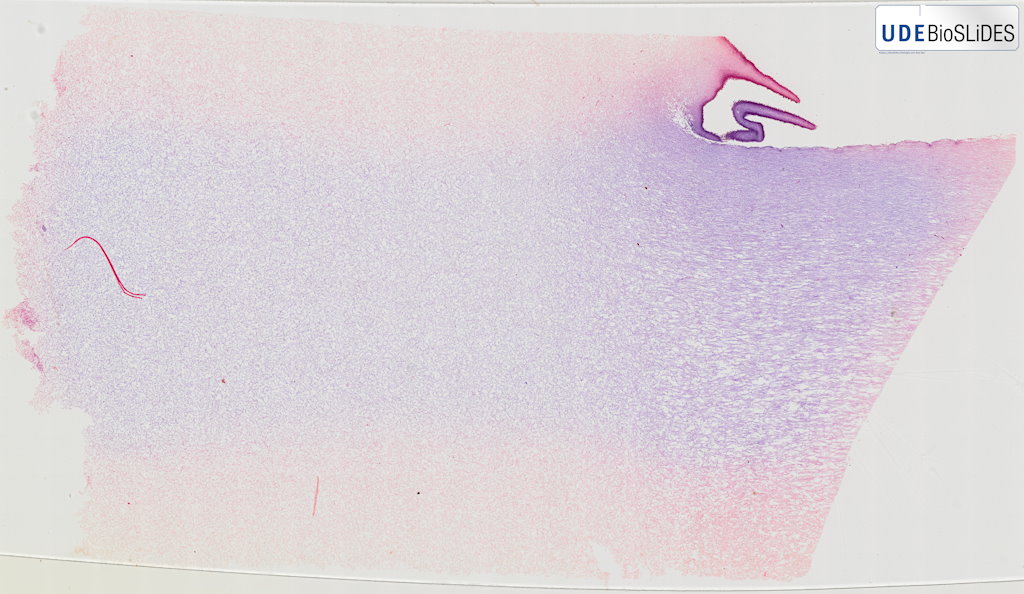

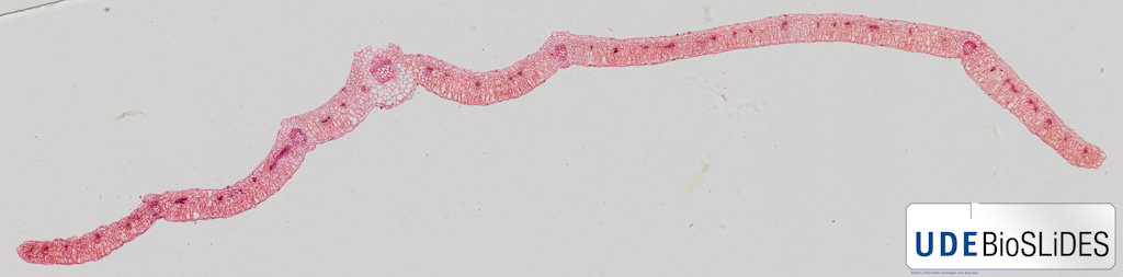


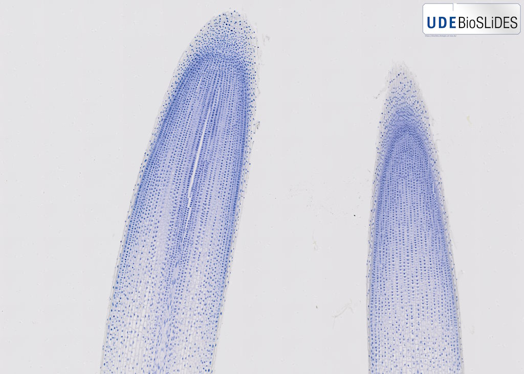

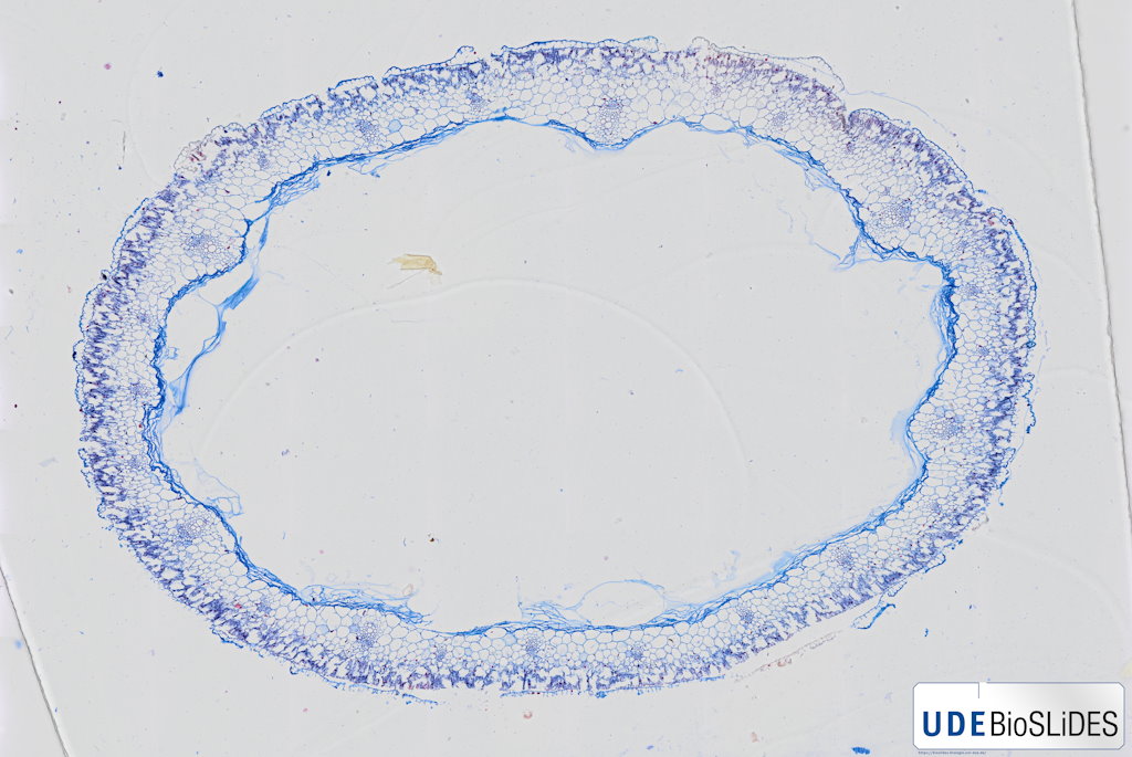

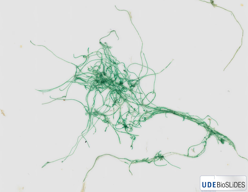


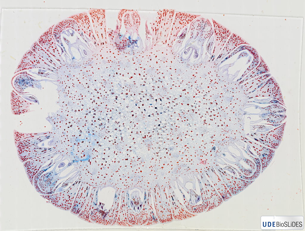
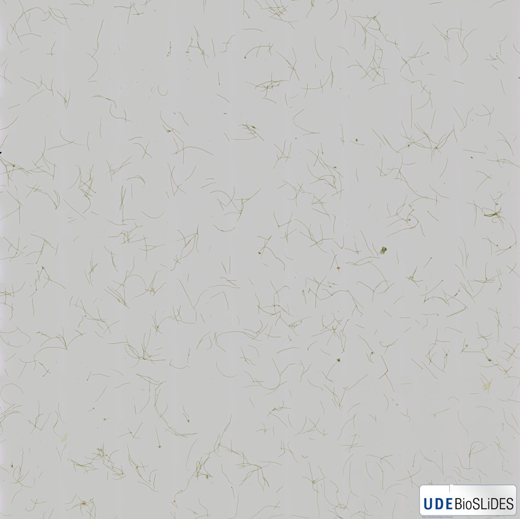


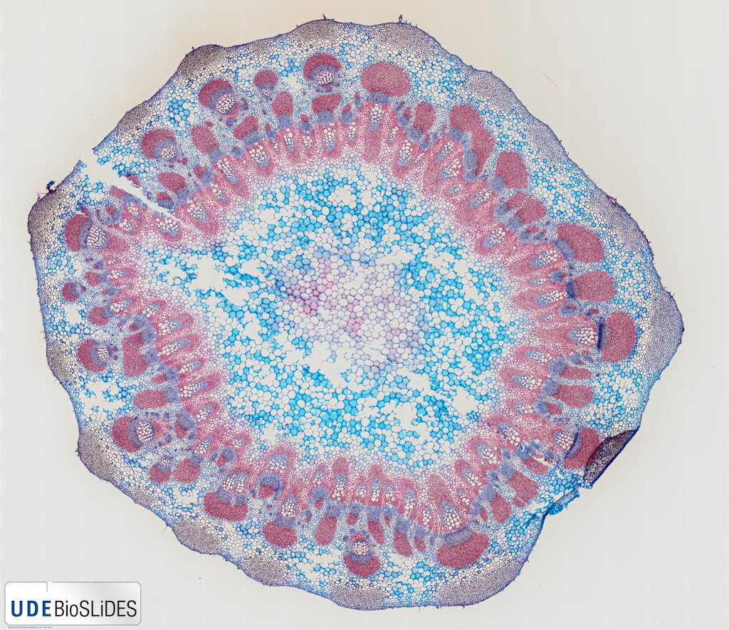


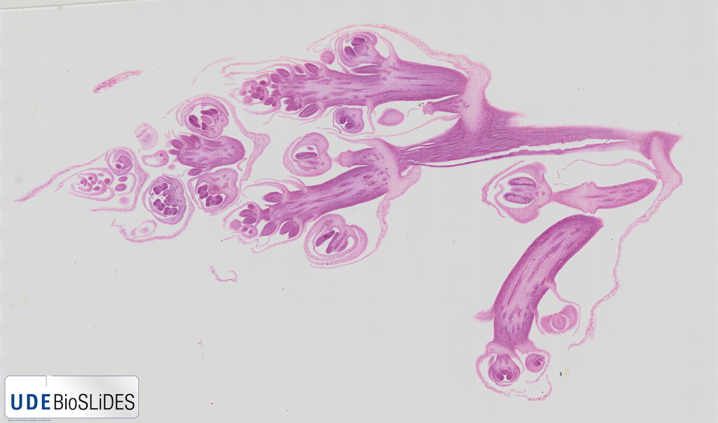


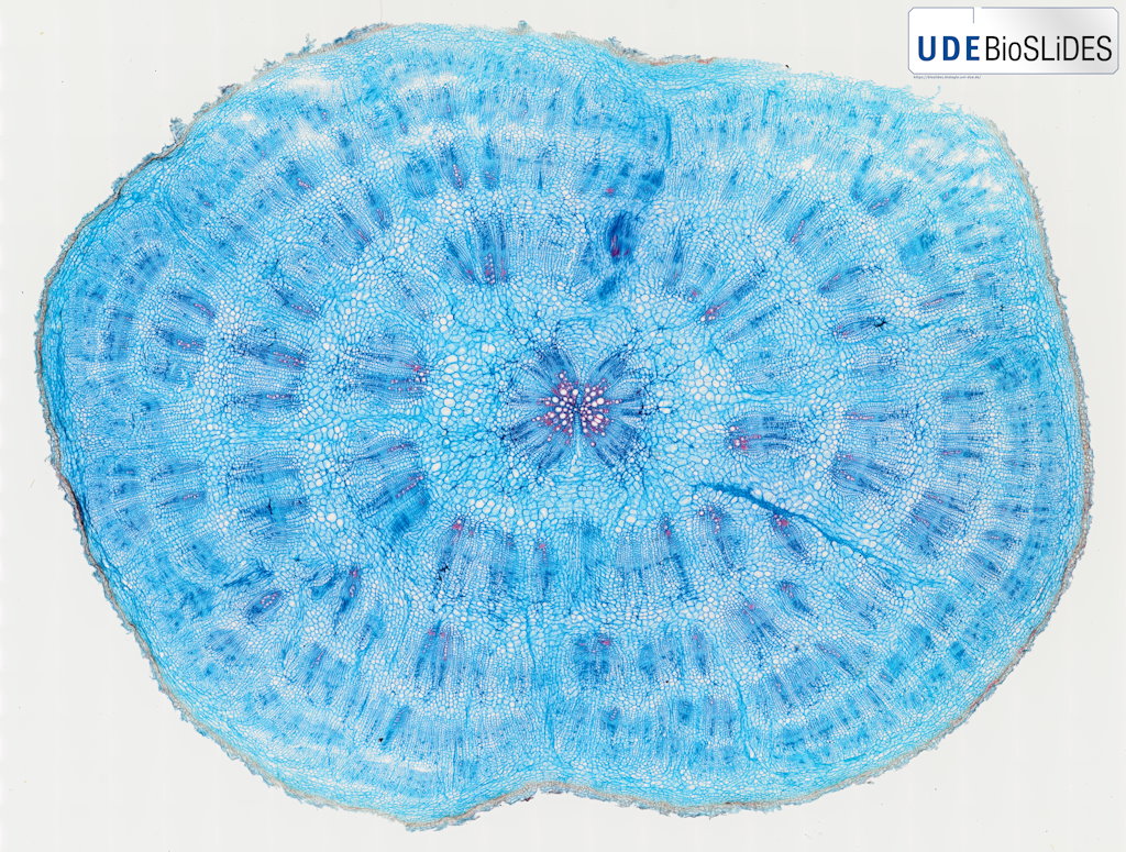

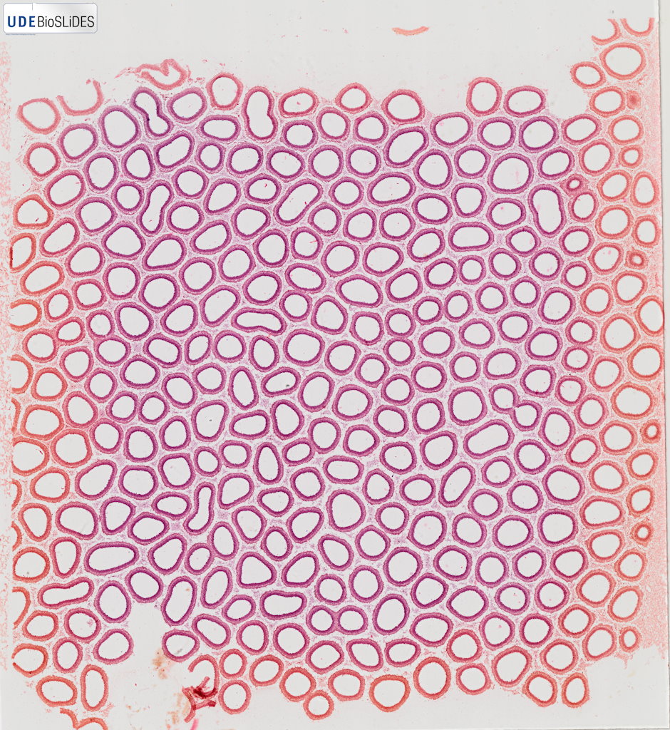


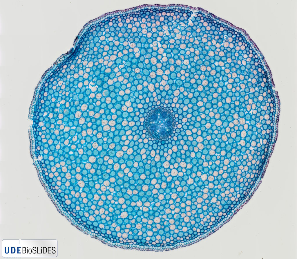
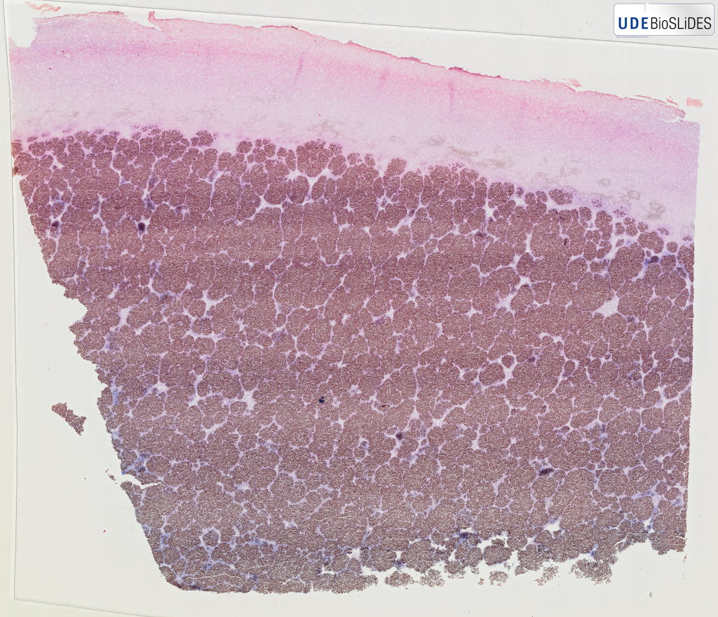


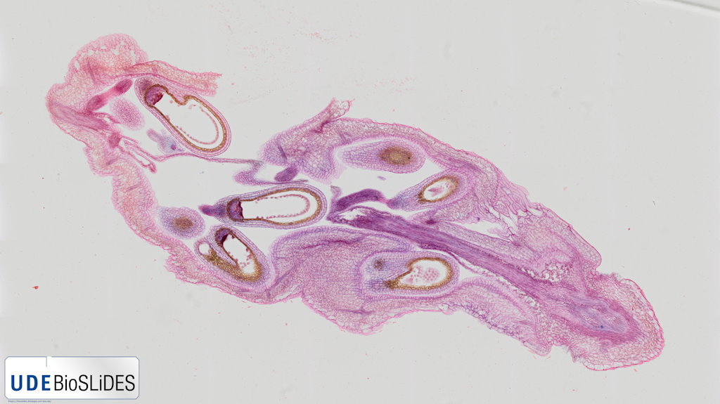


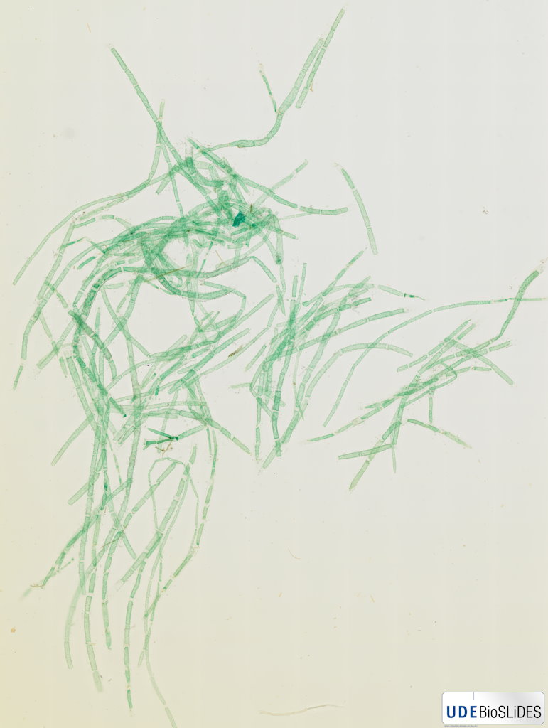
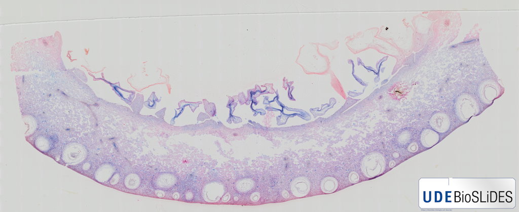

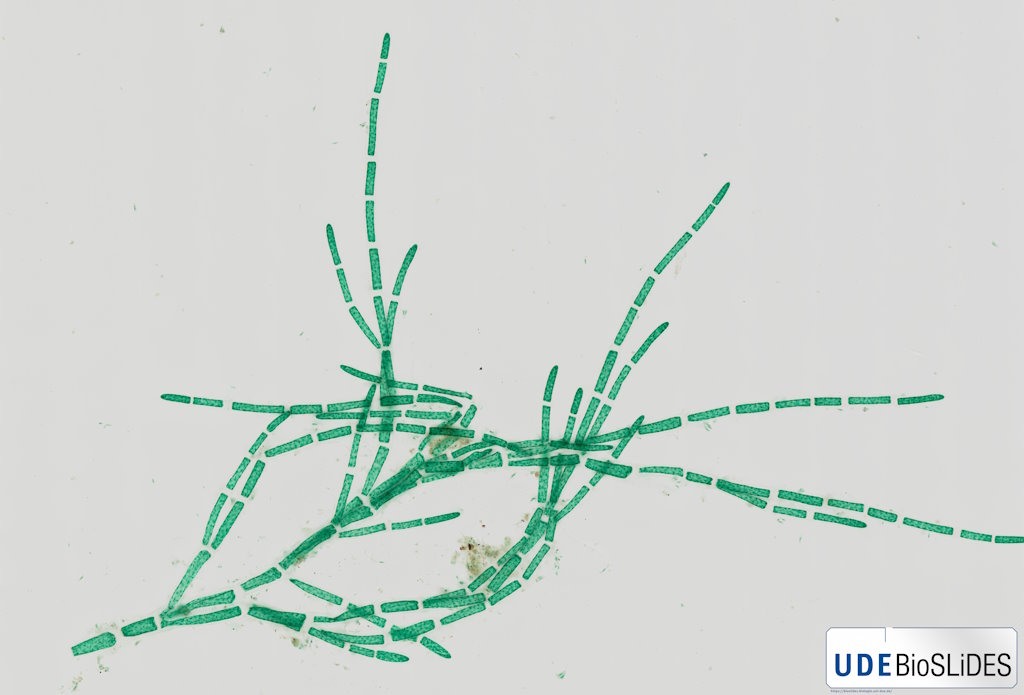
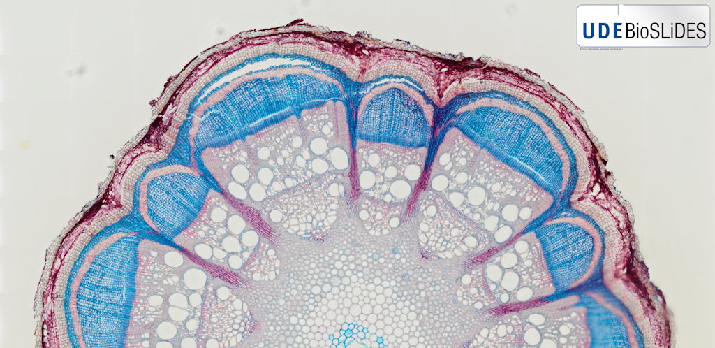
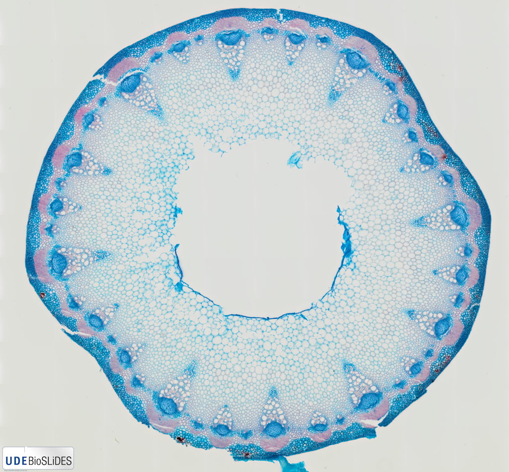




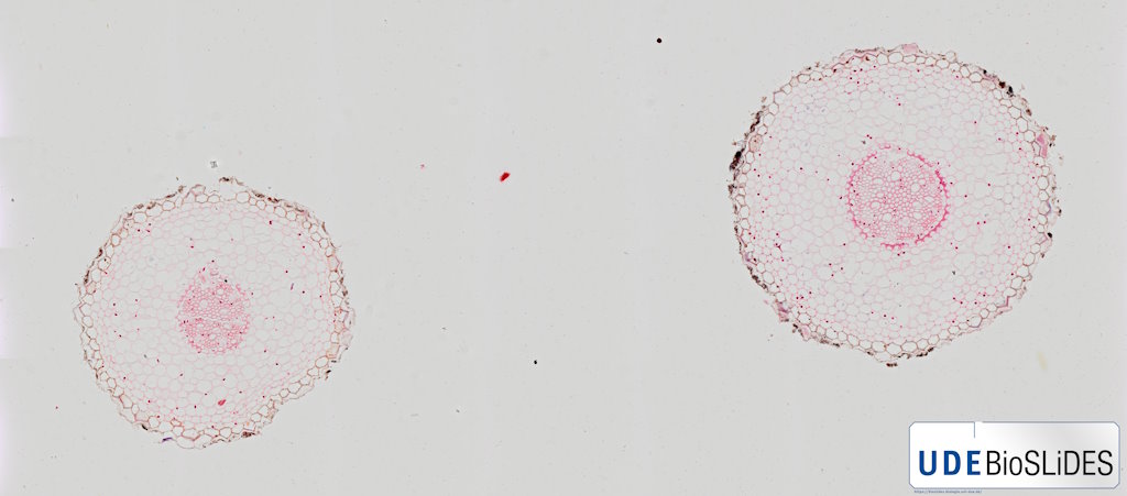
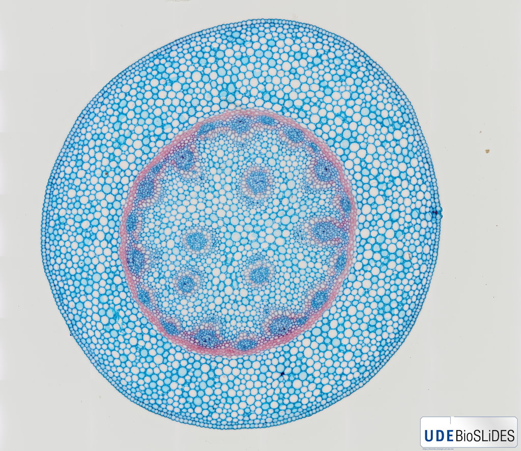
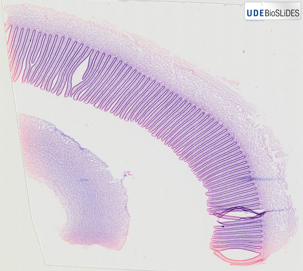


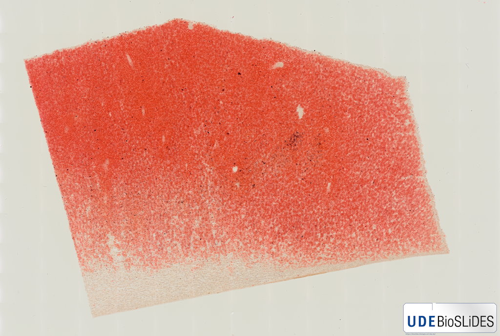
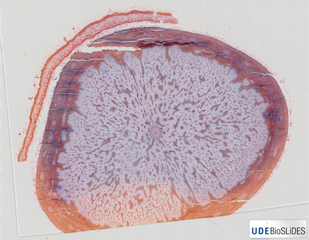


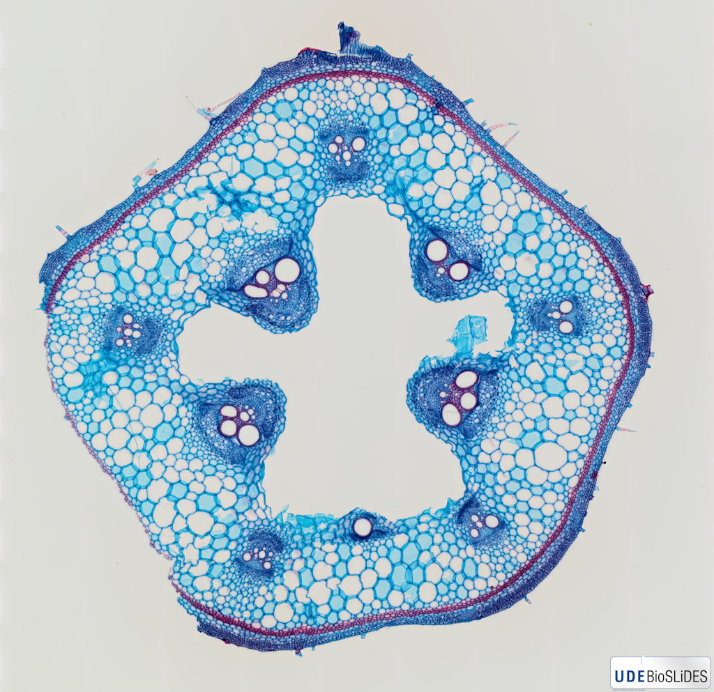


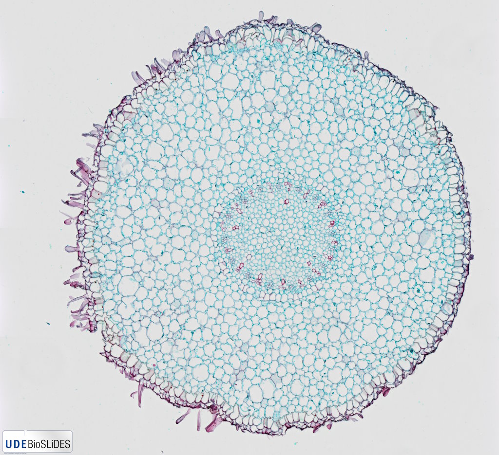


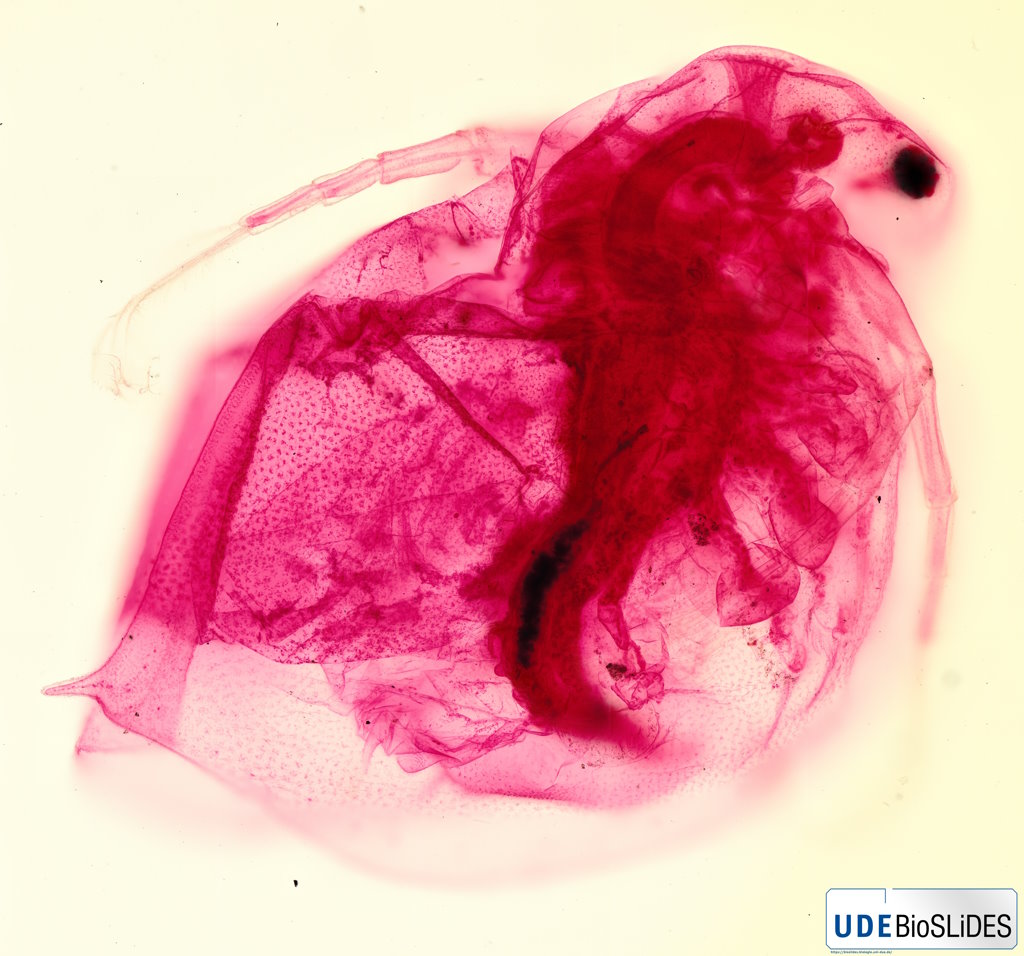


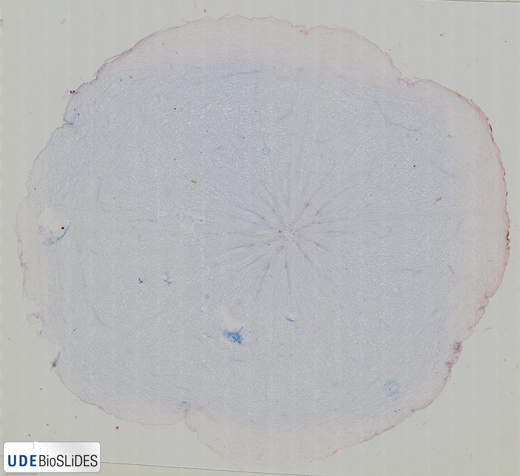

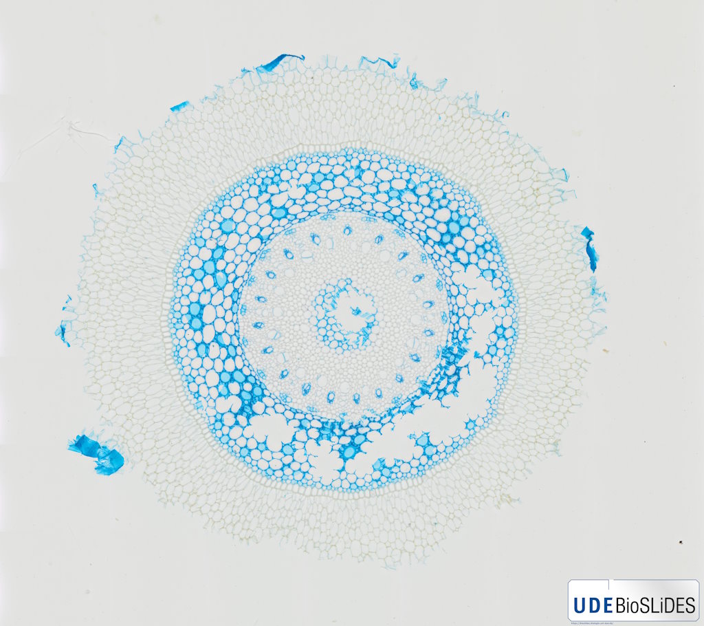





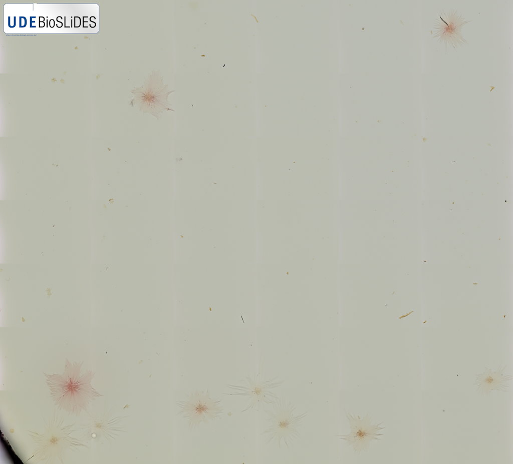
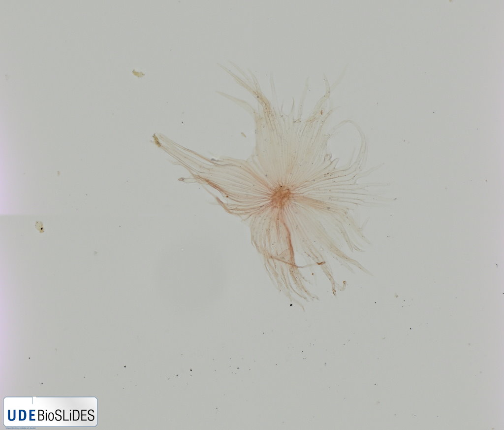
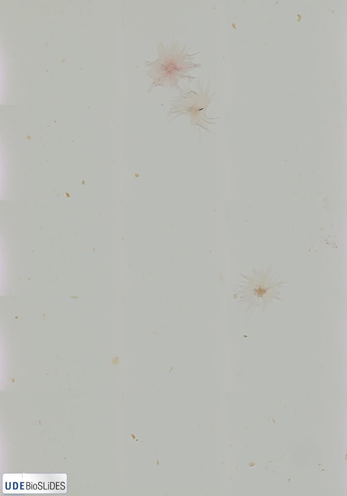
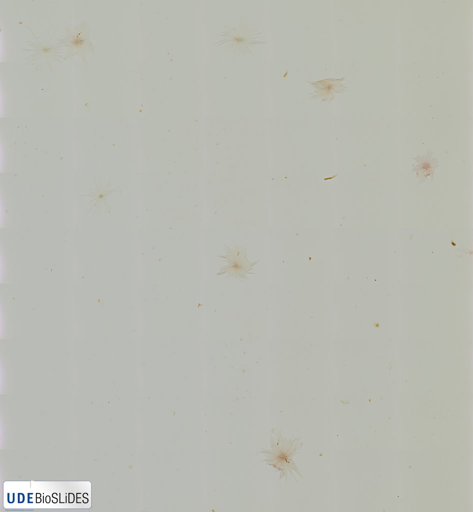
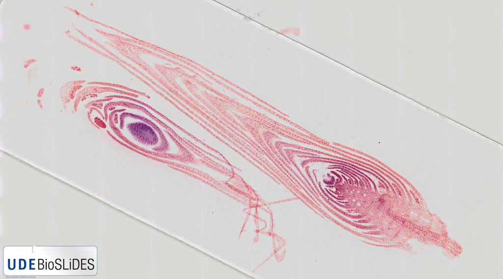

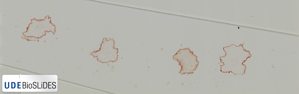
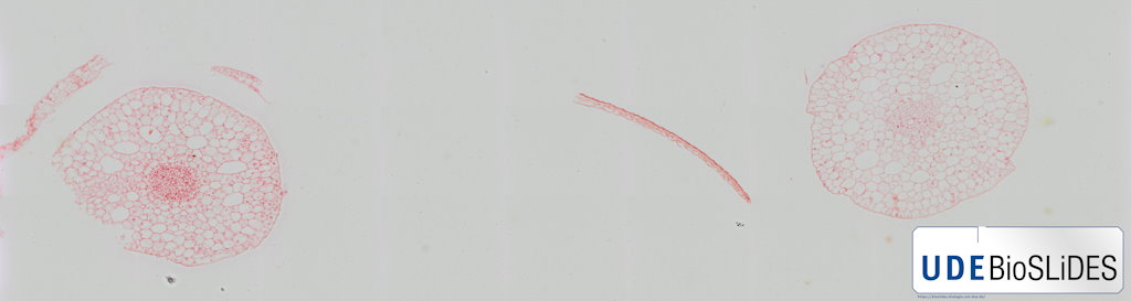


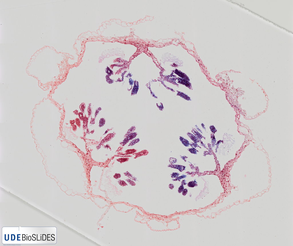


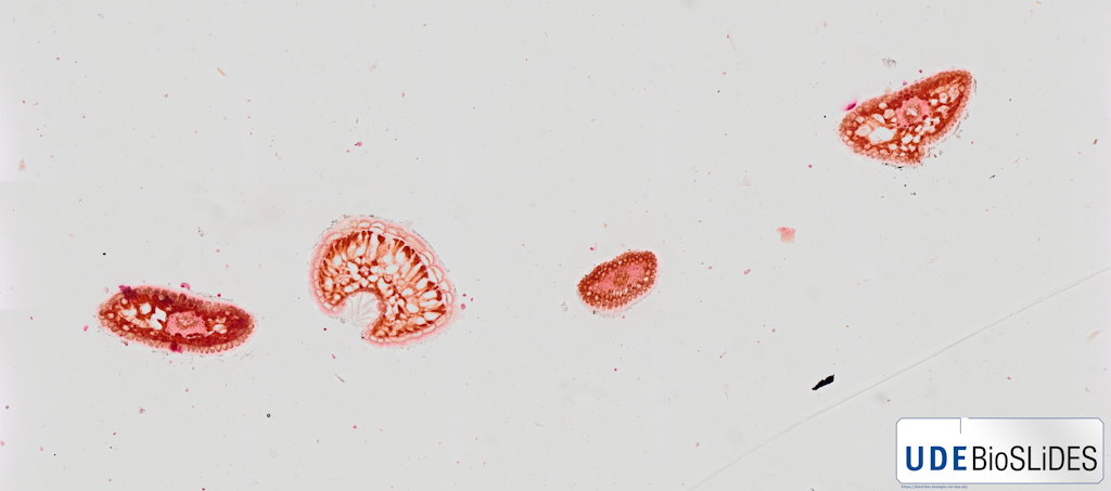





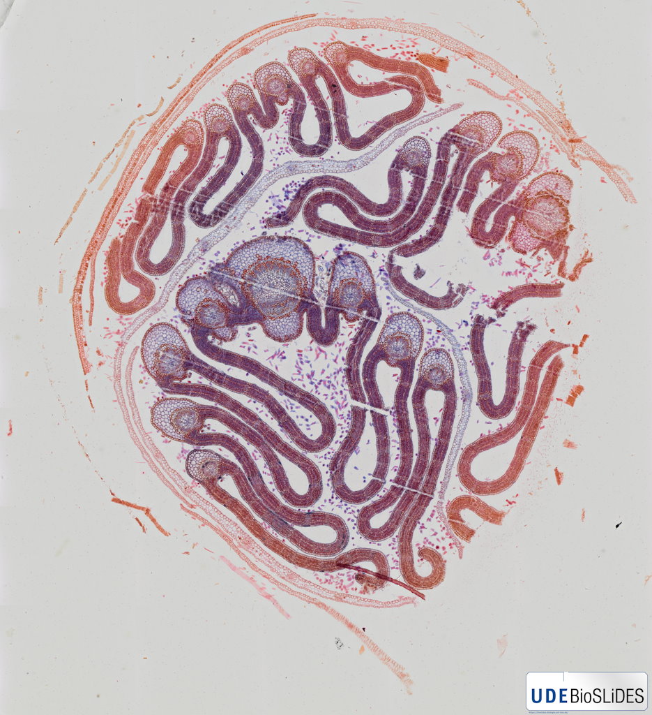







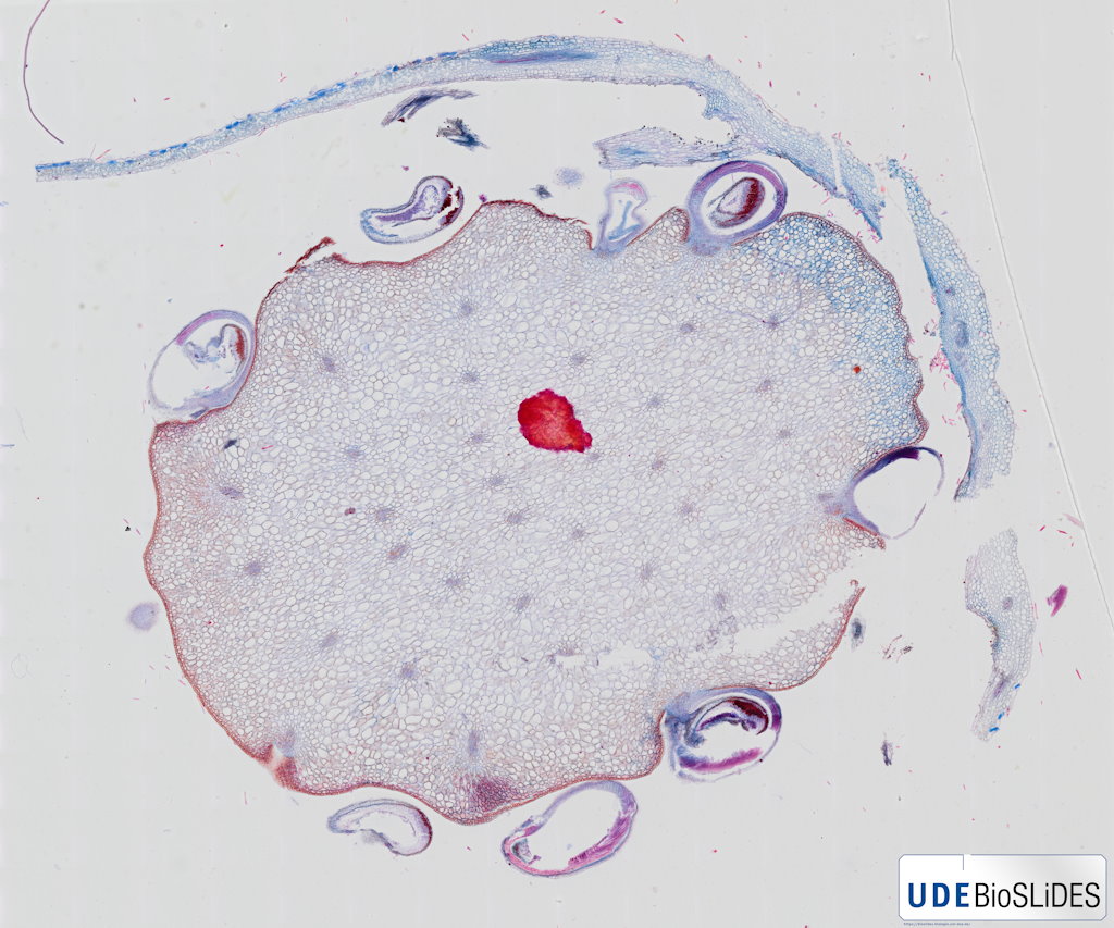


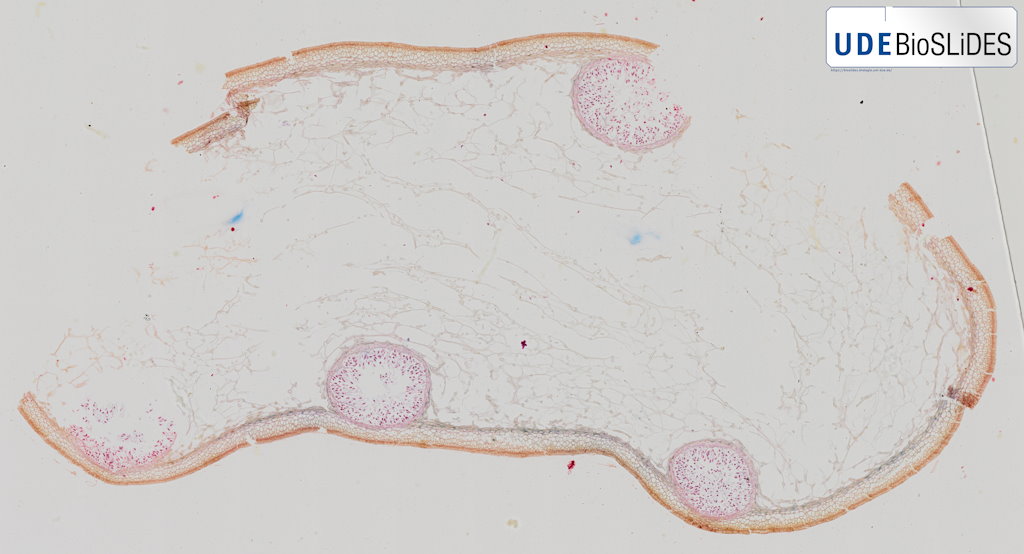
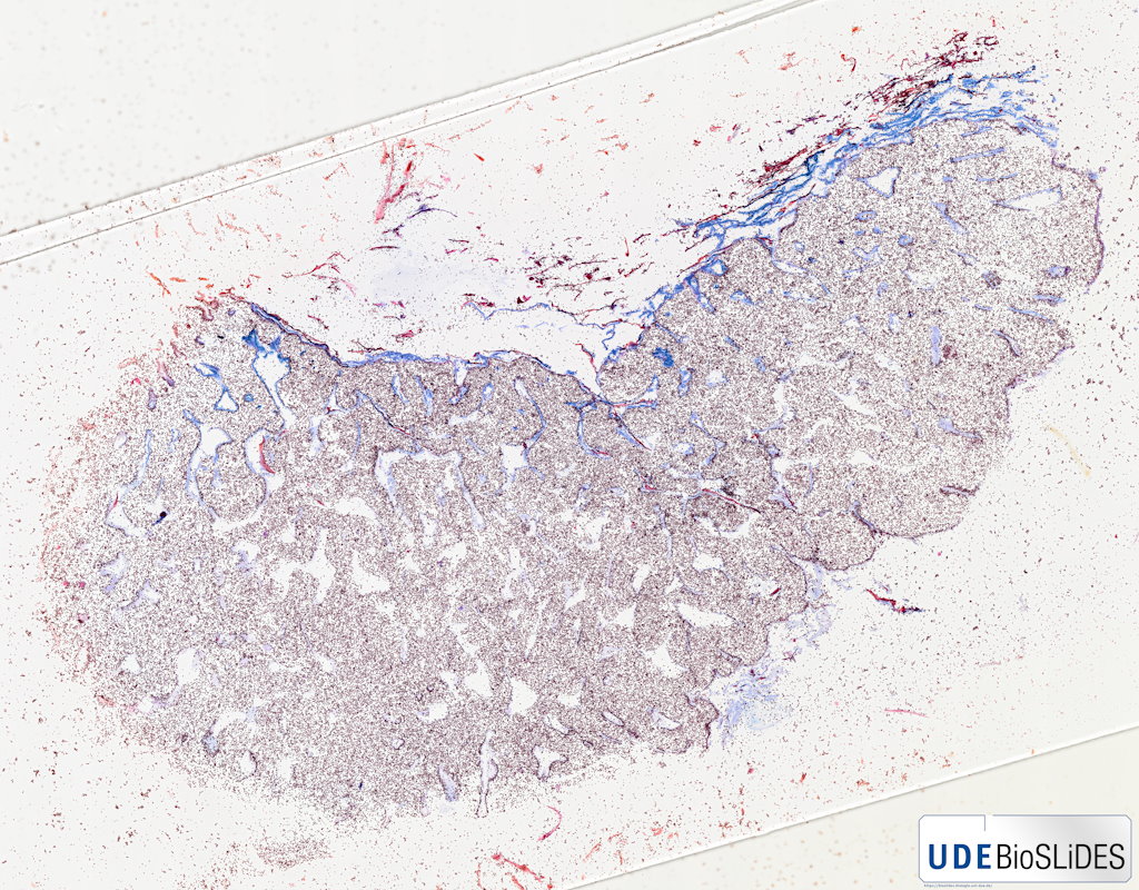
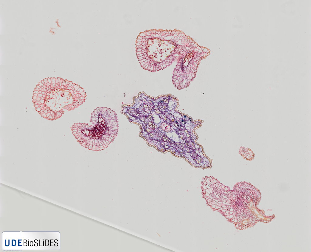
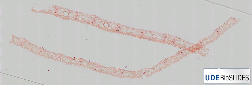
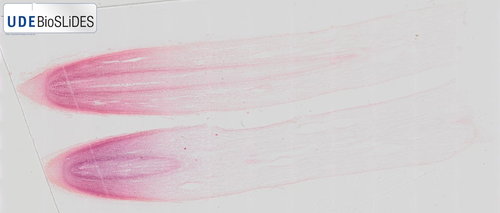
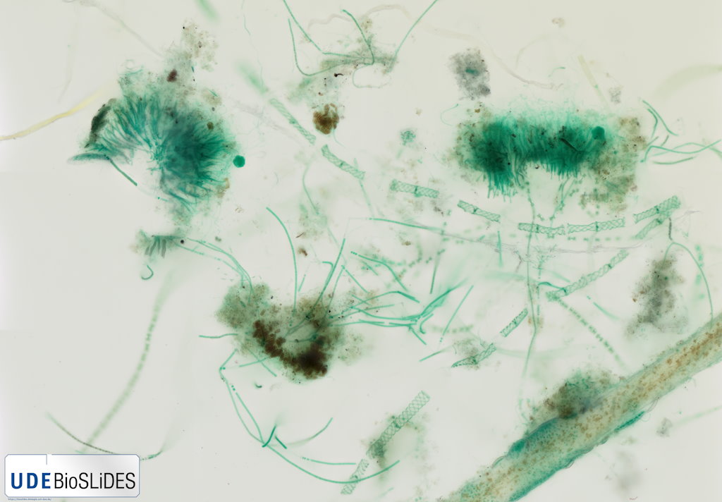


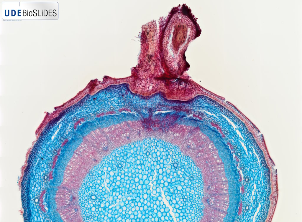


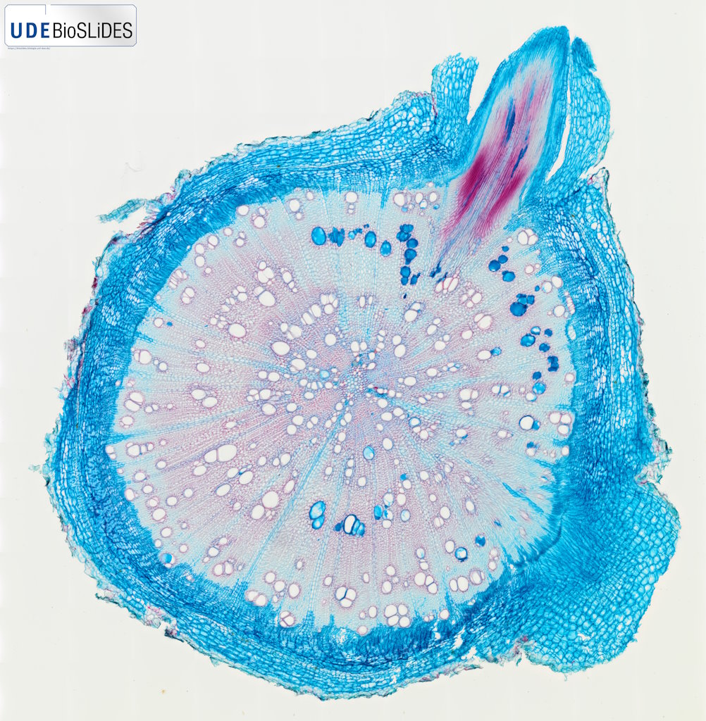
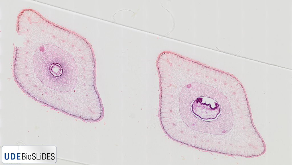


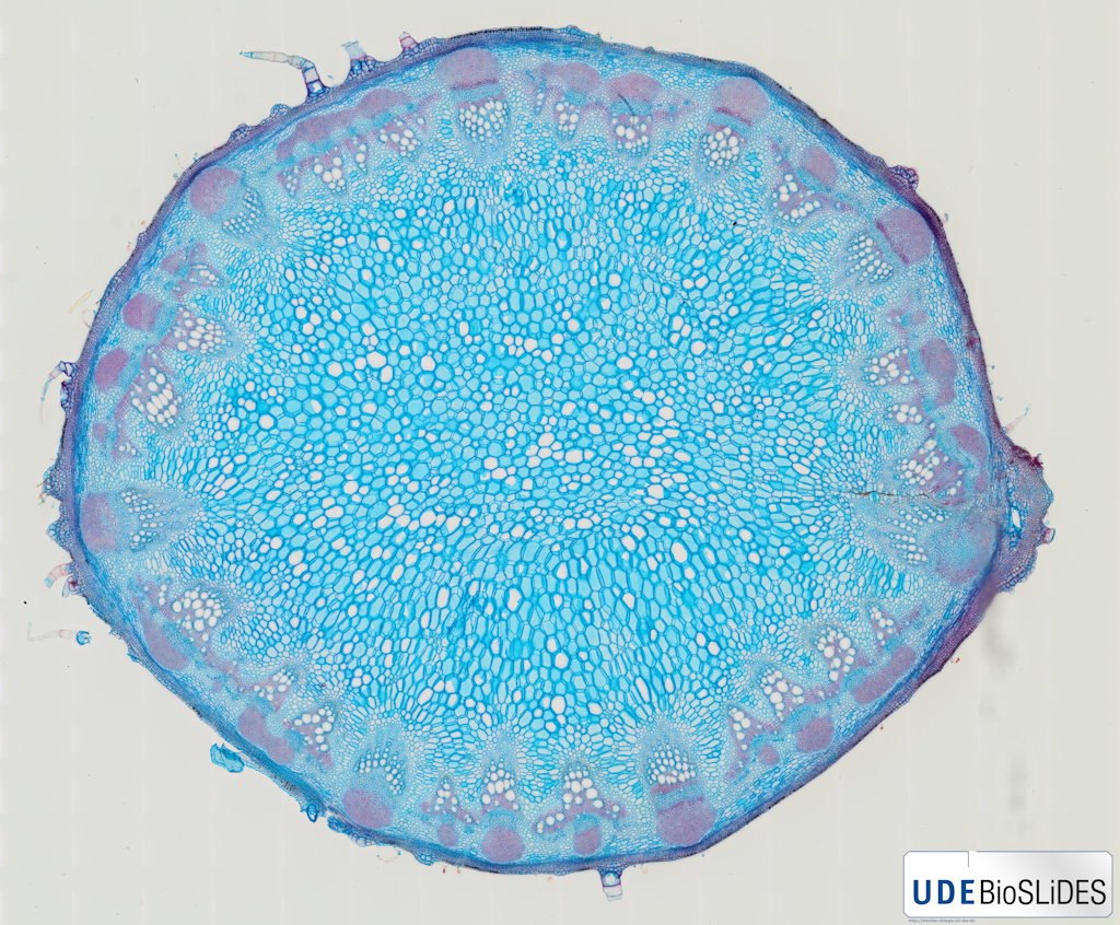







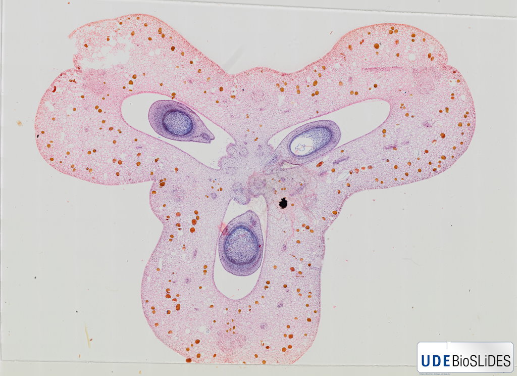
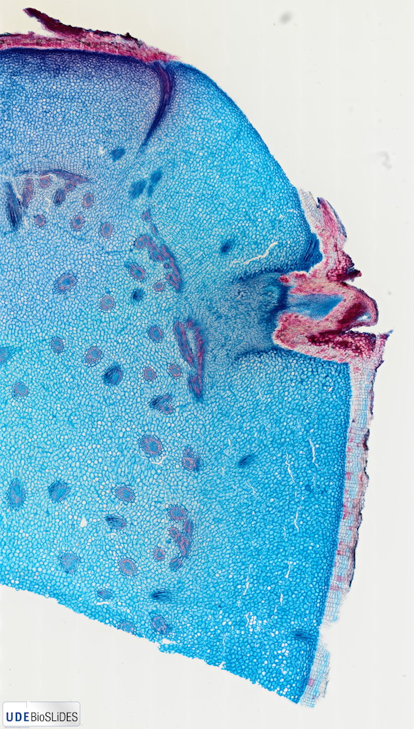
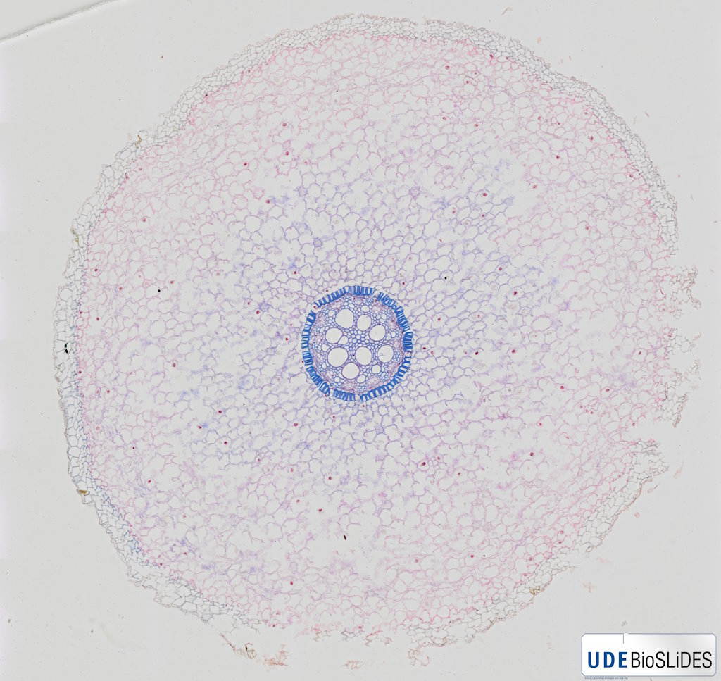


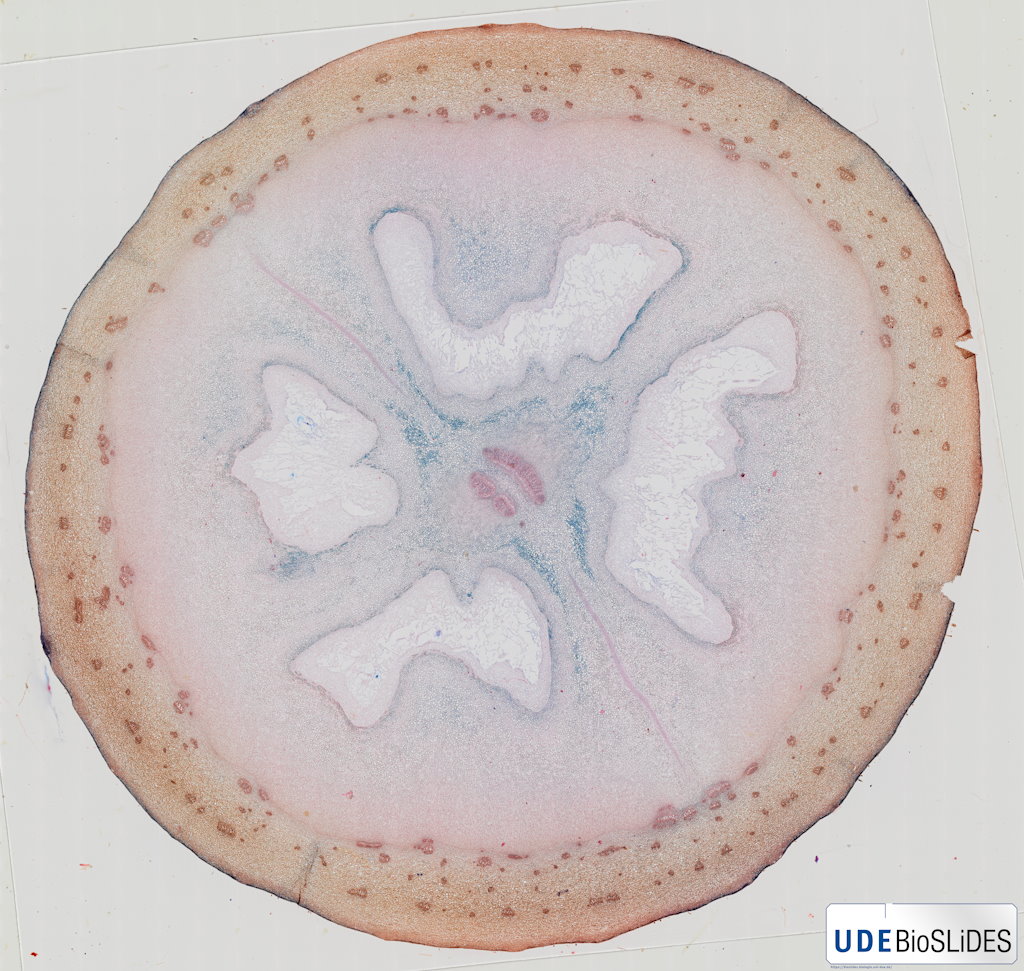


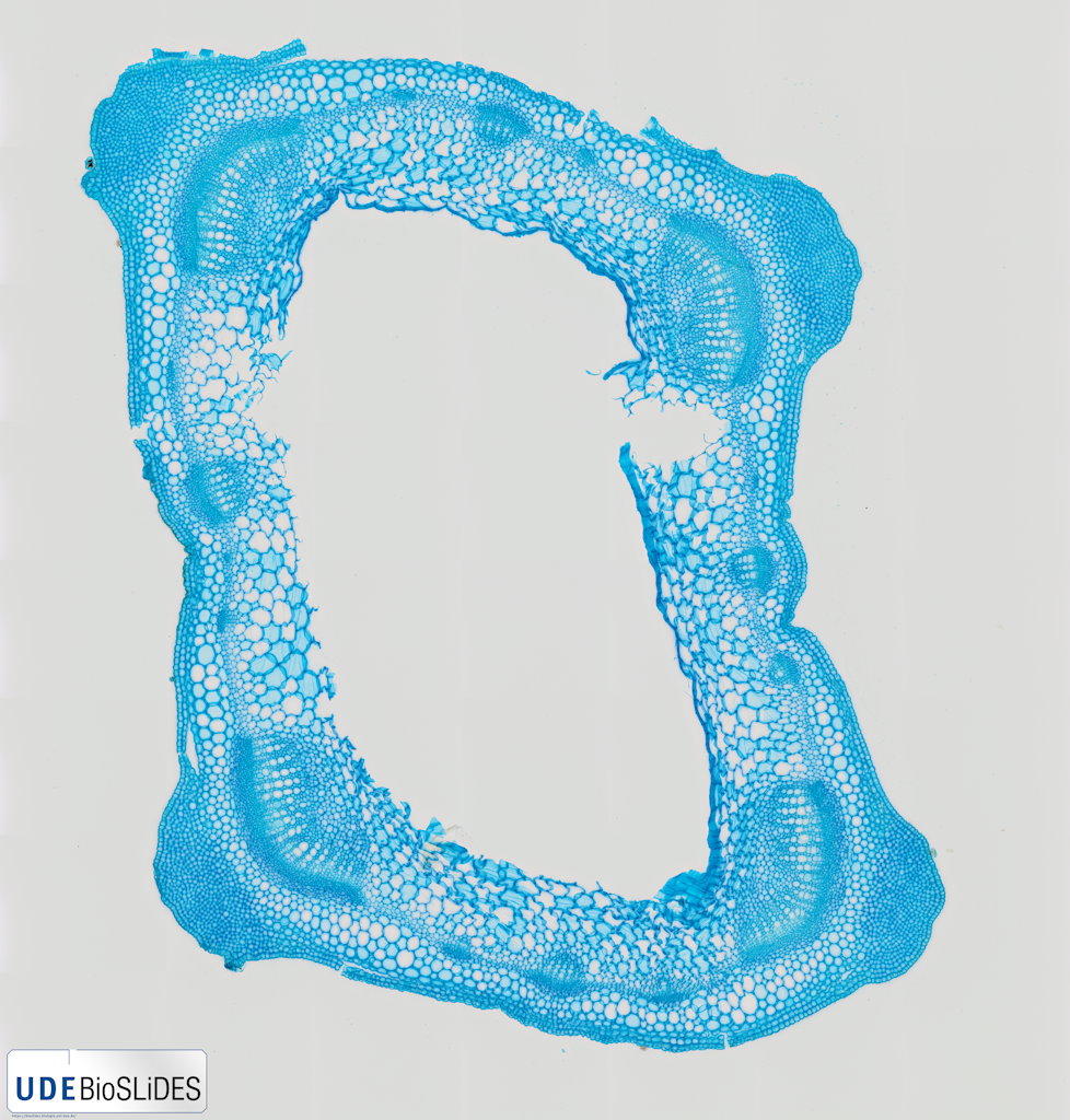


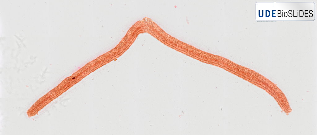
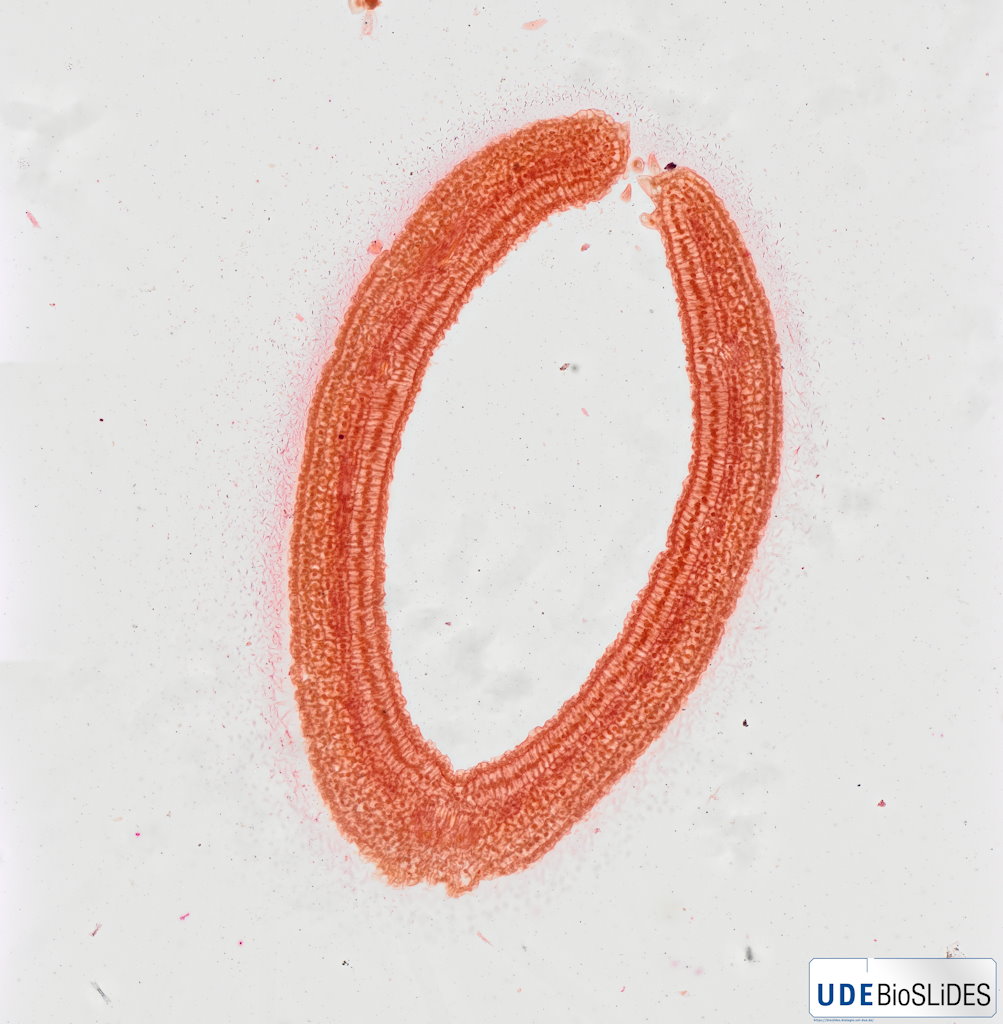
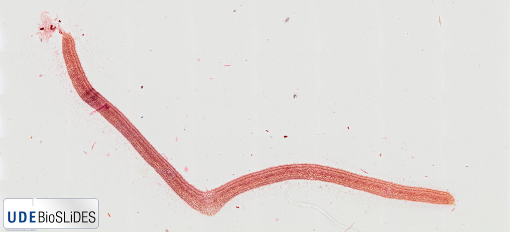


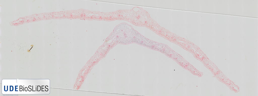
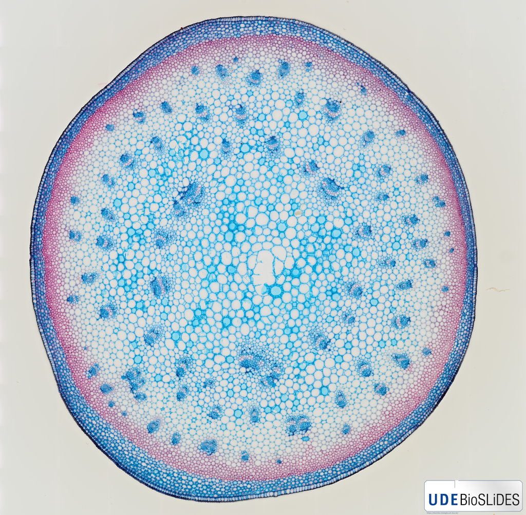
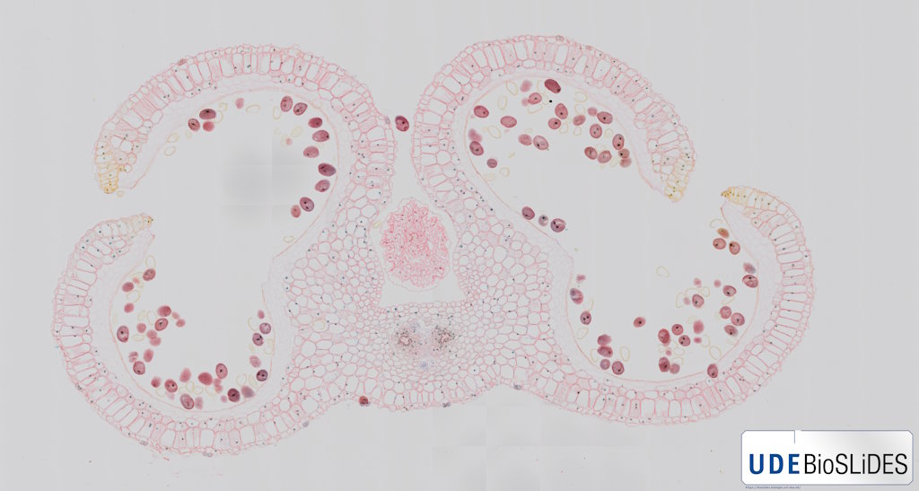
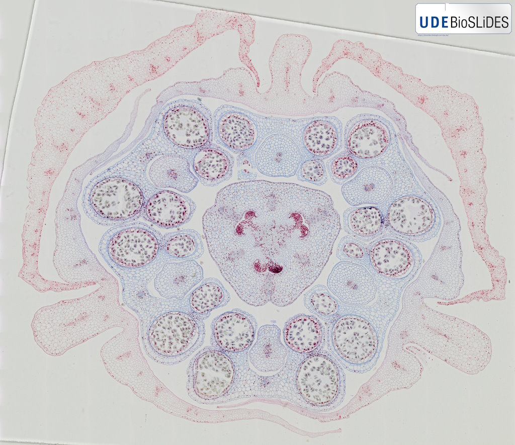
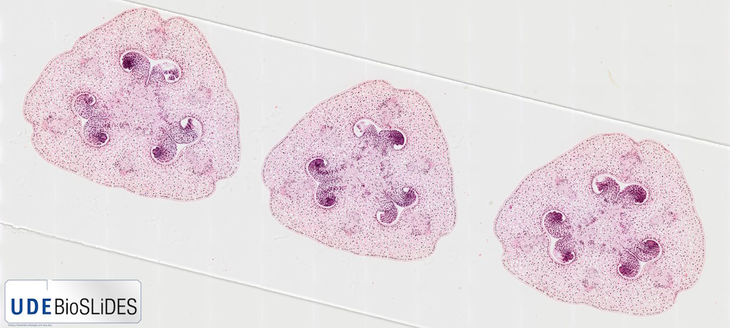
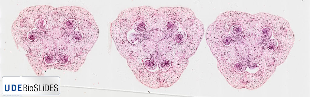
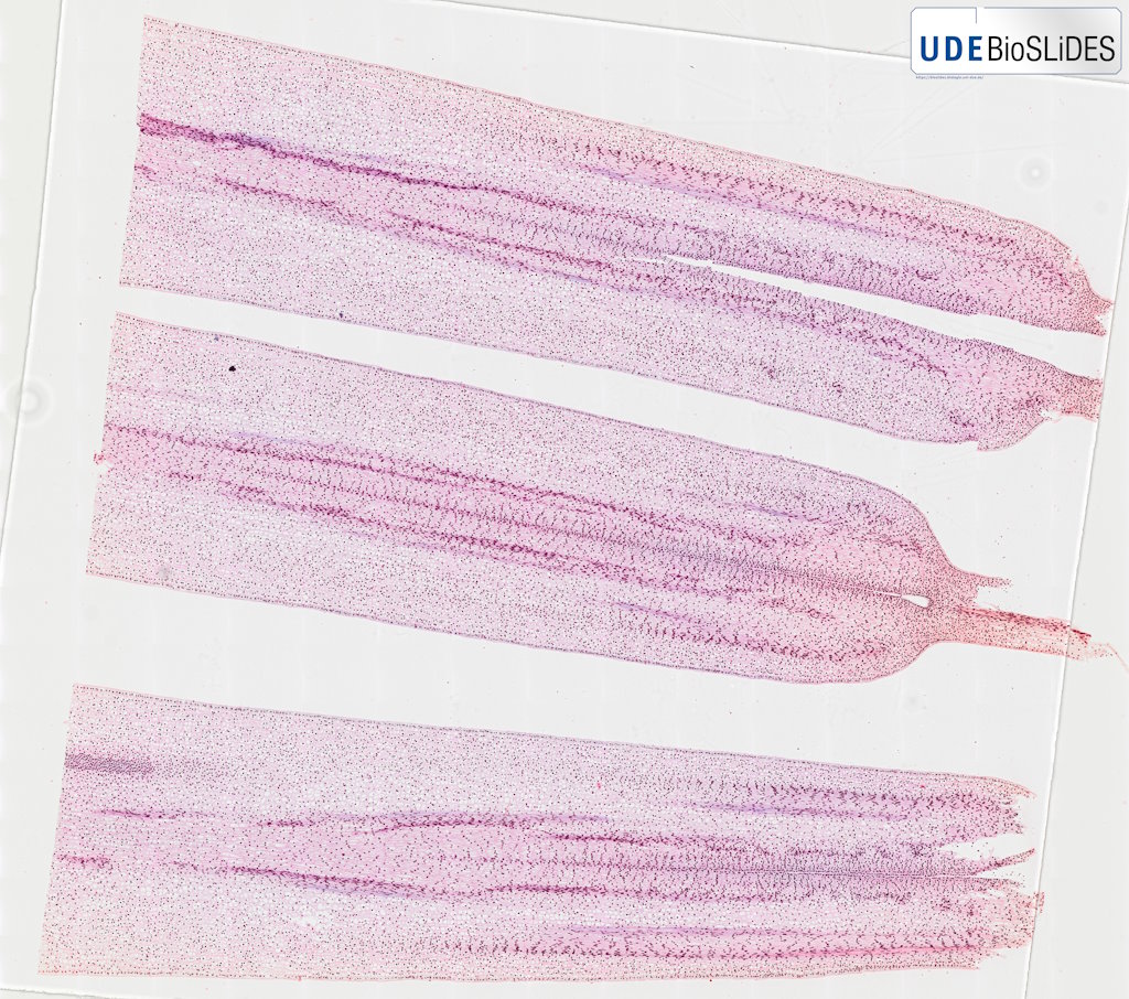
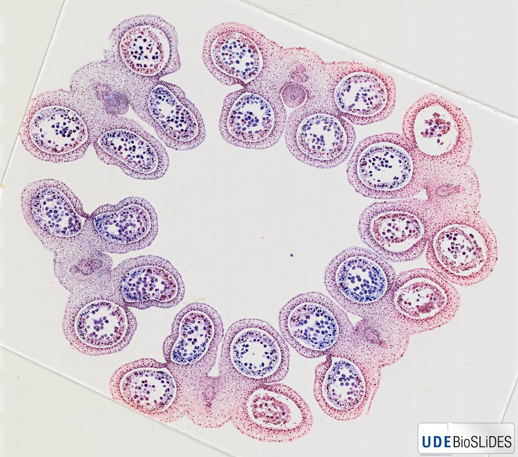
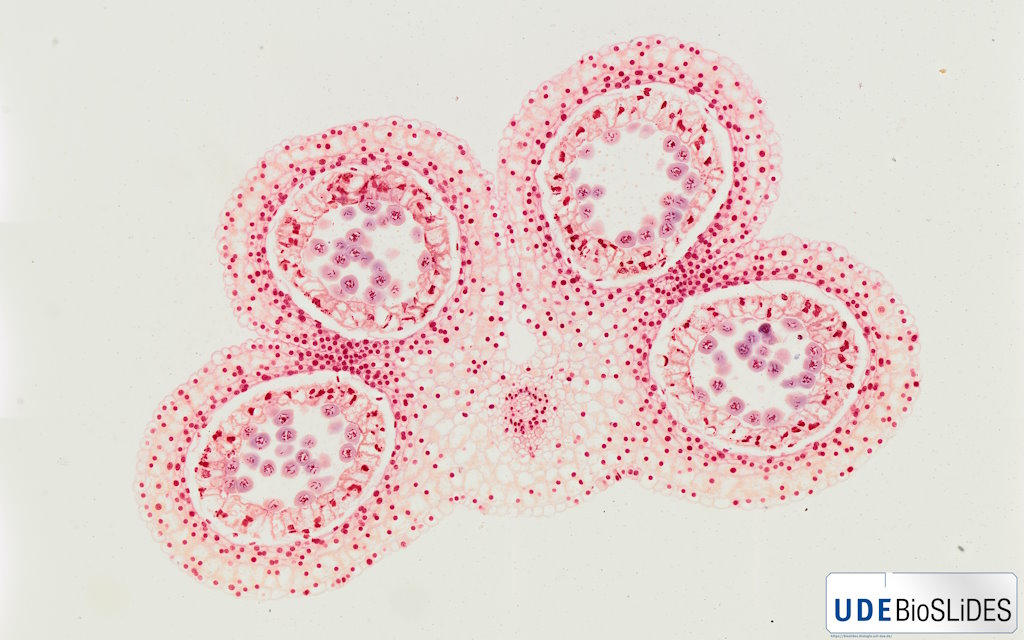
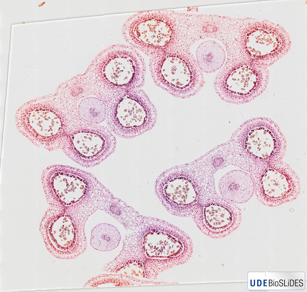

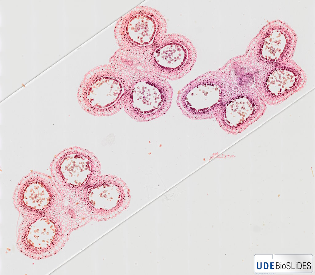
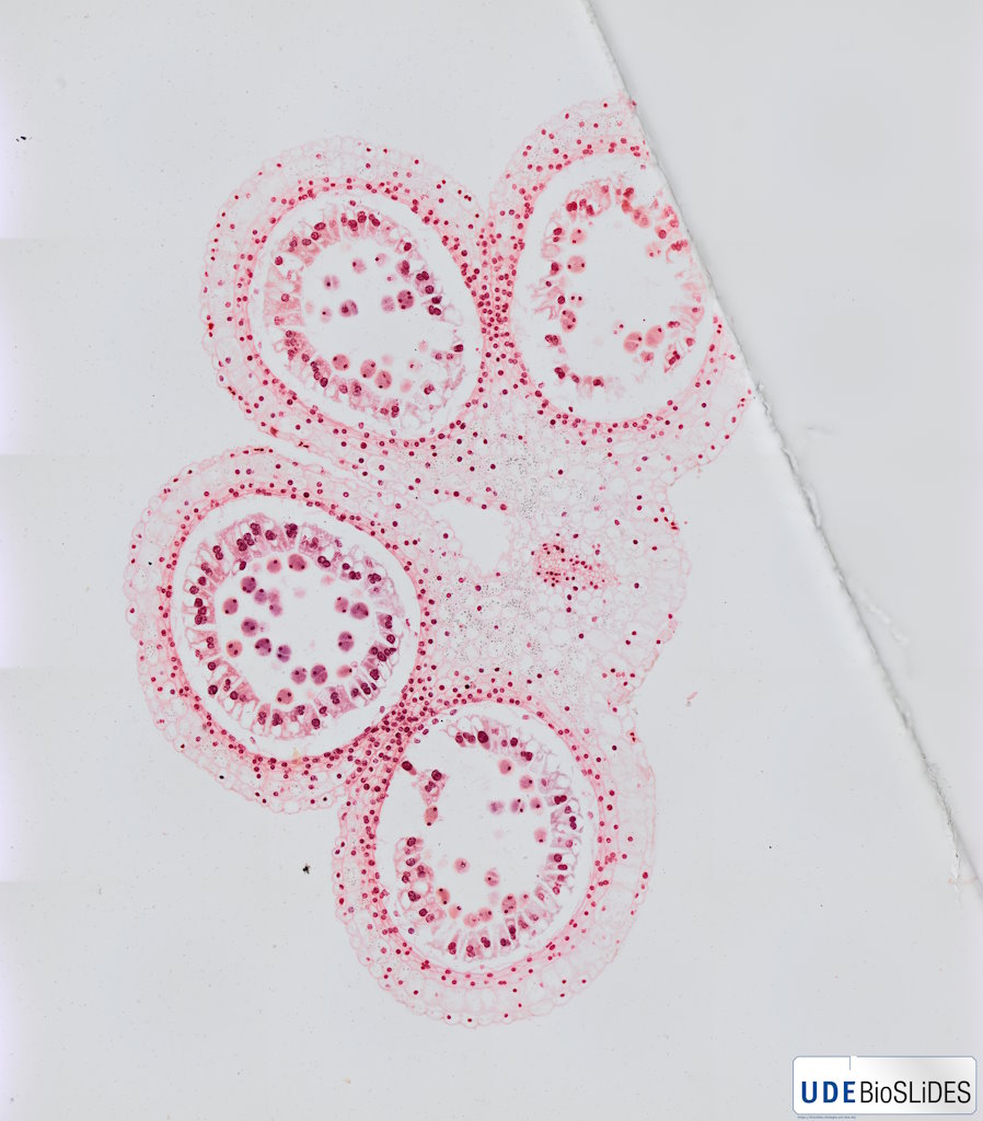
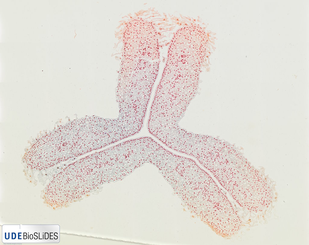


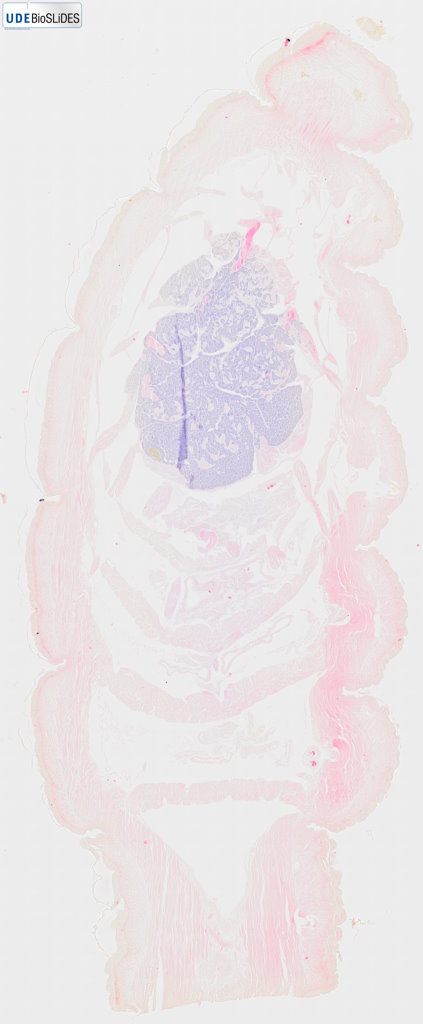
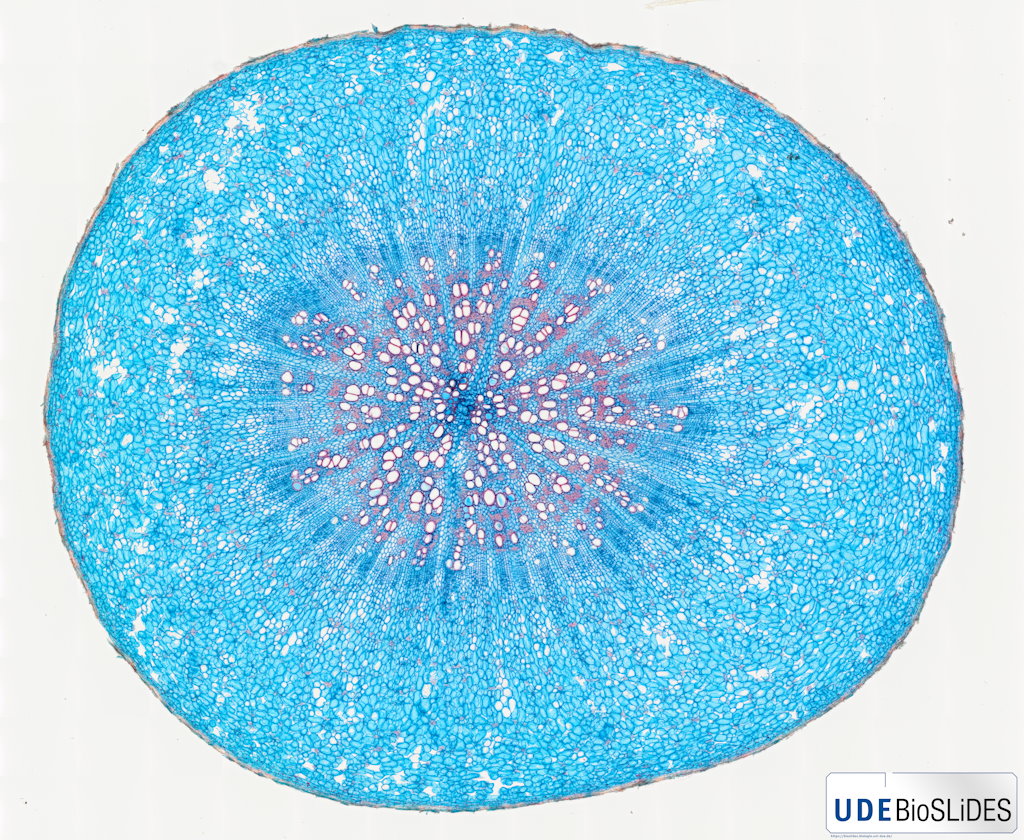


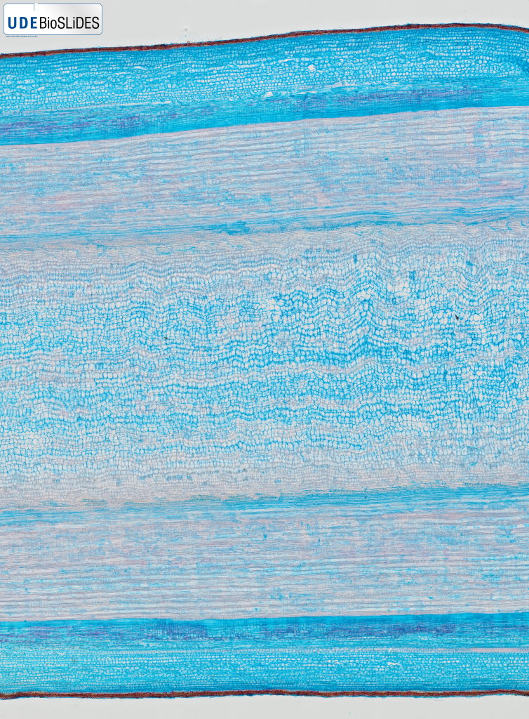

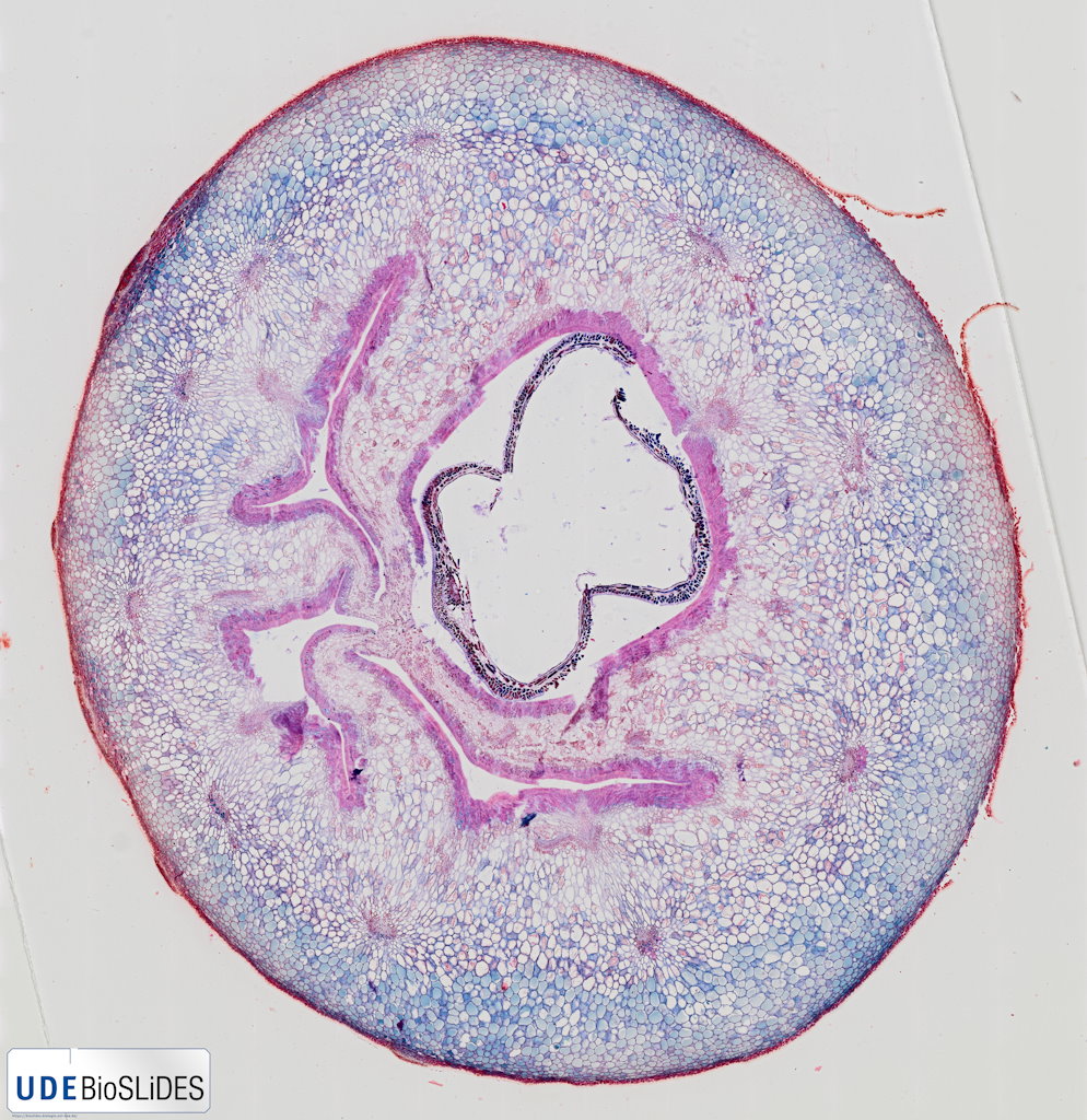


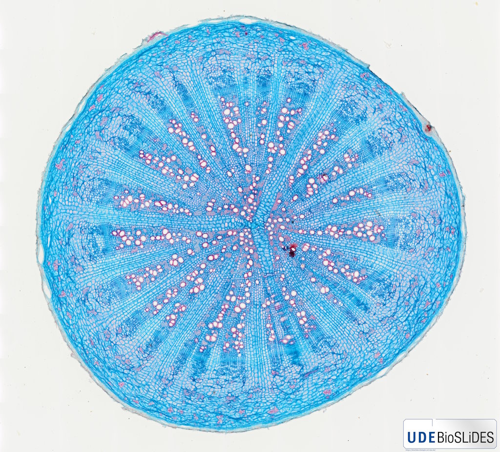


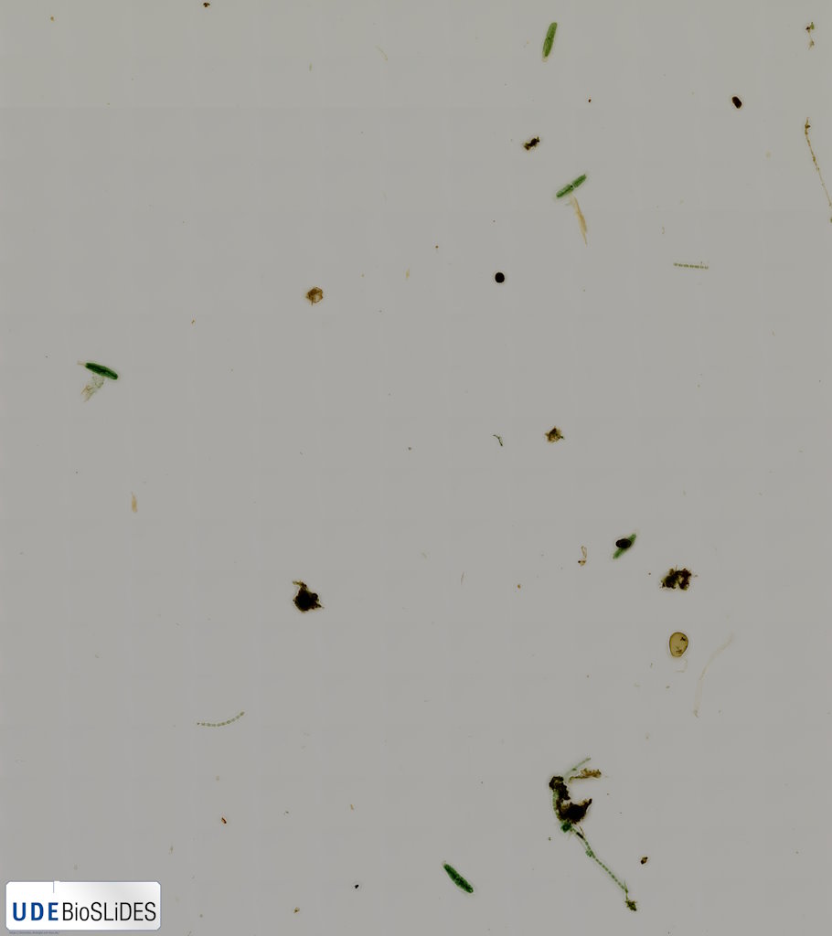





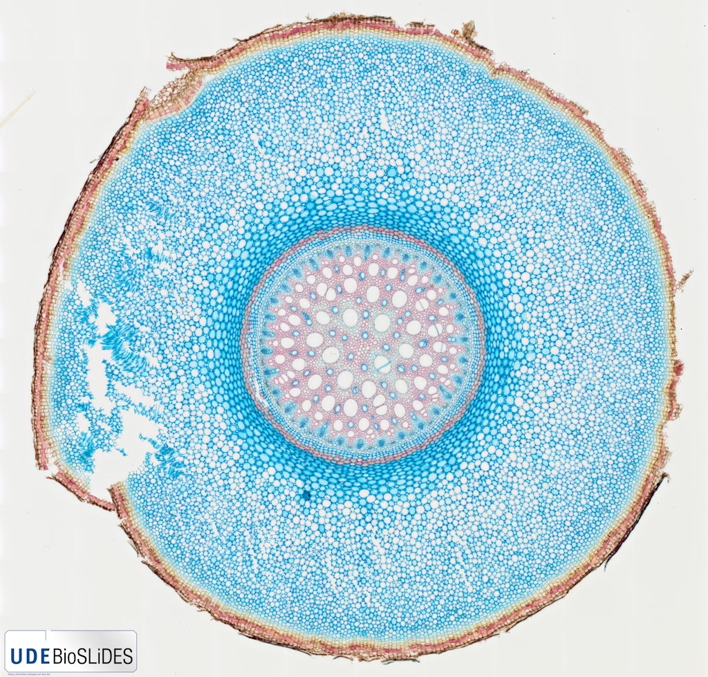

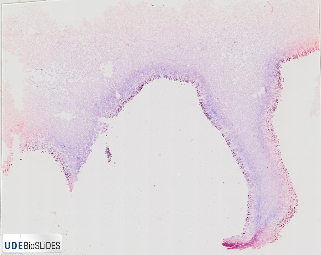
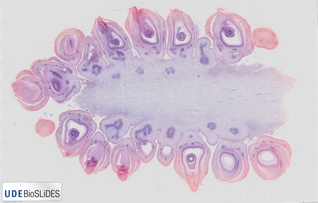


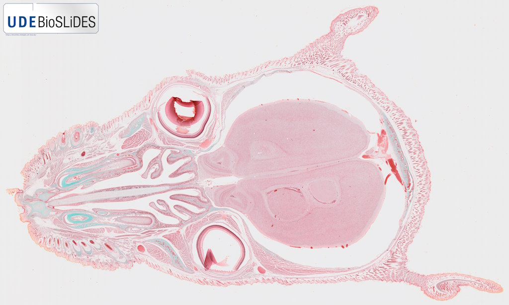


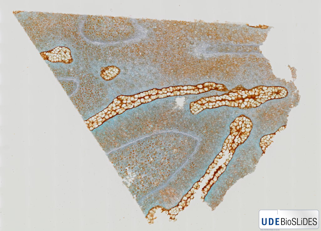


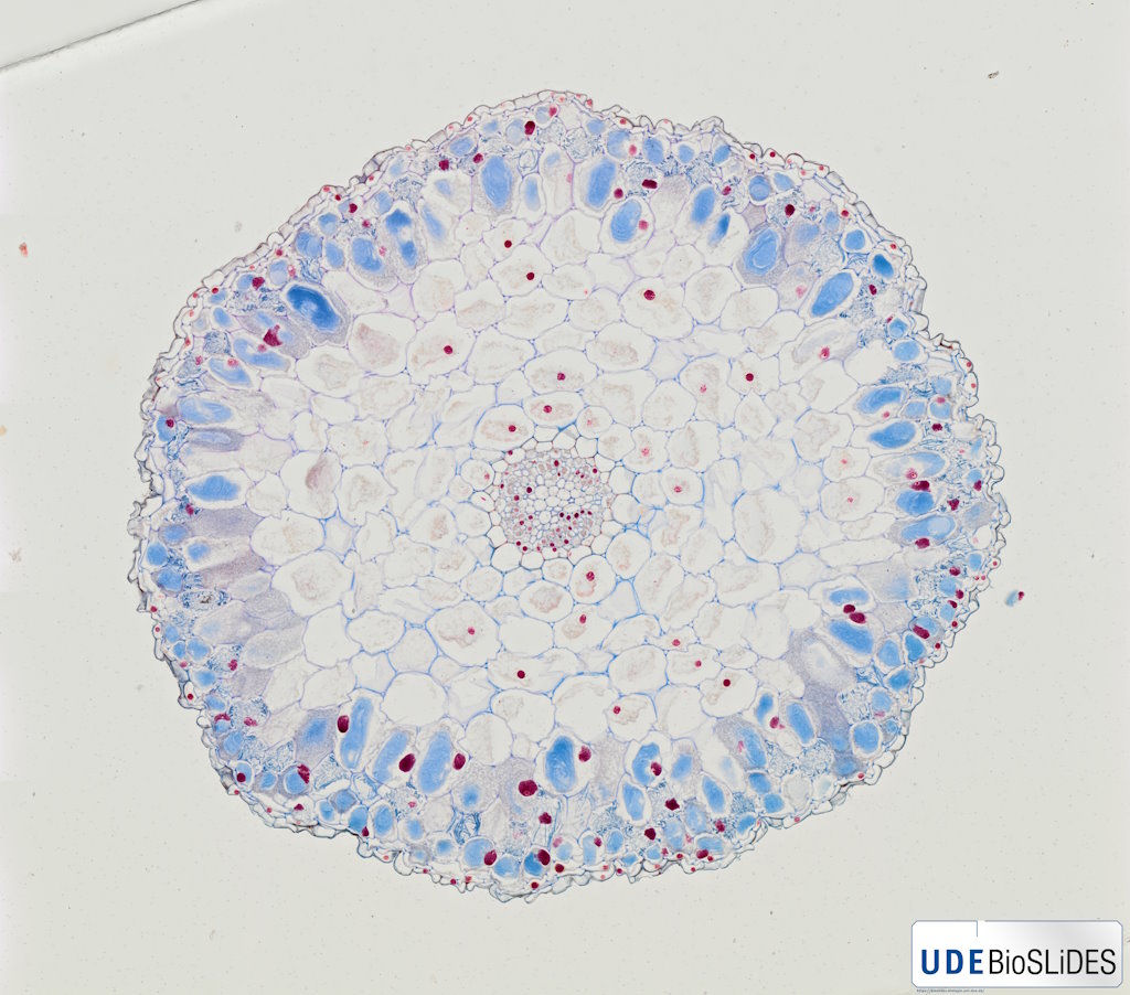

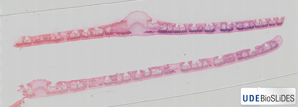

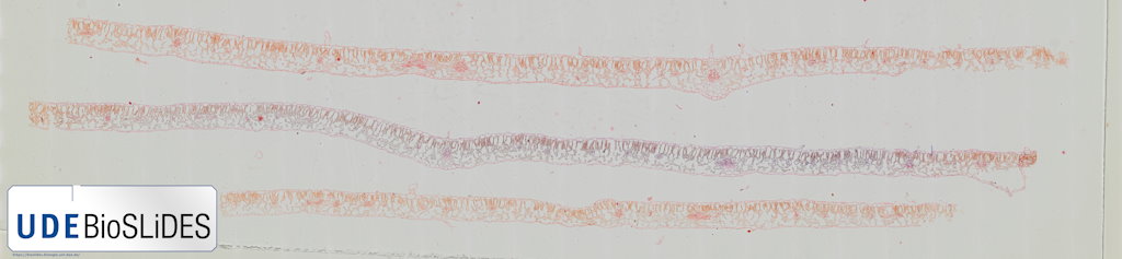

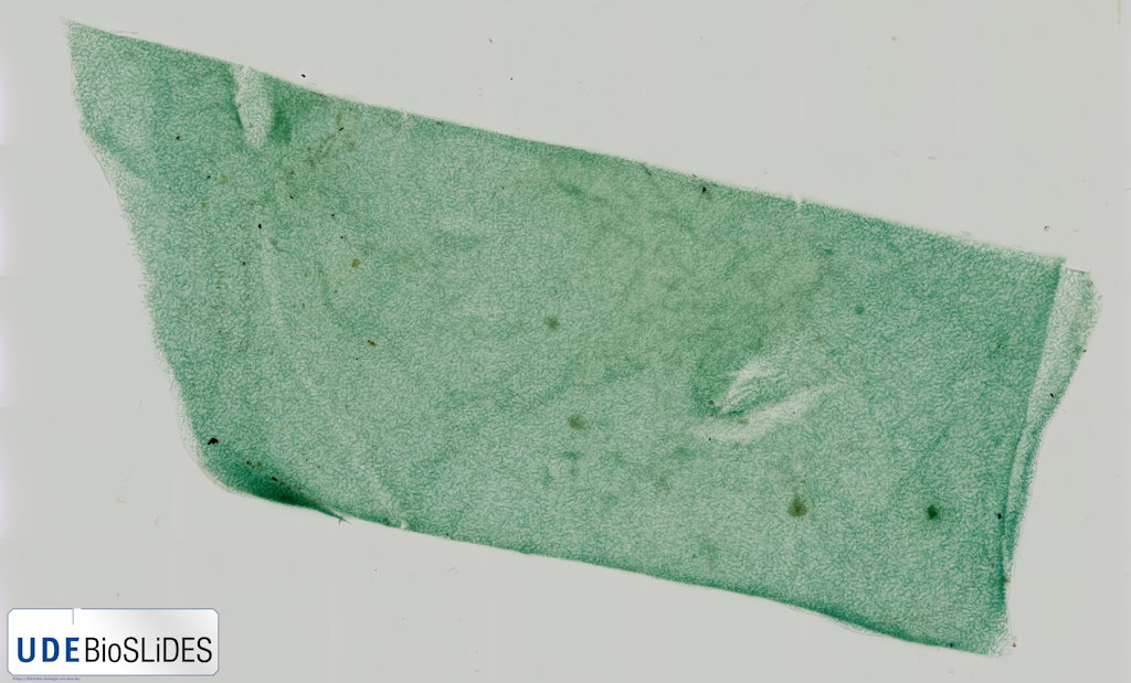
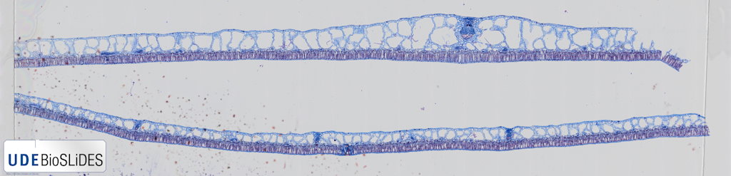
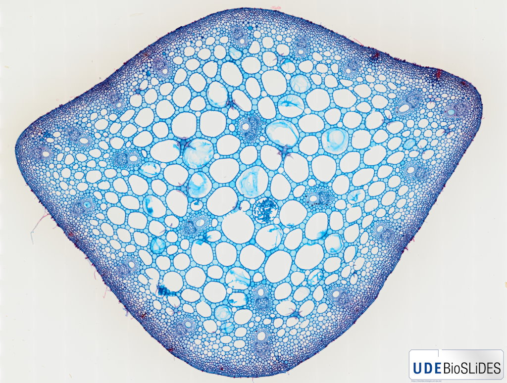


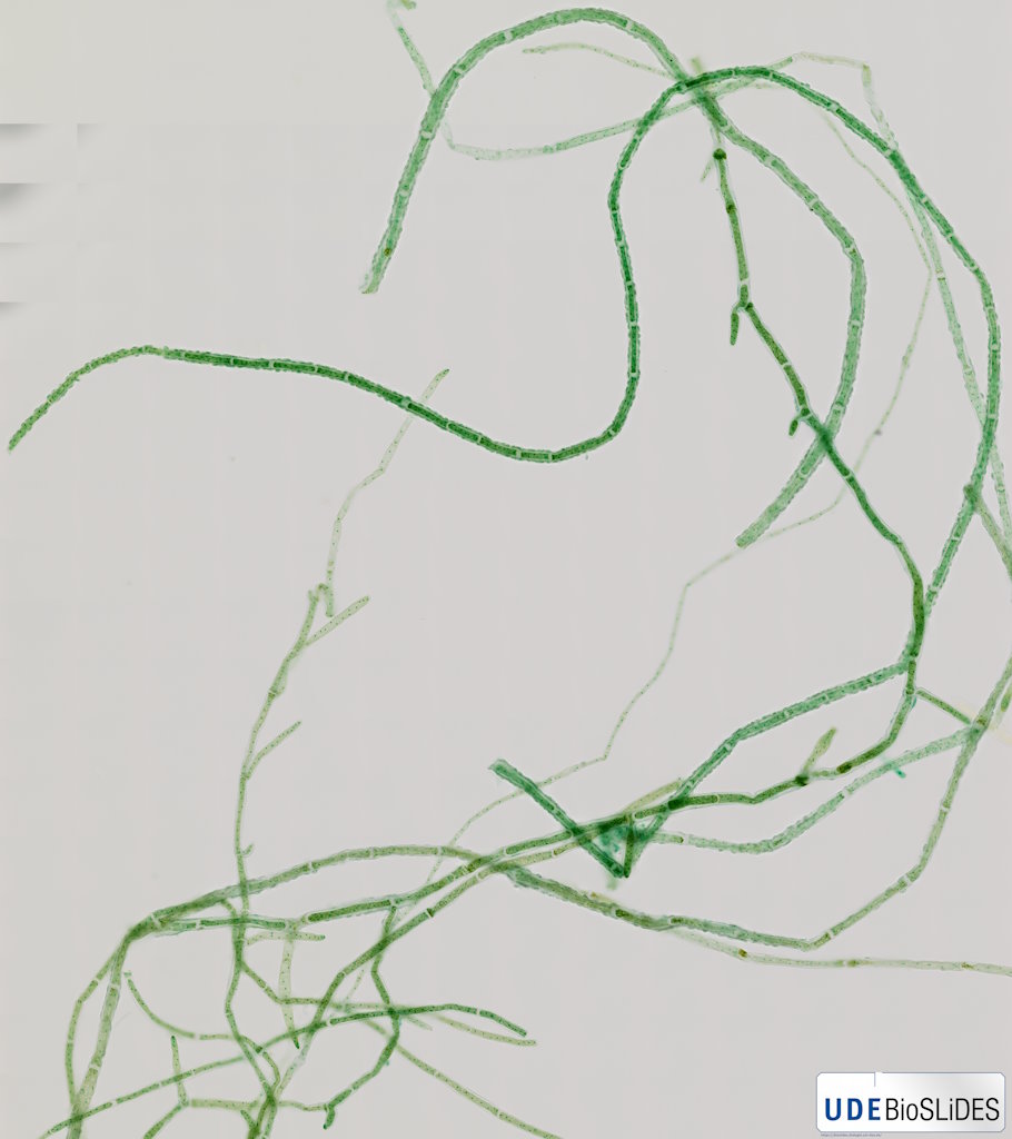


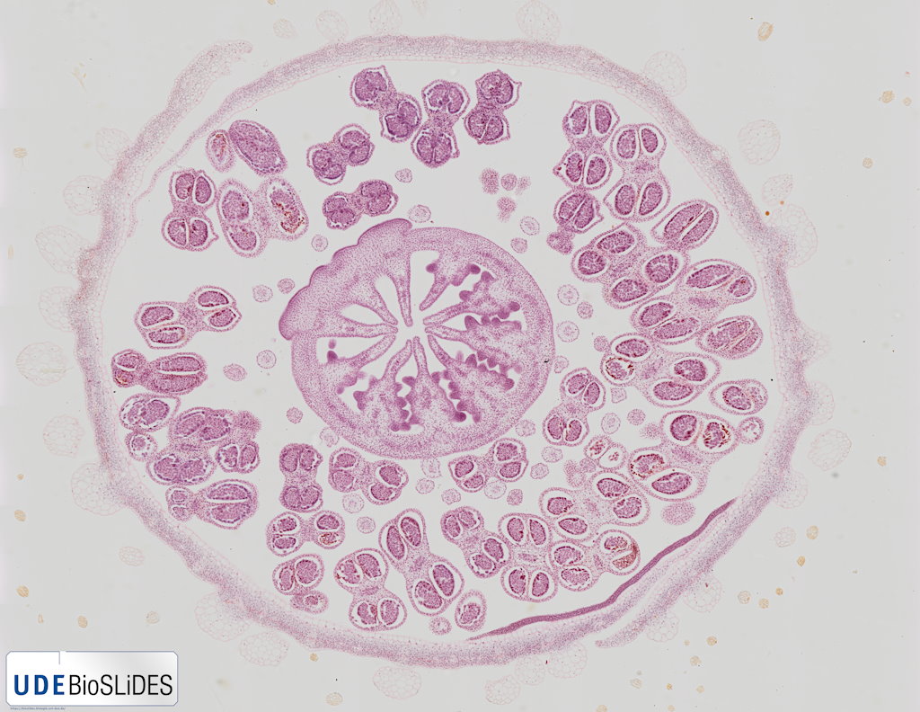


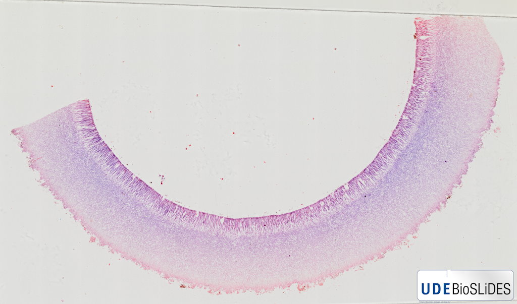
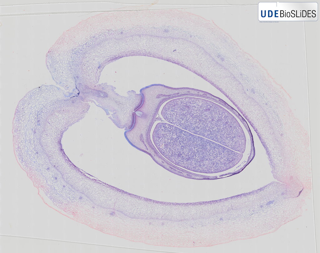
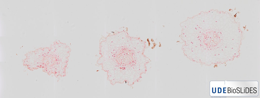


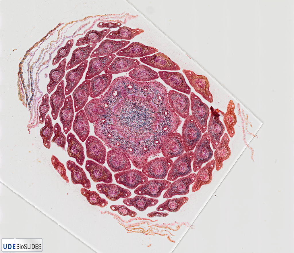
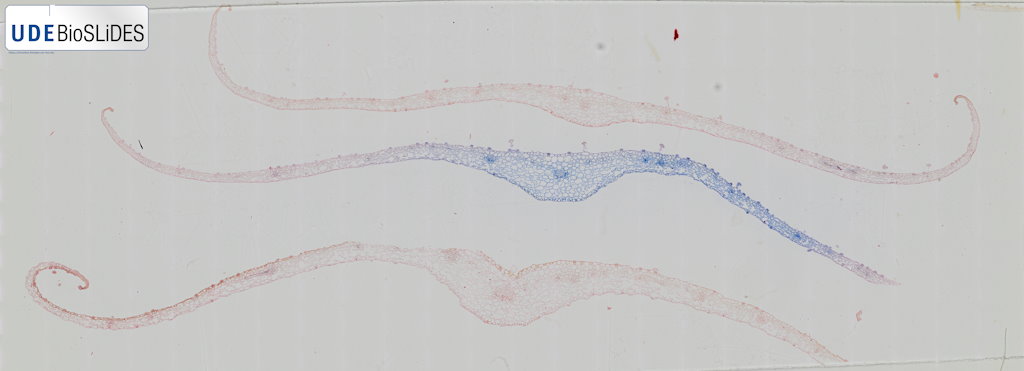


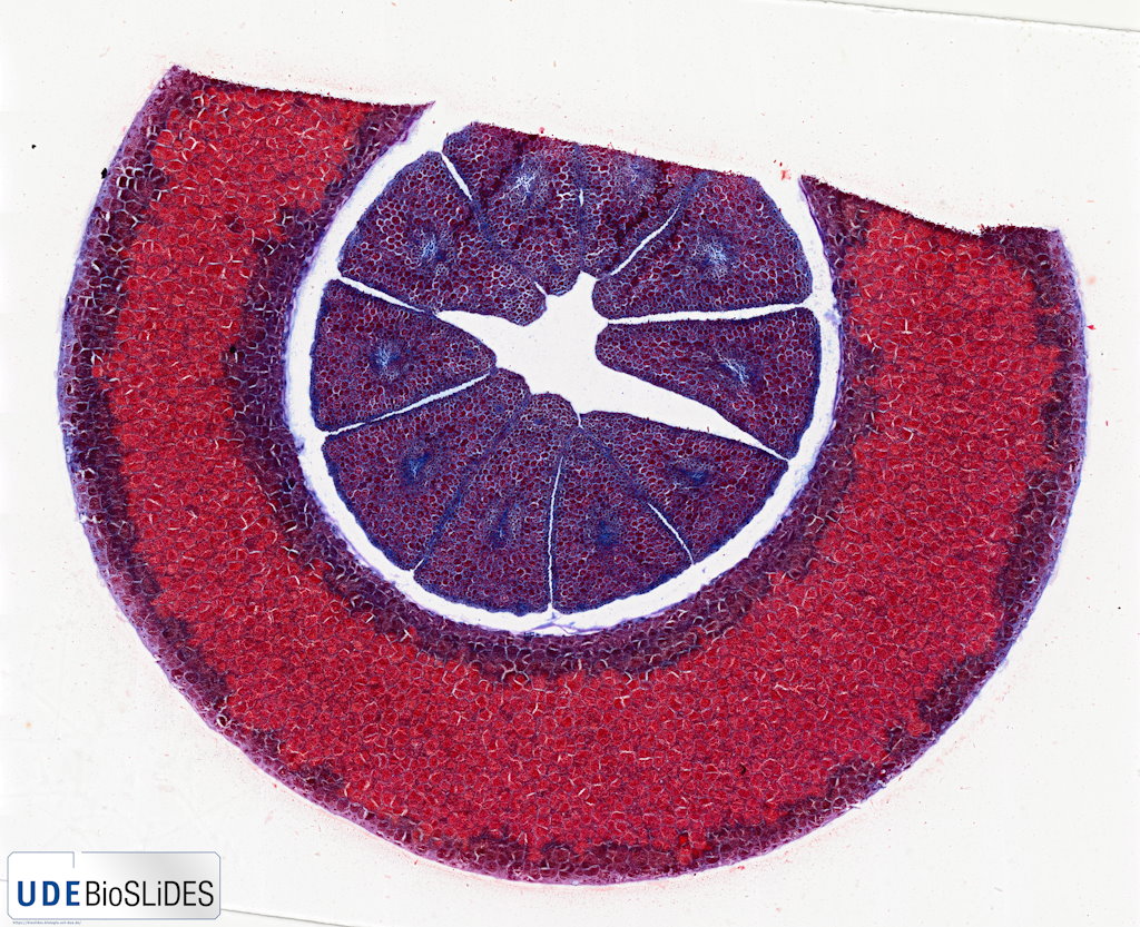
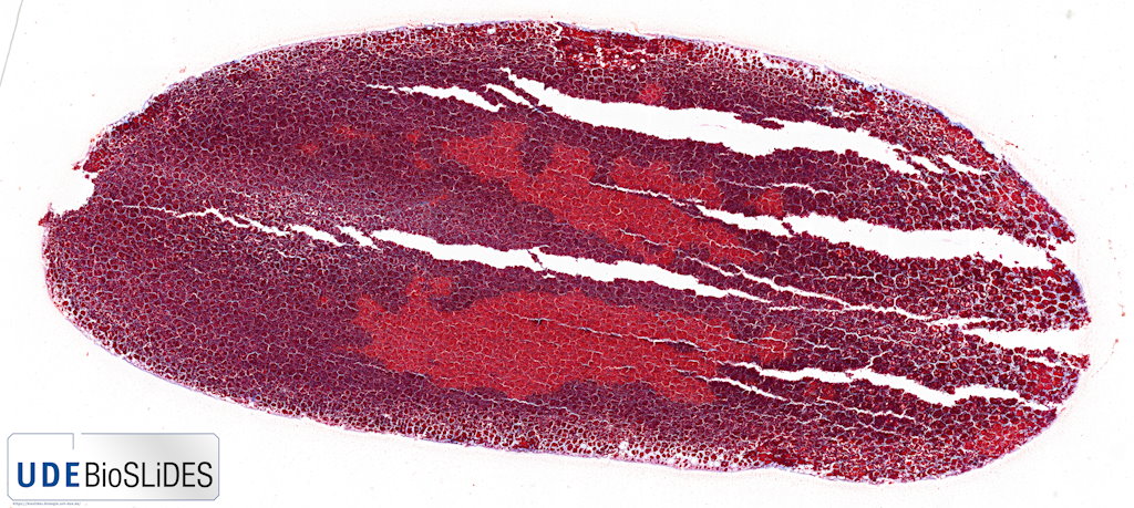
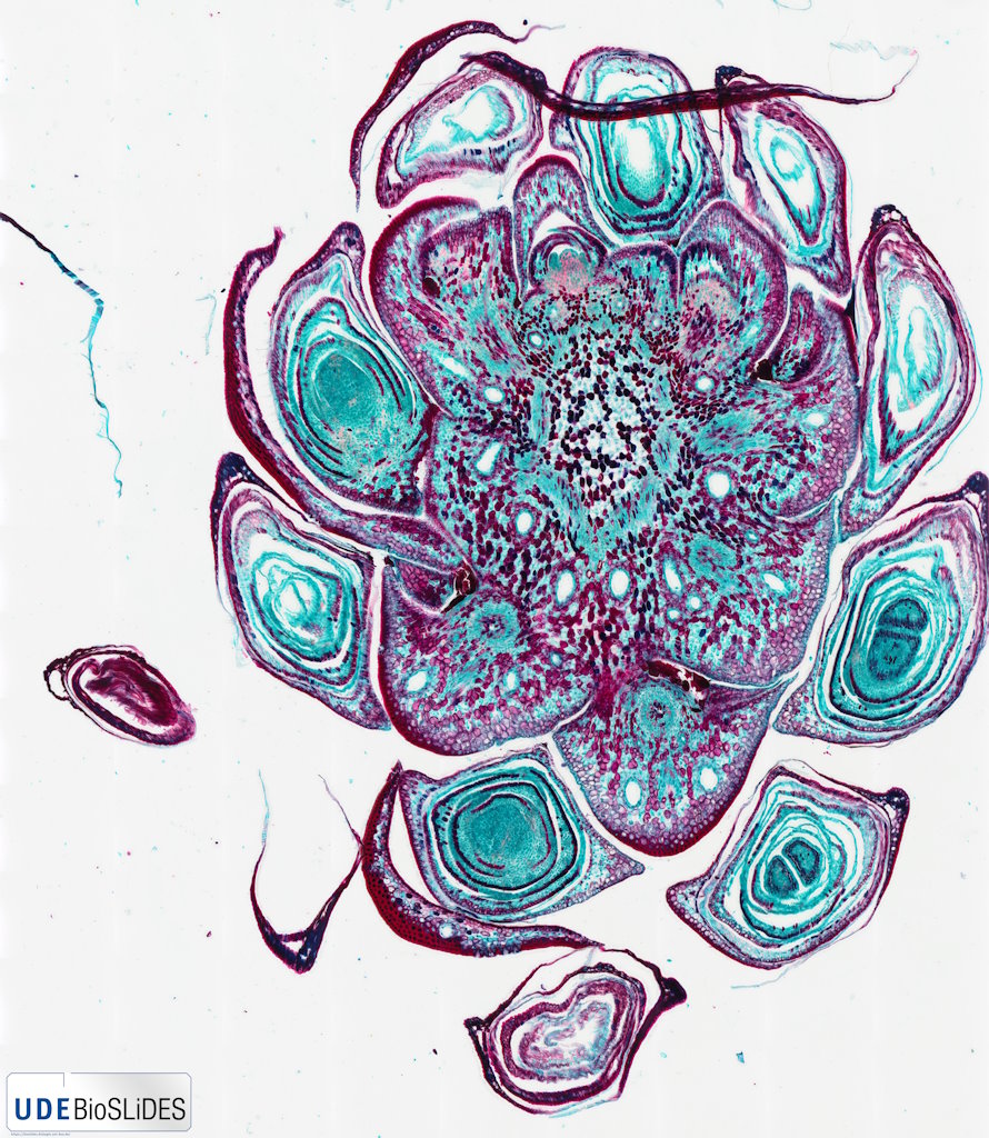

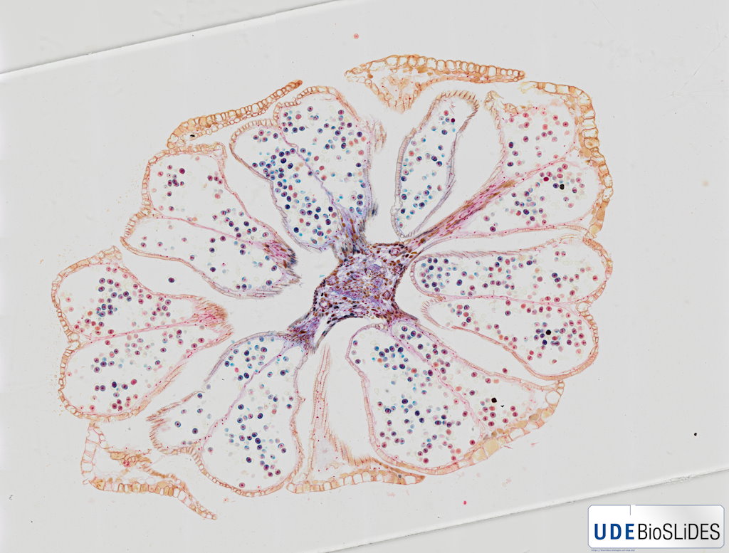
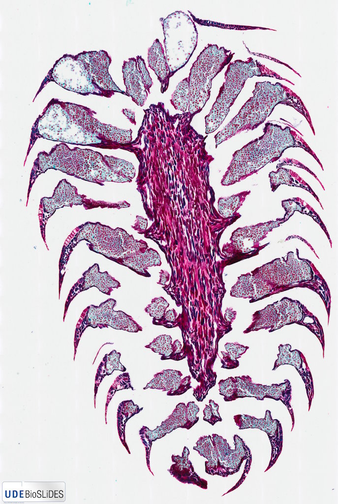


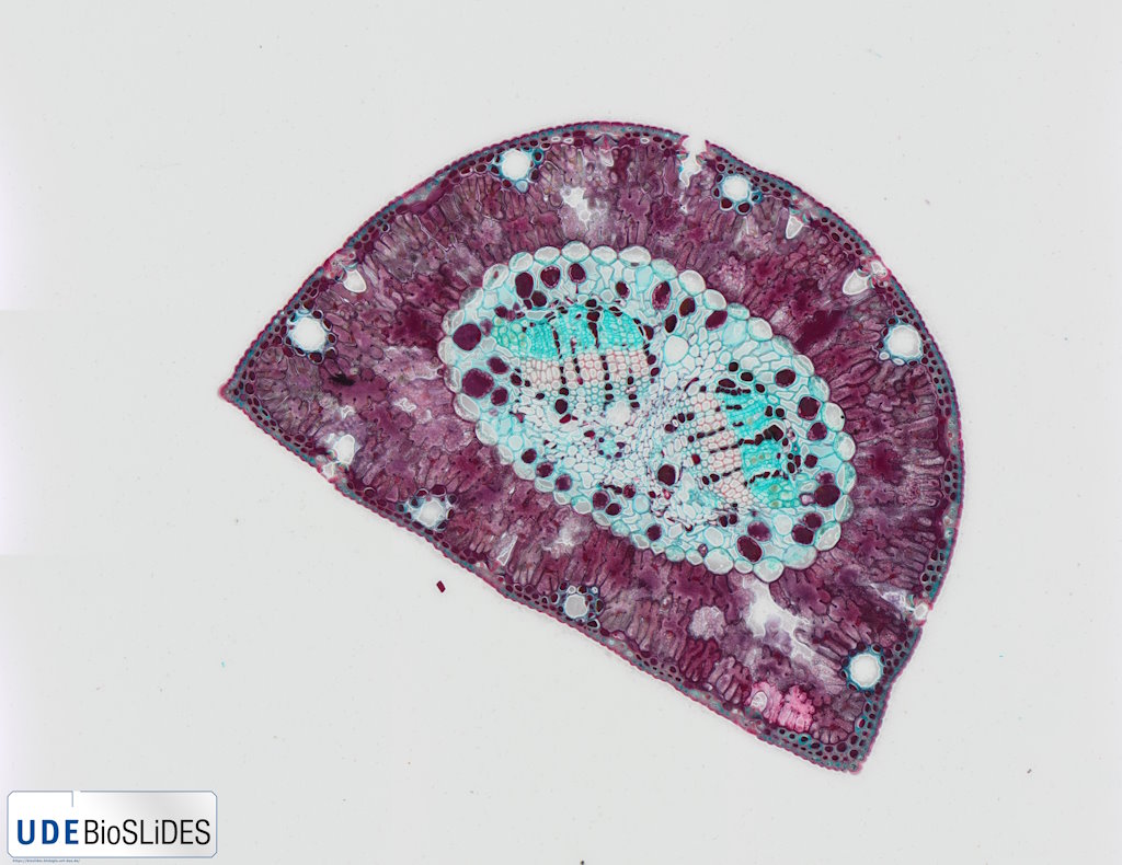
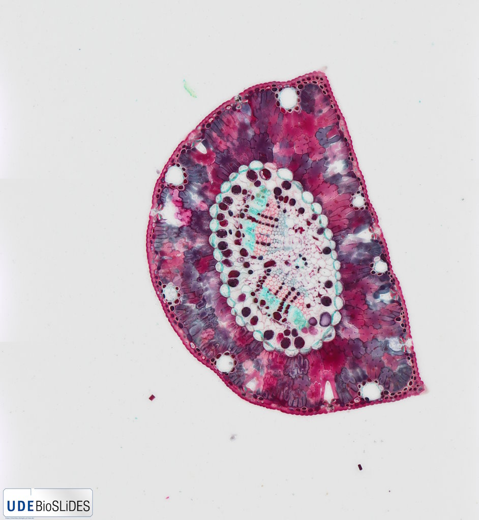


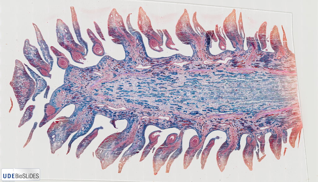
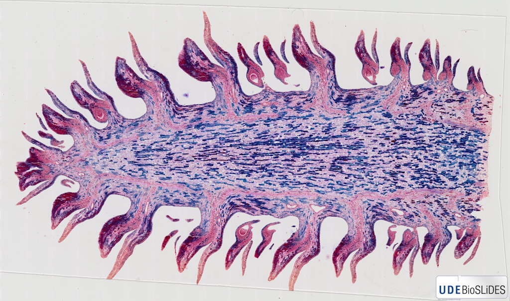
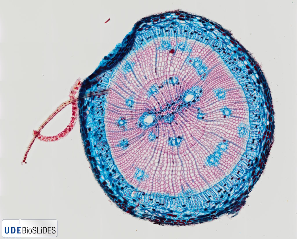


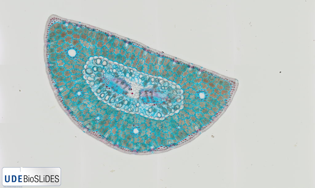


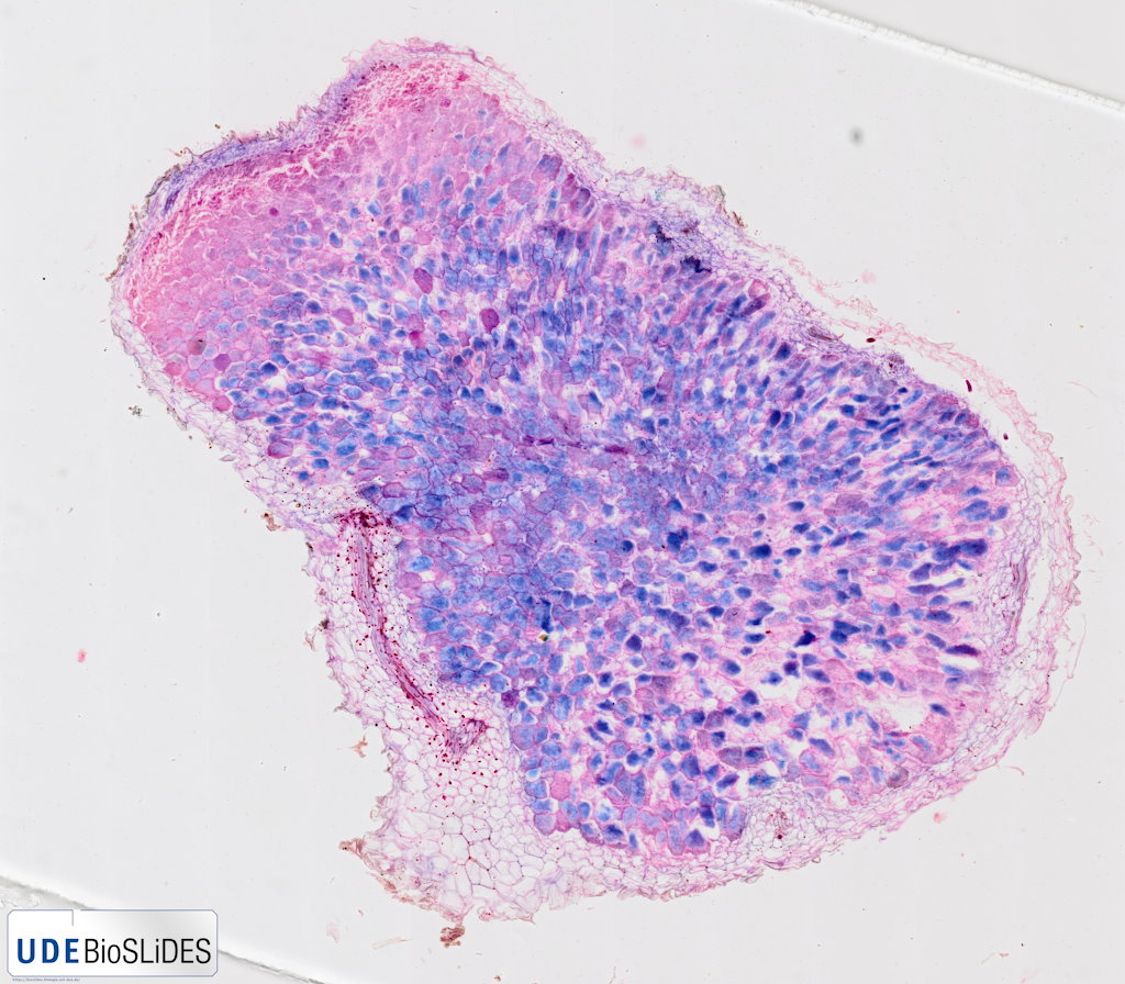
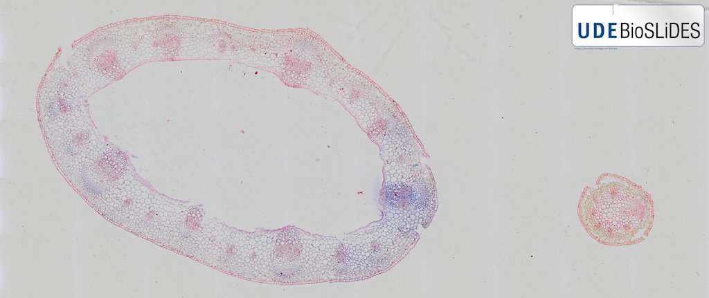
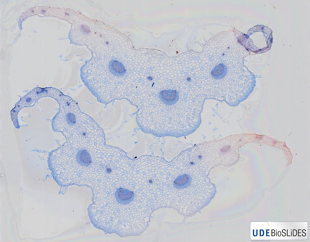


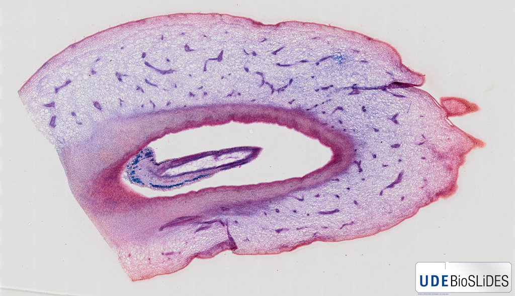
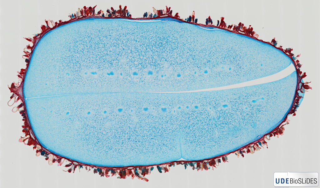


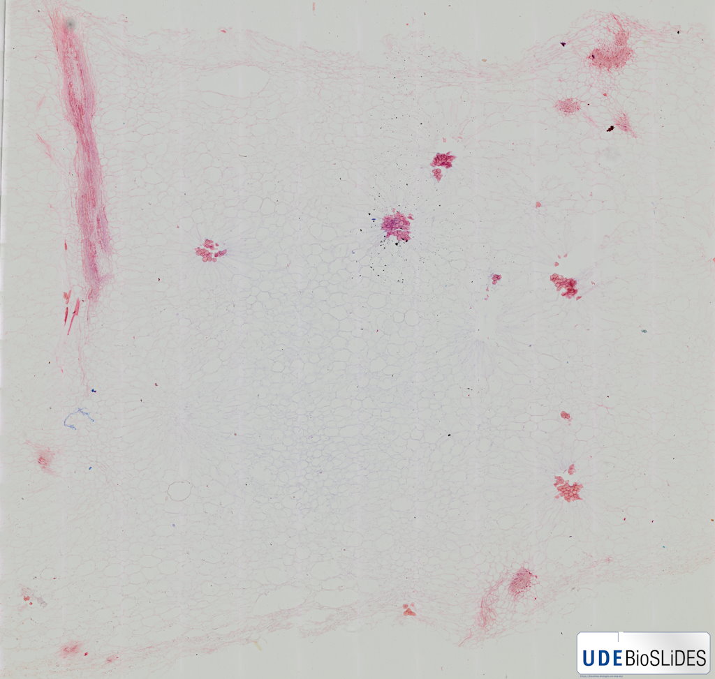


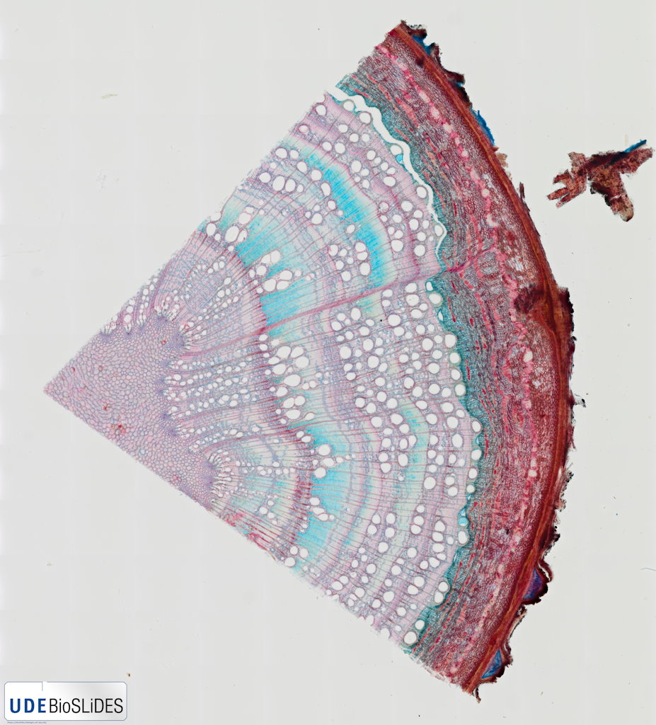


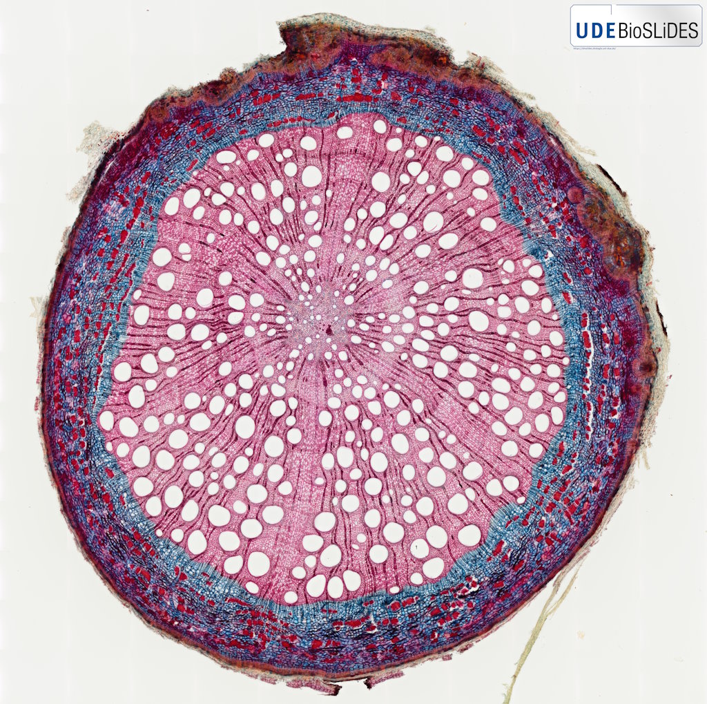


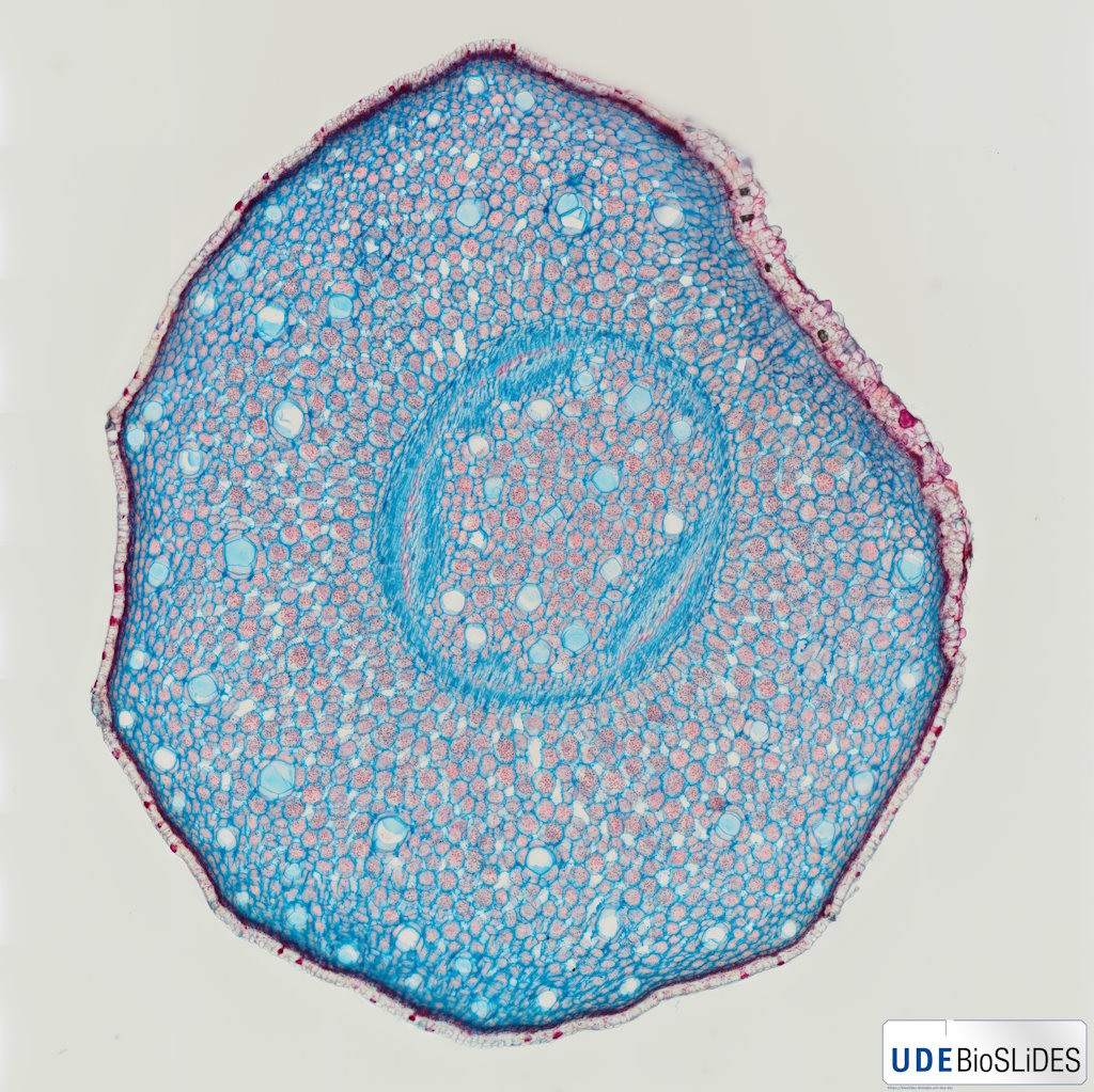
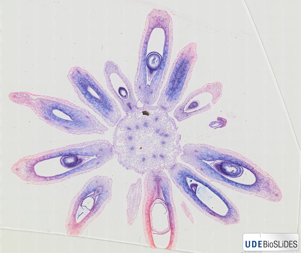
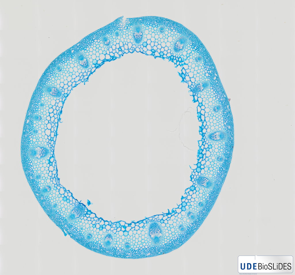
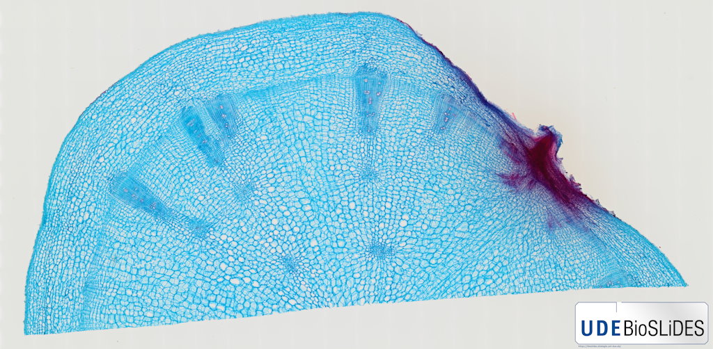


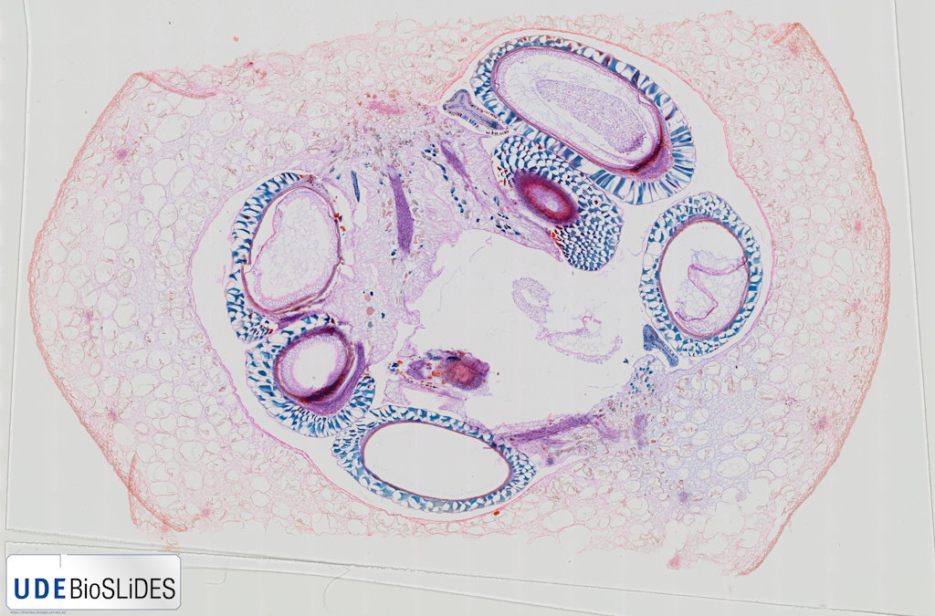


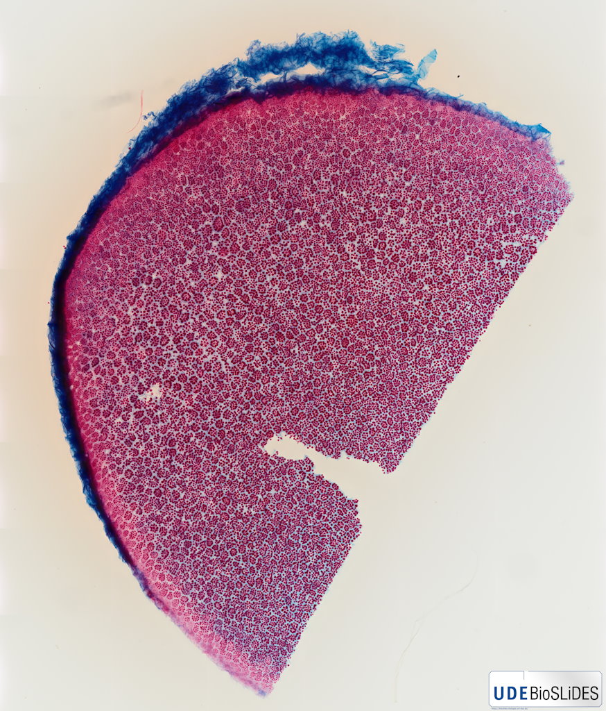


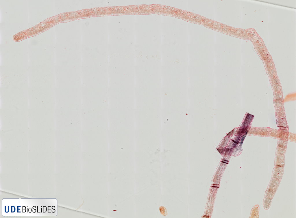


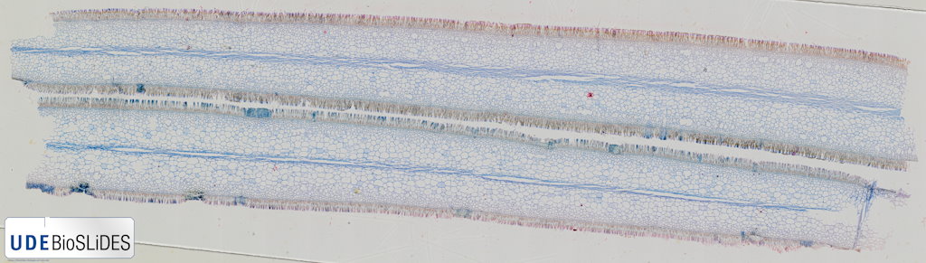


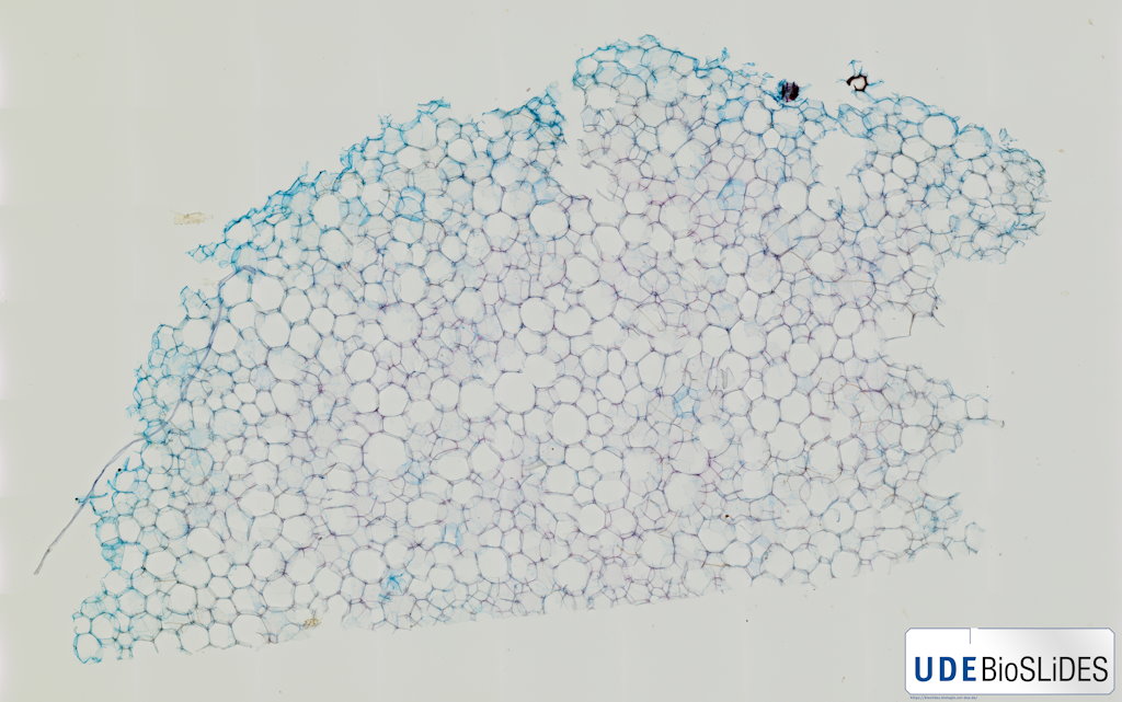
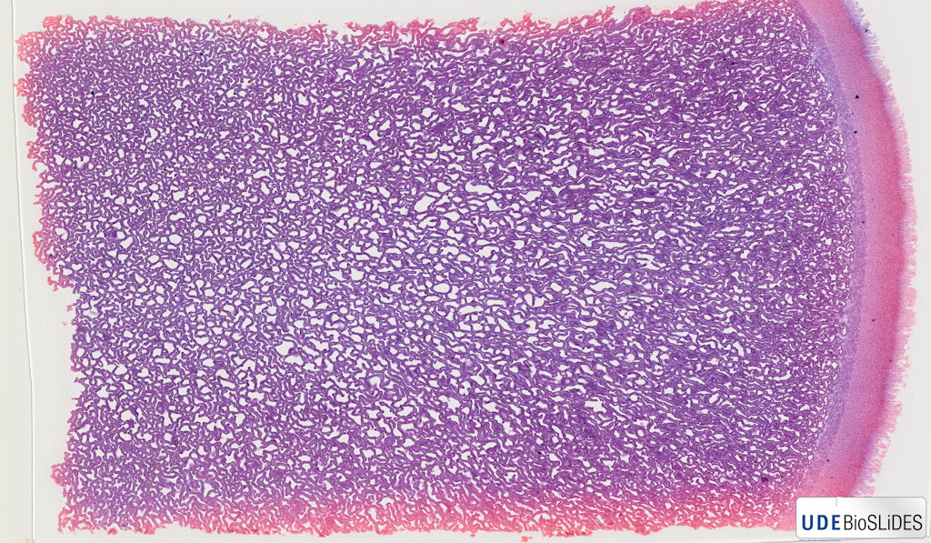

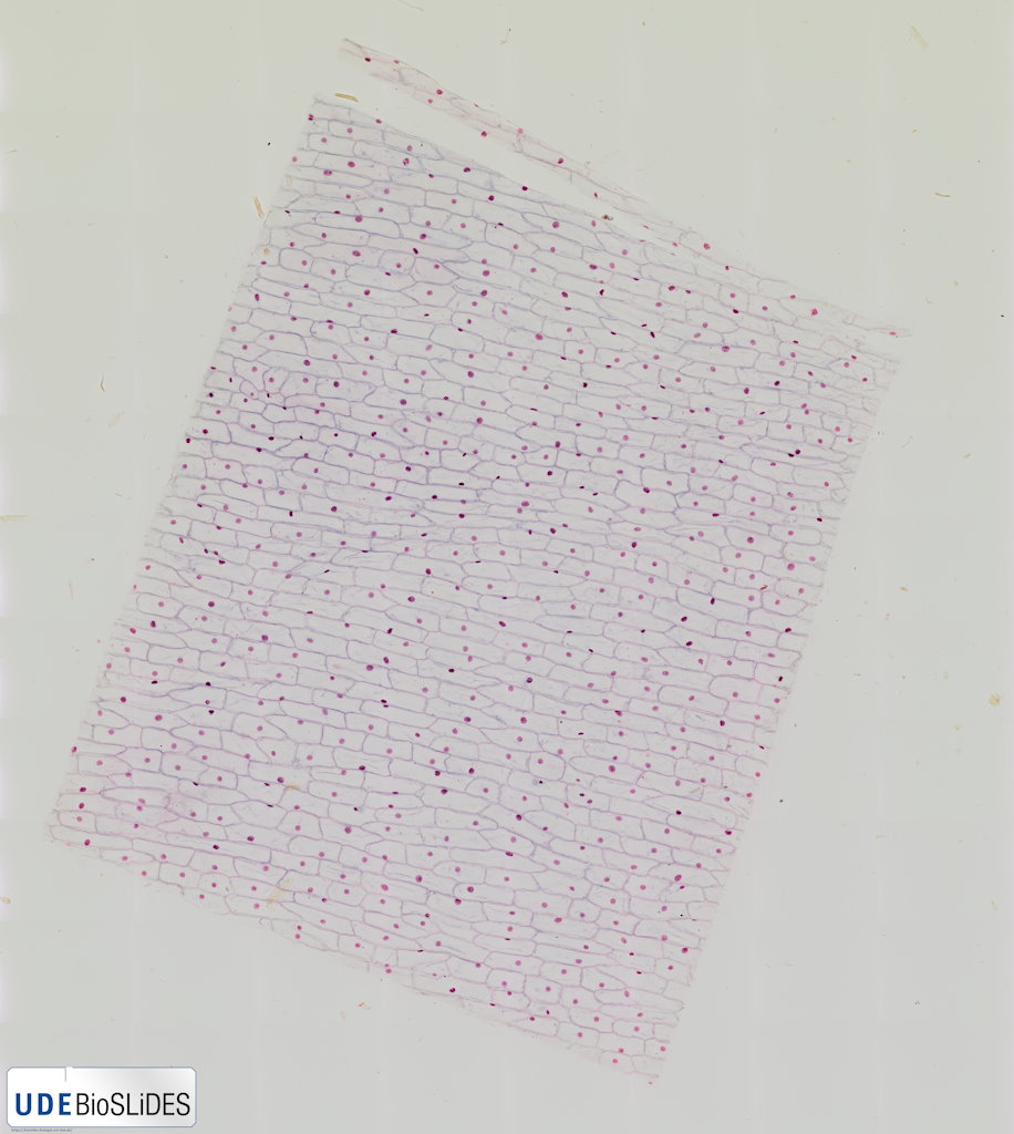
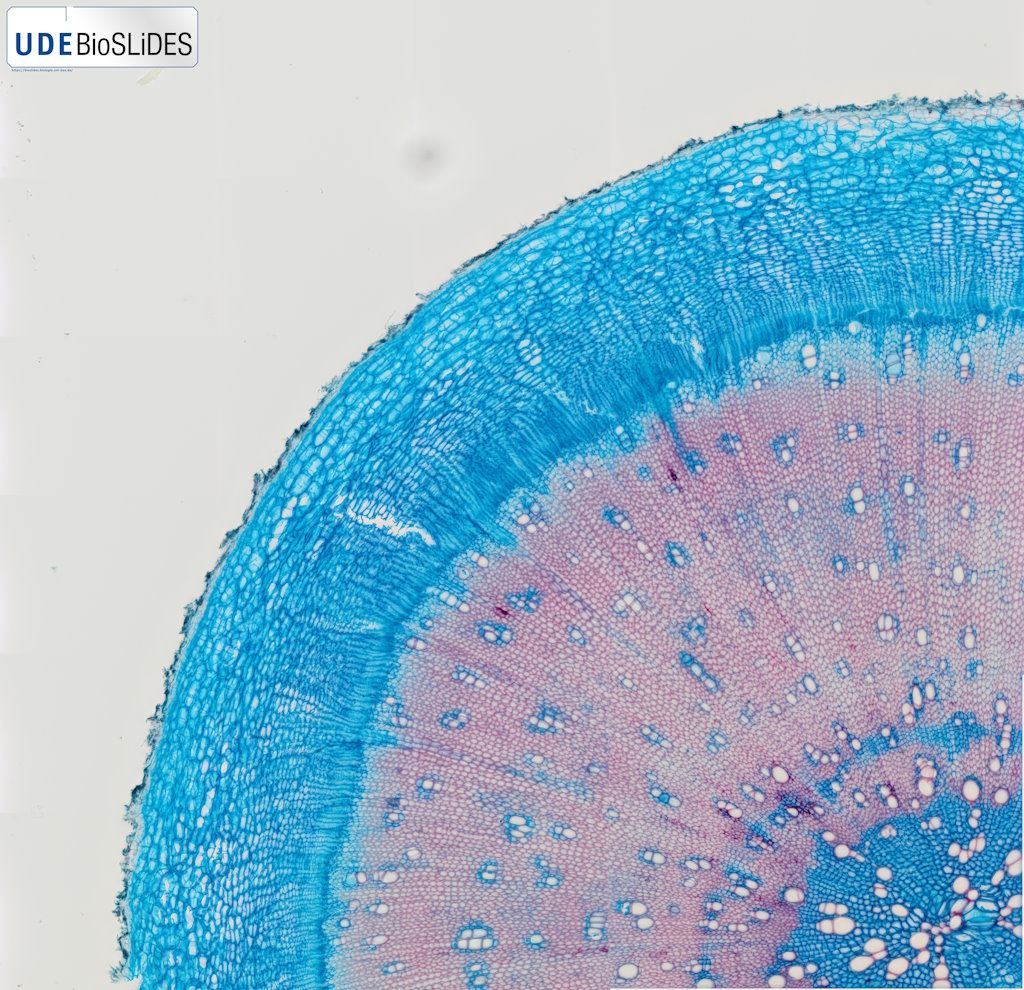

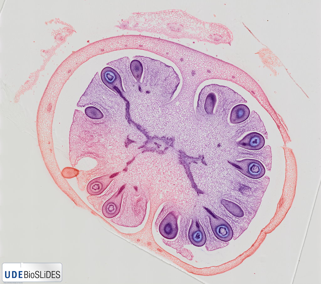
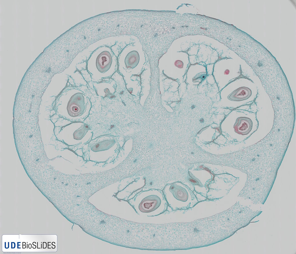
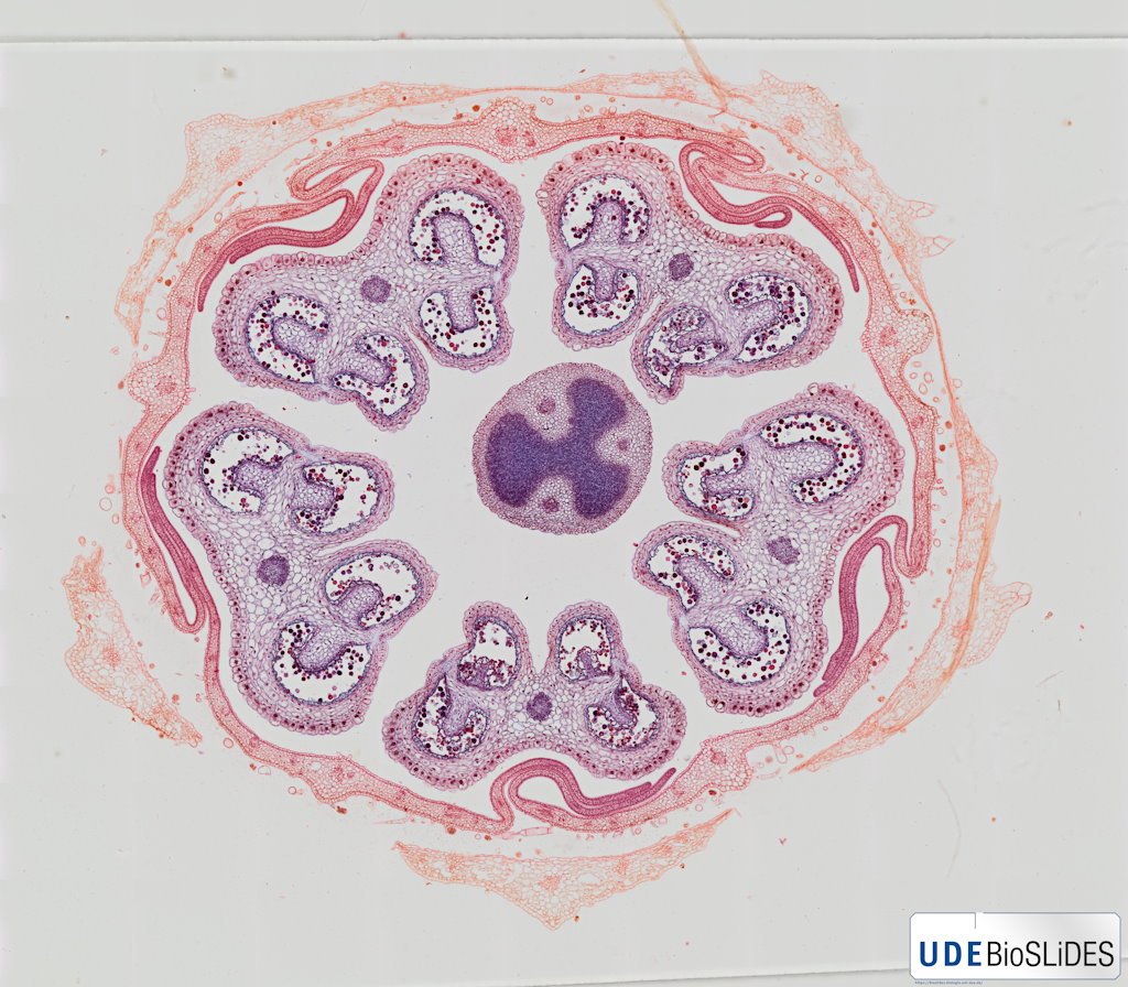

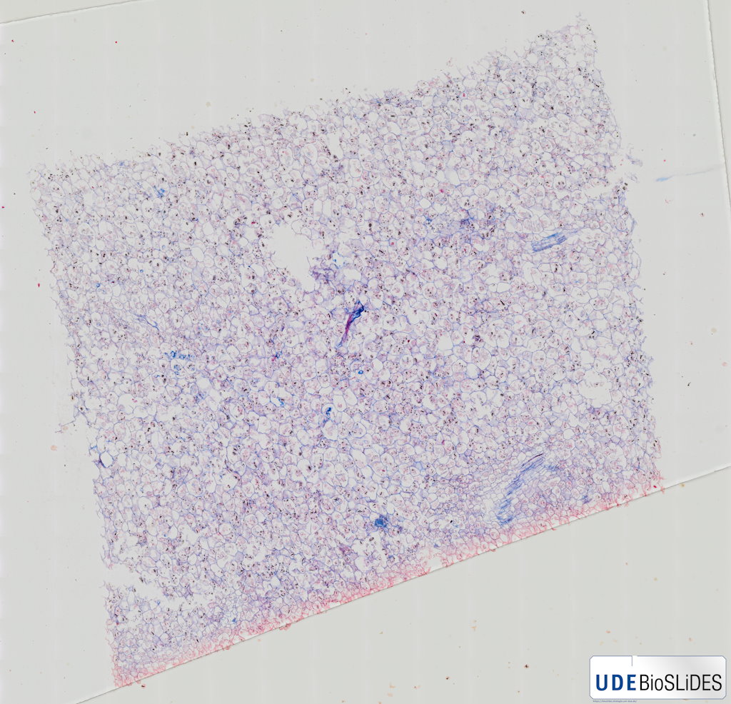
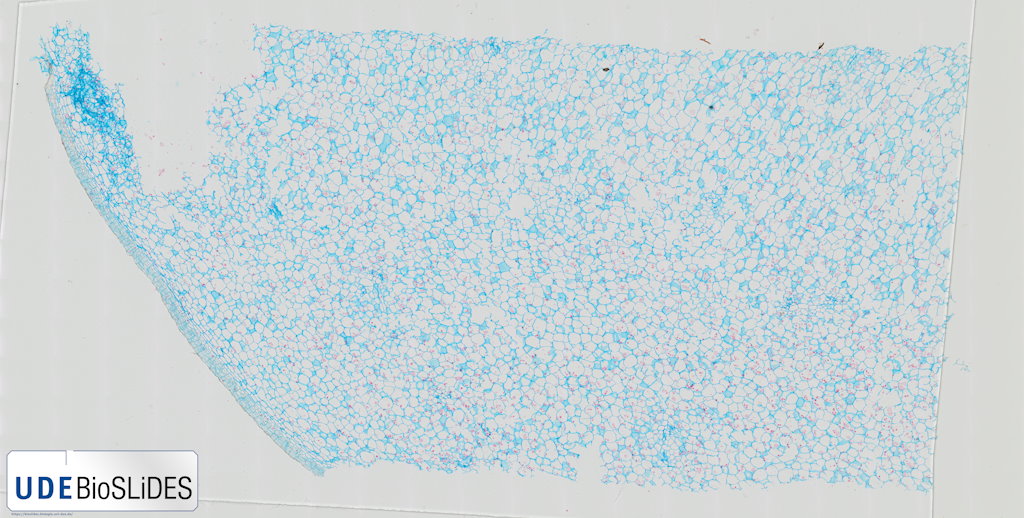


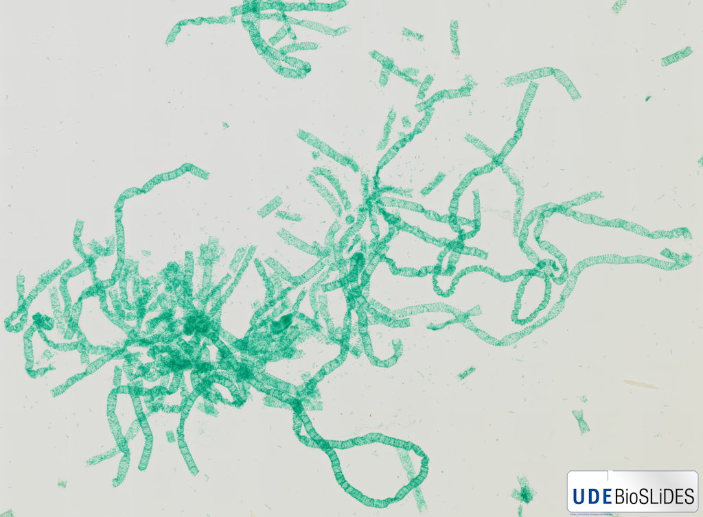

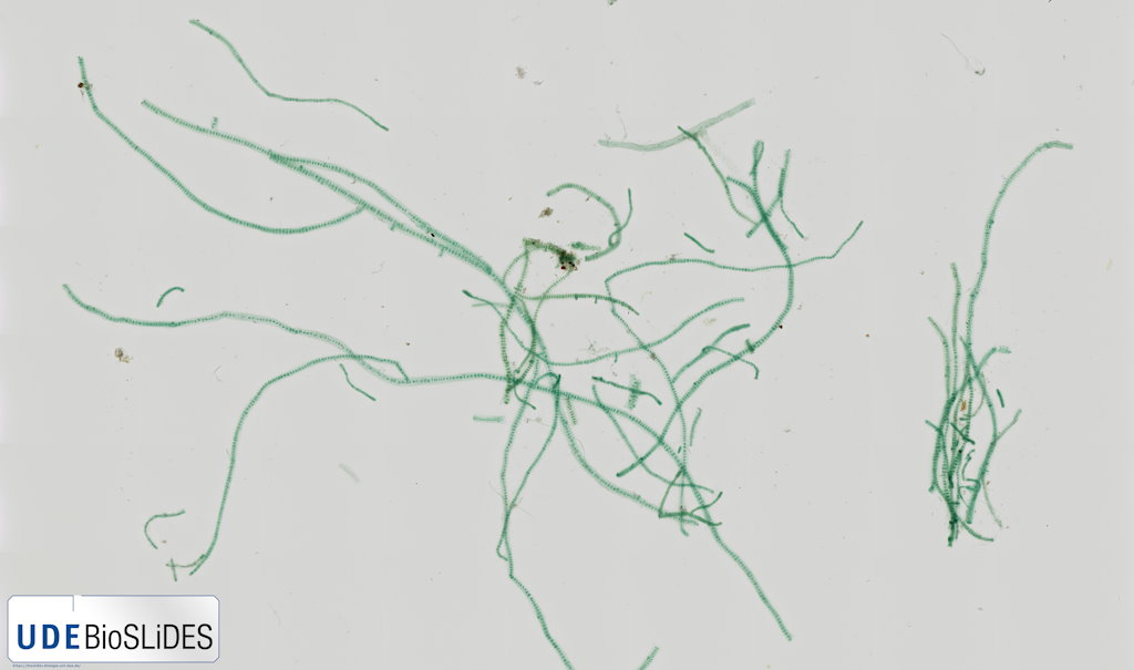


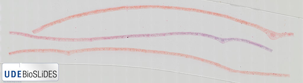


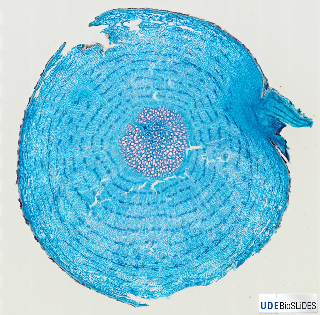
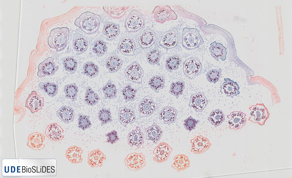
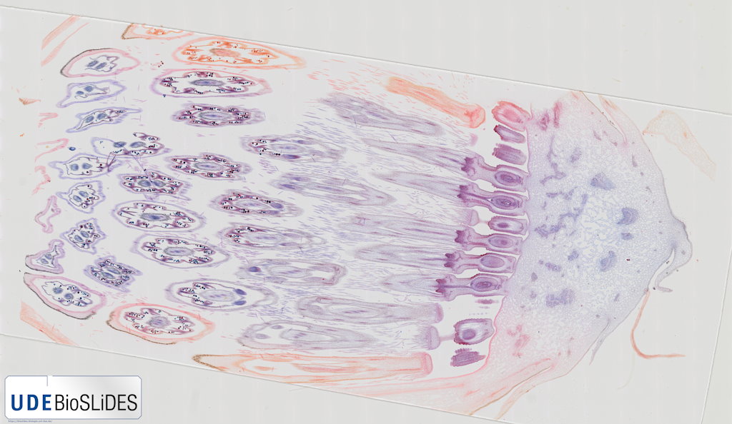


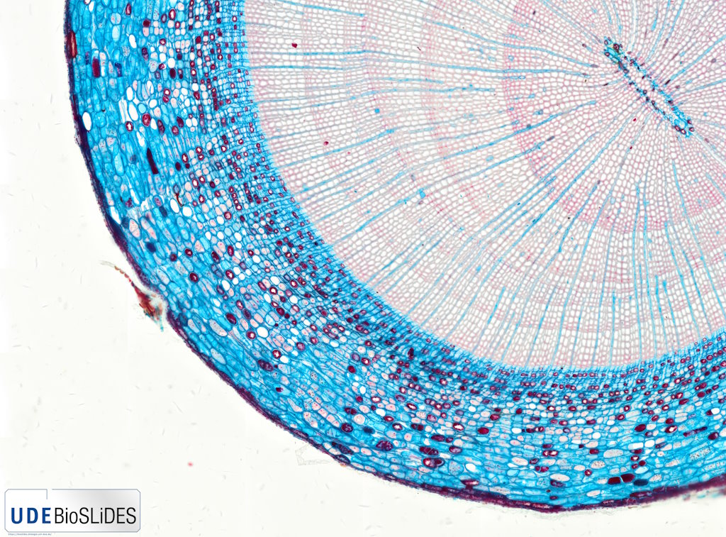
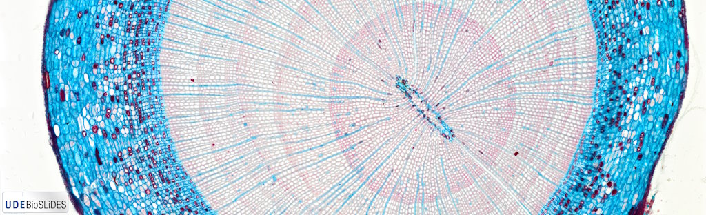


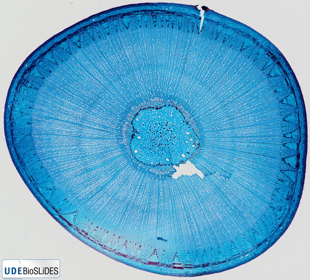


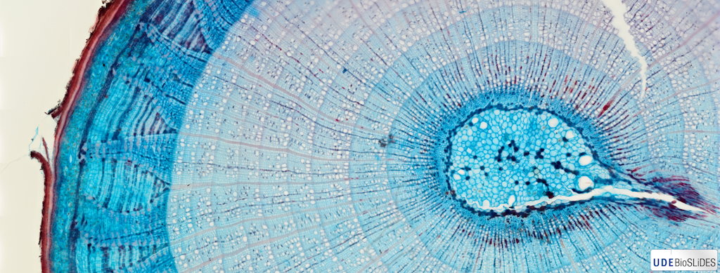
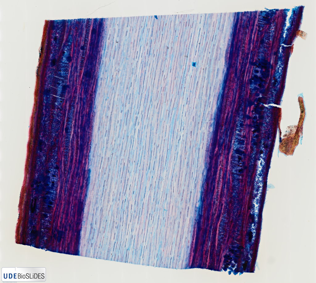
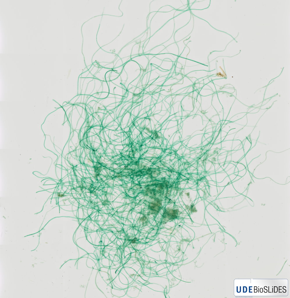


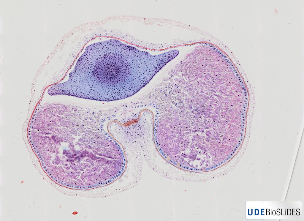



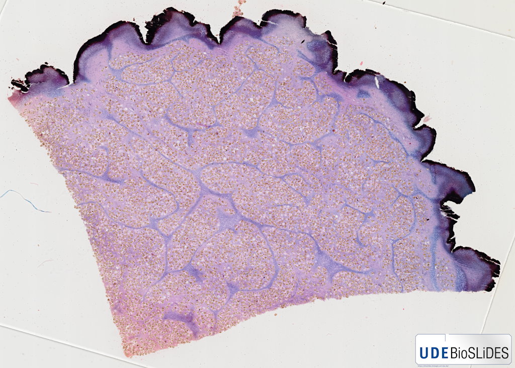
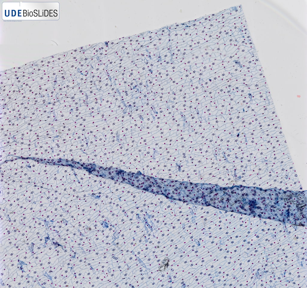


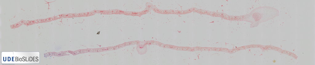
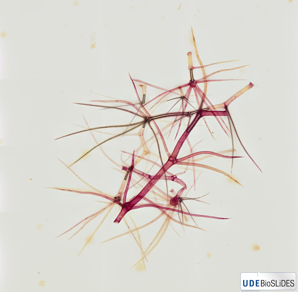

 unguiculata - Spross quer_Gallery.jpg)